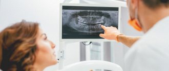
Modern dentistry cannot be imagined without accurate diagnostics. A set of measures to establish a diagnosis is indispensable in the treatment of teeth and gums; it plays a key role in implantation and prosthetics, and is also important during orthodontic treatment.
Diagnostics is carried out at all stages of preventive and therapeutic tasks. The dentist-therapist resorts to full diagnostic measures when the patient comes to the clinic. The specialist also uses operational diagnostic measures as an intermediate stage of treatment after installing a filling. Upon completion of prosthetic procedures, full diagnostics allows you to evaluate the results and correct them in a timely manner.
Dental diagnostics is, without exaggeration, 50% of the success of treatment and prevention.
CT scan
This method, which is based on the exponential law of attenuation of radiation for absorbing media, allows one to obtain many points with different optical densities. This is necessary to assess the condition of hard tissues that cannot be examined visually. During CT scanning, teeth, bone and skin surfaces act as a regulator of optical density.
An even more accurate method, compared to CT, is optical scanning (up to 5 microns, which is much more accurate than 300 microns in tomography). This technology is necessary for visualizing soft tissues, including the gingival surface, in a 3D scene of the dentofacial system.
Radiography
An image is taken of a small area of the jaw, more often to diagnose the condition of one tooth. It allows you to evaluate the characteristics of bone tissue, the tooth itself, the tissues around it and its root (gums, bone, ligaments).
X-rays in dentistry are used to:
- diagnose hidden caries, pulpitis, inflammation or cracks of the root canals;
- collect information about the dental system before implantation, prosthetics or monitor their results;
- assess the volume of bone tissue during sinus lift;
- check the quality of root canal treatment (must be filled without voids or pores);
- assess the structure of the dental system before and after orthodontic treatment;
- perform an accurate diagnosis if the patient’s complaints correspond to several diseases.
There are several types of X-rays used in dentistry.
Intraoral, dental. Performed to obtain an image of a single tooth or small area. The picture is taken using a radiovisiograph - a digital camera that captures an x-ray signal. The technology allows you to obtain high-resolution images: the doctor can see the condition of the canals, hard tissues, etc., and identify diseases at the initial stage. Radiation exposure with digital radiovisiography is minimal, which allows the method to be used for primary and intermediate diagnostics, when assessing the results and quality of treatment. The image is taken in a few minutes: a sensor is placed in the patient’s mouth on the side of the tooth being examined, and a pulsed radiation source is placed near the face. The image is available for viewing immediately.
Panoramic. It is performed to assess the structure and condition of the jaw as a whole, allows you to identify foci of inflammation or infection, pathology or disease of the TMJ, and assess the condition of the maxillary sinus and periodontium. Panoramic photographs are needed in preparation for implantation, prosthetics, and orthodontic treatment. They are performed using a special apparatus. The radiation dose is higher, but remains safe.
CT scan. It is used in planning sinus lifting, bone grafting, implantation, and other surgical interventions, and in diagnosing periodontal diseases. The photographs are taken in certain projections and used to build a three-dimensional model of the jaw. Computed tomography is very informative. The study provides information about the localization of inflammation (including intraosseous), the presence of tumor-like formations, their structure, size, features of the location of the root canals, etc.
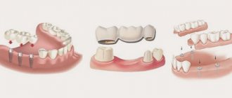
Scanning methods in prosthetic dentistry
There are several 3D scanning techniques, each with its own pros and cons. All of them are divided into two large groups:
- contact or mechanical;
- contactless or optical.
Optical scanners are more in demand in dentistry than mechanical ones, since they spend less time scanning and work more efficiently with large objects. In addition, they allow you to obtain a three-dimensional texture - the characteristic color of an object.
Study of muscle activity using a myograph
A myograph is a device that helps to study the bioelectrical activity of the muscular system. We use a Protens device, which is both a myograph and a tens device. That is, it allows not only to obtain data on the state of certain muscle groups in the facial area, but also to adjust activity and reduce tension.
The device is very miniature - it is fixed on the neck and has sensors that are located in the area of the temples and temporomandibular joints. To take data, you just need to move your lower jaw for a few minutes and just talk to the doctor. The device reads the data and transfers it to the computer, loading it into a special program.
Photogrammetry
This technology stands somewhat apart from optical scanning. It offers high performance because it measures the image, not the object. Other benefits include:
- high accuracy;
- strict methods of processing measurement results;
- the ability to study stationary and moving objects;
- remote nature of measurements.
Monophotogrammetry with structured illumination is most often used. The latter represents a certain known pattern projected onto an object. To build a three-dimensional model, they take the distortion when projecting onto the object.
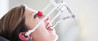
Removing bite parameters - axiography
Afterwards, the state of the bite is determined, that is, the position of the jaws relative to each other, and even the trajectories of movement of the lower jaw relative to the upper are read. To do this, we use the Proarc digital facebow – separately or in combination with the Proaxis optical axiograph.
The facebow consists of a bite fork and a central sensor. This device is less bulky than classic face bows. It is used both independently (to obtain digital data and use it with an electronic articulator) and in combination with a traditional arch and impression material for bite registration. In the second case, after taking impressions, plaster models of the patient’s jaws are created for further work with mechanical (classical) articulators.
On the left is a conventional articulator, on the right is a digital one
An optical axiograph is a hoop with a camera that is placed on the patient's head. The device is complemented by two small sensors with markings, which the patient bites on - this is how they are “attached” to the upper and lower jaws.
To set the initial position of the teeth, the digital facebow is first activated in the mouth. Then it is removed, and the doctor asks the patient to open and close his mouth, move his lower jaw in certain directions, say a short phrase, or even chew something. A camera directed along the face tracks the position of the sensors and, based on these movements, draws trajectories of movement of the teeth and articular heads.
For the patient, these “dashes and lines” are unclear, but they can tell the doctor a lot about the condition of the temporomandibular joint. And also, in combination with bite parameters, suggest a new correct position of the lower jaw. These data can be freely recalculated relative to any plane, taking into account the anatomical landmarks of the skull, and both virtual and real articulators can be programmed for further development of the shape of the prosthesis.
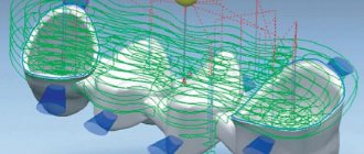
Optical strip scanning
This technology shows better results compared to the above. A special grid or stripes are projected onto the object. Based on their distortions, the relief of the scanned surface is determined. In this case, it is enough to determine the coordinates of the object’s surface using one camera and additional structured lighting. This approach is most often used in modern orthopedic dentistry.
Structured light can be different - laser, infrared, visible. The main condition is automatic recognition of these lines by software. The known distance between the projected stripes allows you to calculate the coordinates of the surface points. The more lines, the more accurate the result will be. The more cameras are used, the higher the scanning speed will be.
Scanning facial parameters using the Proface scanner
The final stage with the participation of the patient is scanning of facial parameters. There are two options here - use a facial scanner and/or a special application for Mac OS on the iPad.
The Proface scanner or program creates a three-dimensional model of the patient's appearance. For this purpose, a specially developed craniometric analysis algorithm is used (craniometry is a technique for measuring the skull). That is, many points of the skull are calculated - due to this, a copy of the patient’s face and all the bones of the head is transferred to the computer.
In addition, photometry is carried out - photographing the face is necessary to evaluate the result “live” and maintain a digital photo record.
An example of photometry is photographing the face from all angles before and after dental implantation with the installation of a fixed prosthesis immediately
We approach diagnostics wisely! We use the latest developments and artificial intelligence! We make an accurate diagnosis and find the best solution to your dental problems.
Sign up for a consultation
Types of scanners
- Stationary, or extraoral. They appeared earlier than the others and became widespread. The scanning field is 80 x 90 or 90 x 90 mm, which covers the plaster model of the entire dentition. Some modifications have limited scanning height. Accuracy – 5-15 microns.
- Intraoral. They appeared later, but are actively replacing their extraoral counterparts. Such scanners save the dentist’s time, since with their arrival in the dental laboratory there is no longer a need to stir the plaster and wait for it to harden, install pins, make collapsible models, and plaster them for scanning. However, intraoral scanners are still inferior to stationary scanners in accuracy (their indicator is from 30 microns).
- Facial. These devices stand apart from the previous ones and allow you to obtain a three-dimensional image of the face for planning the treatment of pulpitis, periodontitis, caries, taking into account aesthetic requirements.
The emergence of new diagnostic devices in orthopedic dentistry has simplified the doctor’s task and also reduced the time required to make a diagnosis and draw up a treatment regimen. Three-dimensional technologies have significantly increased the accuracy of therapeutic and orthopedic manipulations at all stages of exposure.
How complex digital diagnostics are carried out
The most popular area for digital diagnostics is complex dental implantation with the installation of a complete fixed bridge prosthesis 1-3 days after surgery. Getting teeth back so quickly is quite a difficult task. After all, as a rule, the remains of patients’ own teeth are in poor condition or missing, and the bite is disturbed. The implant surgeon and orthopedist need to re-create the balance of the entire jaw system. But it is impossible to do this right away; muscles and joints need time to rebuild. That is why there is a so-called adaptation period or adaptation prosthesis, through which orthopedists set the vectors of change in combination with the necessary balance of load on the implants. As a result, in addition to aesthetics, prostheses must normalize the position of the joint and the functioning of the facial muscles - and such a large-scale task cannot be solved without careful functional diagnostics and planning.
Comprehensive functional diagnostics is important not only when installing an adaptive fixed prosthesis, but especially when replacing prosthetics. A permanent prosthesis on implants lasts for decades, but only if it functions without overload.
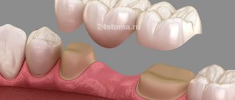
However, a detailed analysis of the initial situation is useful in any orthopedic treatment: when installing veneers, crowns, bridges, etc. Any intervention in the shape of the teeth shifts the balance - the chewing load is distributed slightly differently, the facial muscles are tensed differently, and the movement of the joints changes. Will these changes be for the better or will they lead to structural failure and complications? This can be done
Periodontal diagnostics
Diagnosis of periodontal diseases is aimed at assessing the condition of not only hard but also soft tissues surrounding the tooth. For this purpose, a comprehensive study is used, which consists of radiography and measurement of the depth of periodontal pockets. The choice of type of x-ray examination depends on the clinical situation.
Periodontal diagnostics is necessary for the main periodontal diseases - periodontitis and periodontal disease. It helps to predict with great accuracy the occurrence of tooth mobility at the stage when this problem can be prevented.
The modern diagnostic equipment of our clinic allows us to diagnose dental diseases effectively and safely. An accurate and timely diagnosis is the key to successful treatment!
How long does a comprehensive diagnosis take?
Diagnostics using digital equipment and preliminary analysis of the results last from 30 to 60 minutes. All data is instantly transferred to the computer memory with simultaneous calculation and construction of virtual models.
Depending on the manipulation, clinical picture and stage of treatment, the set of diagnostic data may vary. For example, during complex restoration of teeth on implants, data is collected gradually, since there is a period of adaptation, and the creation of a full-fledged axiography is advisable at the stage of installing a permanent prosthesis.
However, basic parameters are taken BEFORE treatment. Thanks to them, the doctor will show preliminary results on the computer screen: whether there are any abnormalities in the bite and functioning of the joint. Optimal treatment options can also be assessed. This is a fairly fundamental question for many patients. After all, the doctor sees the clinical picture and the permissible “corridor” of manipulations, and can say whether, for example, it will be necessary to remove or move teeth if we are talking about partial prosthetics. This is also important for those patients whose teeth have been removed a long time ago and need to gradually restore obvious disorders of the jaw joint and bite relationships.
Are there any disadvantages to digital diagnostics?
The disadvantages of high-precision digital solutions include their low prevalence due to the high cost of the equipment, as well as the time spent by the doctor. The technologies are relatively new and are just beginning to come into practice. There are still few doctors who have a full range of equipment and can carry out the analysis inside and out. Most are just learning new products and using only individual devices - for example, myographs or face arcs. That's why you won't see them in every clinic.
Smile-at-Once is so far the only clinic in Moscow with a full diagnostic range of devices and programs for planning orthopedic treatment and creating prosthetics.
Teleradiography of the skull
Diagnostic methods in dentistry include examination of the skull. Before orthodontic treatment, the doctor needs to see the angle of inclination of the teeth and the degree of their displacement. Using TRG, the doctor can examine in detail all parts of the skull from all sides by rotating the image. During teleradiography, the patient does not touch the device.
Teleradiography of the skull is performed in three projections: frontal, axial and lateral. Frontal projection is a basic photograph of the head, which shows the condition of the teeth, bones, jaws, symmetry or asymmetry, and inflammatory processes. The axial or chin projection helps the implantologist evaluate the structure of the nasal cavity, cheekbones, and sinuses of the upper jaw before implanting the upper teeth.
In Dental Guru clinics, the orthodontist performs teleradiography of the skull in a lateral projection to determine the bite, plan further strategies and tactics for correcting it. After the examination, diagnostic models will be made that will allow you to more accurately plan your treatment and even show the final result.
Development of a prosthesis model in the Proart program
All information obtained during complex functional diagnostics is loaded into a single virtual space and combined into a common model. This may include a CT image of the jaw system (computed tomography data), an axiogram, scans of the face and oral cavity from the inside, virtual prototypes of prostheses, cephalometric analysis data (that is, parameters of the facial skeleton) and much more.
It turns out to be a kind of virtual double of the patient. But, unlike a real person, we can look “under the skin” without any harm and see how the teeth and jaws move and interact, where there are problems and what will change after installing the prosthesis.
We also use the additional Prosthetics software module, which is designed specifically for the orthopedic field. With its help, we can flexibly plan treatment and send complete data about the future prosthesis to our laboratory.
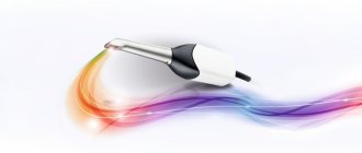
Diagnocam (DIAGNOСAM) - laser diagnostics of caries without irradiation
The compact laser diagnostic device DIAGNOCAM was developed by our business partners - the German company KaVo.
Using Diagnocam, dentists can perform the following in real time without x-rays:
- Visual diagnosis of caries at the earliest stages;
- Effective diagnosis of supragingival tooth surfaces and secondary caries;
- Detection of enamel cracks without the use of a dental microscope, etc.
Diagnosticam can be used to diagnose caries in weakened patients with concomitant chronic diseases and in pregnant women.
Spot X-ray
An X-ray picture of a separate tooth is most often necessary in order to monitor the treatment process when an accurate diagnosis has already been established.
In our clinic, targeted images are performed using a radiovisiograph - a modern digital X-ray machine, which differs from its predecessors in a much lower radiation dose and the ability to display the obtained data directly on the monitor screen. This allows X-ray examinations to be performed as many times as necessary (up to 60 times a year).
The results of the study are saved in the patient’s electronic record - this allows you to monitor the dynamics of treatment of the disease, and they can also be copied to any digital medium.
The study is carried out as follows: a miniature sensor is placed in the patient's mouth, which looks like a tiny plate and is connected to a computer using a wire. The research process takes only a few seconds. The data instantly appears on the monitor screen, and if necessary, you can immediately take a second photo. Thanks to computer processing, it is possible to enlarge or highlight individual fragments of the image and determine the density of bone tissue.
How much does such research cost?
Equipment for functional computer diagnostics of the jaw system is quite expensive. This also increases the price for patients - clinics charge a lot of money for individual procedures even without further treatment. This can also be attributed to the disadvantages of technology. Accordingly, many doctors and laboratories prefer to work with more familiar and accessible mechanical equipment.
In our center, such diagnostics are included in the cost of comprehensive dental restoration on implants, when installing veneers, as well as extended dental bridges. There is no need to pay anything separately - we make sure that our patients receive a guaranteed treatment result and are satisfied with the new prostheses, both in terms of aesthetics and their functional side. If you need functional diagnostics as a separate service, then its cost varies from 15,0000 to 30,000 rubles, depending on the amount of data required.