Chapter 8. SURFACE FORMATIONS ON TEETH
8.1. CUTICLE. PELLICULA. PLAQUE
Under physiological conditions, a cuticle and pellicle are formed on the surface of the enamel.
Cuticle
Cuticle
in the form of a thin shell covers the surface of the enamel of teeth after their eruption.
It consists of two layers, referred to as the primary and secondary cuticle. The primary cuticle
is an internal thin (about 0.5-1.5 µm) homogeneous layer of glycoproteins, which is the last secretory product of enameloblasts.
The secondary cuticle
is formed by the outer (about 10 µm) layer of the reduced epithelium of the enamel organ.
During chewing, the cuticle is erased and may partially remain on the proximal surfaces.
Unlike the cuticle, tooth pellicle and dental plaque can form on the surface of the enamel throughout life.
Pellicle
Tooth enamel pellicle
- is a thin protein layer with a thickness of 0.5 to 1 nm, which is formed from salivary glycoproteins on the surface of the tooth. It is a structureless formation that does not contain bacteria. The pellicle is formed within 20-30 minutes after eating, and its formation begins with the adsorption of specific salivary proteins on enamel hydroxyapatites.
The pellicle has selective permeability and ensures the diffusion of ions into the surface layer of enamel, and also protects tooth enamel from exposure to chemical agents. In this process
glycosylated proteins rich in proline are involved. The dental pellicle not only regulates the processes of mineralization and demineralization of enamel, but also controls the composition of the microbial flora. After mechanical cleaning of the teeth, the pellicle is restored to the enamel surface within several hours.
The formation of pellicles involves amino sugars, monosaccharides, acidic and glycosylated proteins rich in proline, lactoferrin, lactoperoxidase, cystatin SAIII and, in lower concentrations, lysozyme and histatin-5. These proteins have hydroxyl or carboxyl groups in their amino acid composition, which allows them to bind calcium ions. Acidic proteins rich in proline, which are part of the tooth pellicle, bind to the protein statherin and prevent the interaction of this protein with enamel hydroxyapatite at acidic pH values. Ionic bonds and hydrophobic interactions are formed between the enamel surface and the deposited proteins. They thereby delay the demineralization of tooth enamel and inhibit excessive mineral deposition. It has been established that histatin-1, which is a powerful inhibitor of the growth of hydroxyapatite crystals, is also involved in the formation of acquired tooth pellicle.
Nature of formations
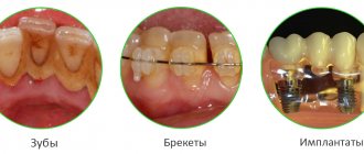
All surface formations on teeth are divided into physiological, determined by their structure, and pathological.
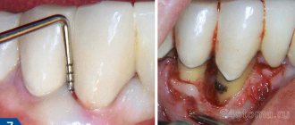
The latter are formed due to bacteria accumulating in the oral cavity, as well as food debris. This is the result of improper oral care and neglect of preventive measures to improve dental health.
Pathological deposits are of particular interest, since physiological deposits, in most cases, do not provoke dental diseases.
Such deposits develop gradually: first they take the form of a soft coating, and then, as the crystals grow, they turn into hardened masses and stones.
Features of the structure of the enamel of baby teeth
The main distinguishing feature of the enamel layer of children's teeth is that it is less durable and also much thinner than the enamel of permanent teeth. This is explained by the lower content of mineral compounds in teeth in relation to water and organic substances. Considering these features, if you examine baby teeth and their enamel layer under a microscope, you will notice the following differences:
- Due to the fact that the service life, as well as periods of mineralization and the tendency towards this process are shorter, the Retzius lines are much less pronounced in the structure of primary dental units.
- If in permanent teeth enamel prisms are located apically, then in milk teeth their direction is completely different, they are located horizontally.
- In children's primary teeth, the final enamel layer is much less pronounced; prisms are clearly visible on its surface, while its structure is much more porous, with microscopic cracks present.
Under the influence of each of these features, tooth enamel in children is more susceptible to wear and damage. For this reason, children develop caries much more often and it progresses faster, which is why it is important to visit the dentist regularly and have their teeth treated in a timely manner.
Strengthening baby teeth enamel
As was said earlier in relation to baby teeth, their enamel is more vulnerable. To protect it, saving the child from premature loss of dental units and problems in the future, doctors perform the following actions to provide temporary protection:
- Fluoridation involves treating teeth with special fluoride-based compounds; it is recommended to repeat this procedure 2-3 times a year.
- Fissure sealing - the dentist performs the procedure of filling the recesses and grooves of chewing teeth with temporary filling material, protecting the dental structures from the negative effects of harmful microorganisms and other unfavorable factors.
- Application gels and preventive mouth guards for teeth - the method is based on enriching the enamel layer with useful components (fluorine, calcium, vitamins) through the use of special products.
How tartar and other deposits are removed:
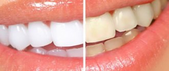
Professional teeth cleaning in Tula is performed using the Air Flow device. This procedure helps to avoid problems such as plaque and tartar, reduces the risk of caries, pulpitis, periodontitis and other diseases of the teeth and gums. The procedure returns teeth to their true color, which is often perceived as teeth whitening. The dentist will clean your teeth of plaque and remove tartar, which inevitably accumulates on our teeth and which cannot be removed at home. Most dental diseases begin with plaque and tartar.
When cleaning with the Air Flow device, dental plaque is “knocked down” using a solution of water with abrasive powder, which is supplied from the tip of the device in a thin stream under very high pressure.
What should you do after cleaning?
Protect your teeth from disease with enamel fluoridation and fissure sealants. Special preparations are applied to the teeth to make the tooth enamel stronger and prevent the formation of caries. We can also make custom mouth guards for you, which you just need to wear at night. A special gel in a mouth guard will saturate your teeth with useful microelements and make them stronger. For home use, we recommend the unique DENTAL RESOURSE enamel remineralization system, which includes a deep fluoridation procedure in the doctor's office and a fluoride rinse, which is dispensed at home.
Type protozoa
Protozoa are single-celled organisms. Of course, there can be no talk of any tissues or organs - but this does not mean at all that protozoa do not have processes of gas exchange, excretion, and transport of nutrients - they all occur, but in a special way.
In protozoa, one cell performs all the functions of the whole organism, so the cells have a complex structure. Cells have all the basic vital functions: irritability, reproduction, metabolism.
The structure of a protozoan cell
The shape of the protozoan cell is constant, surrounded by a pellicle - an outer, compacted layer of cytoplasm that maintains a constant shape. In some protozoa (amoeba, in the figure above), the pellicle is absent and the cell shape is inconsistent, spreading.
The protozoan cell is eukaryotic - it has a formed nucleus, separated by a nuclear membrane from the cytoplasm. In the cytoplasm of many protozoa, ectoplasm (the peripheral outer, denser layer of the cytoplasm) and endoplasm (the inner granular layer of the cytoplasm, less dense, mobile) are distinguished.
Typical for eukaryotes is a set of organelles in the cell: mitochondria, endoplasmic reticulum (network), Golgi apparatus (complex), reserve nutrients (glycogen, fatty inclusions), ribosomes, lysosomes.
Contractile vacuoles
A structural feature is the presence in the cell of the simplest contractile vacuoles, which serve to maintain osmotic pressure. An excess of water constantly enters the protozoan cell, and to prevent the cell from bursting due to increased pressure, water is constantly removed from the cell. Thus, the secretion function is performed by contractile vacuoles.
The work of the contractile vacuole is subject to a specific mechanism. First, radiant tubules located around the vacuole accumulate water. When a sufficiently large amount of water accumulates in them, they pour it into the central cavity - the contractile vacuole. The vacuole contracts and excess water is removed from the cell into the external environment, thus preventing cell rupture.
Chemotaxis
Since there is no nervous system, irritability in protozoa is carried out using chemotaxis. Chemotaxis is the movement of mobile organisms under the influence of unilateral irritation by chemicals. Chemotaxis can be positive (movement towards a chemical) or negative (movement in the opposite direction, away from a chemical).
The digestive system is also absent; its function is transferred to the digestive vacuoles. The type of nutrition is intracellular, carried out using phagocytosis (from the Greek phago - eat) - the capture and digestion of solid food particles, and pinocytosis (from the Greek pino - drink) - the capture and transportation of liquid.
The figure below shows the stages of phagocytosis. Phagocytosis was discovered by I.I. Mechnikov, the creator of the phagocytic theory of immunity. I note that adhesion (from the Latin adhaesio - sticking) is the adhesion between a cell and a solid food particle (another cell, for example a bacterium) that it is about to absorb.
Breath
Obviously, protozoa do not have respiratory organs. Protozoa breathe across the entire surface of the cell.
Reproduction
In protozoa, asexual and sexual reproduction is possible. Asexuality occurs through division (mitosis), schizogony, and sporulation (meiosis). Sexuality - through copulation and conjugation.
Schizogony (from the Greek schizo - divide) is multiple asexual reproduction in which, due to division without rupture of the cytoplasmic membrane, the cell becomes multinucleated and then breaks up into many daughter cells (corresponding to the number of nuclei).
Copulation (from Latin copulatio - copulation) is the fusion of both plasma and the nuclei of both copulating haploid (n) individuals.
Conjugation (from Latin conjugatio - connection) is a temporary connection of two individuals, which at the same time exchange parts of their nuclear apparatus and cytoplasm. In ciliates, the generative nuclei fuse. After conjugation, vigorous division of individuals occurs.
The meaning of protozoa
Protozoa are a link in the food chain. Phytoplankton (producers) are creators of organic substances that serve as food for many organisms. Zooplankton (consumers) - feed on phytoplankton and themselves serve as food for other organisms. Some protozoa are the causes of many parasitic diseases of humans, plants and animals.
Source: https://studarium.ru/article/39
Strengthening the enamel of molars
There are more methods to preserve molars and maintain the condition of their enamel layer. Firstly, this is due to fewer contraindications for adults. Secondly, molars require long-term strengthening.
The main methods of strengthening the enamel of permanent teeth include:
- Drug therapy is based on the use of vitamin complexes containing vitamins of groups B6, B12, D. In addition, the patient is selected drugs that promote better absorption of calcium and fluoride by the body.
- Special gels and oral hygiene products – this technique uses specialized toothpastes and gels containing components necessary for teeth to strengthen and maintain the condition of the enamel layer. Also, teeth are subjected to unimaginative cleaning in a dental office.
- Mineralization and preventive cleaning – mineralization is performed using special means to increase the strength of the enamel and reduce its susceptibility to a number of negative factors. As for cleaning, such procedures are performed by dentists in the clinic using special equipment. During cleaning, plaque and tartar are eliminated , pathogenic bacteria and microorganisms that can harm the enamel layer are removed.
- Home prevention - to maintain healthy teeth and enamel, patients are advised to perform a light massage of the gums, enrich the diet with fresh vegetables and fruits rich in vitamins.
Author: Zhukov M.A.
Physiological and pathological formations
Surface films of a physiological nature include the cuticle and pellicle.
Pathological deposits are dental plaque, tartar, plaque of various types.
Formations of a pathological nature are formed due to:
- poor oral care;
- the predominance of too soft foods in the diet;
- malocclusion;
- inflammatory diseases of the oral cavity;
- damage to the enamel structure.
Since the enamel, even under normal conditions, has a rough structure and is covered with microscopic cracks, external factors contribute to its uneven expansion. This creates conditions for the accumulation of minerals on the teeth, which over time form a dense structure.
Such deposits provoke the destruction of enamel, gum disease, caries, and bad breath. In addition, because of them, teeth do not look aesthetically pleasing.
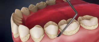
Plaque
In many places, the pellicle of the tooth may be covered with a layer of plaque.
Perhaps the formation of a pellicle is the initial stage of the formation of dental plaque or, conversely, dental plaque eventually turns into a pellicle. Meckel (1965) considered the pellicle as old dental plaque, which was modified due to the dissolution of the bacteria it contained. Dental plaque was studied using staining, chemical analyses, electrophoresis, electron microscopy and other modern research methods. There are difficulties in obtaining sufficient plaque for chemical analyses. Samples are often contaminated with components of oral fluid. At the same time, it is known that saliva sediment cannot be used for analysis as a substitute for dental plaque.
Mandel et al. (1957) proposed collecting plaque on strips of foil or other material attached to the teeth. Plaque can also be collected on artificial teeth. Per 1 mg of dry mass of dental plaque there are 3.37 mcg of calcium, 8.37 mcg of phosphorus, 4.20 mcg of potassium, 1.30 mcg of sodium. Calcium and phosphorus in dental plaque are mainly formed from saliva, although other sources are possible. Thus, Luoma (1964) found in dental plaque from 14 to 45% of radioactive phosphorus (32P), which entered it from the enamel. In samples of two to three days of dental plaque in young people, the content of phosphorus, sodium and potassium is higher than in saliva.
The concentration of inorganic plaque salts increases over time. About 40% of the dry mass of inorganic plaque material is present in the form of oxyapatite. However, the mechanism and conditions for the synthesis of apatites in dental plaque, as well as the significance of this process for the tooth, have not been sufficiently studied.
Metabolic reactions of phosphorus in dental plaque are largely dependent on carbohydrates. The phosphate content in dental plaque decreases by 1/3 when sugar remains in the mouth for fifteen minutes.
The pH value has a significant influence on the adsorption of phosphorus in dental plaque. Thus, at a pH of 7.0-7.4, the absorption of radioactive phosphorus (32P in experiments with dental plaque cells) is accelerated. The optimal rate of accumulation of this element occurs at pH 6.8–7.0.