Cervical or basal caries is one of the most dangerous forms of the disease. With this diagnosis, the pathological focus is located in the area of the tooth adjacent to the edge of the gum. It is more difficult to treat due to the difficulty of accessing the carious cavity. The putrefactive process quickly spreads to the root and, faster than other forms, ends with a fracture of the base, that is, loss of the tooth.
Features of the disease
The cervical form of caries is particularly aggressive and inconspicuous in its early stages. Symptoms of this form of caries can appear when the tooth appears intact at first glance. According to statistics, more than 70% of cases of cervical caries are detected too late, when the root canals are damaged or the neck of the tooth is fractured.
Dentists call root caries the most aggressive form of the disease. This statement is based on three significant factors.
The first factor is specific localization
The neck of the tooth, connecting the root and crown, has the thinnest layer of enamel. In addition, this part is covered by the gingival margin, which is why more bacteria and substances that contribute to the destruction of the hard surface of the teeth accumulate on its surface.
Since the thickness of the enamel on the neck is not large enough, and the degree of its mineralization is lower than on the crown, destruction occurs faster. As a result, caries deepens into the dentin and pulp in a matter of weeks, that is, many times faster than when the crown is damaged.
The second factor is specific distribution
Cervical caries tends to spread the pathological process under the gingival margin. In this case, the lesion not only goes deeper inside the tooth, but also around its circumference. This leads to the breaking off of part of the crown at an early stage of the disease.
Important! When the carious lesion covers the entire surface of the cervix, we are talking about the transition of cervical caries to circular. In this case, it is almost impossible to save the tooth.
The third factor is a blow to aesthetics or the invisibility of caries
When located on the front teeth, cervical caries cannot be hidden: it is clearly visible when talking and especially when smiling. This has an extremely negative impact on the psychological state of the patient.
Cervical caries located on chewing teeth, on the contrary, is often ignored and left without treatment. Therefore, more than 80% of patients with this diagnosis turn to dentists when the disease is complicated by pulpitis and the spread of the inflammatory process to the periosteum.
Possibility of complications
Complications after caries are varied:
- allergy to a developing infection;
- development of inflammation of the gums and facial tissues;
- an infected tooth affects the condition of the body;
- the appearance of flux is possible;
- A jaw cyst may develop.
Painful sensations do not allow high-quality chewing of food, which harms the stomach and intestines. Damage to a tooth can lead to its removal, and this is a serious stress received by the body. Complications can be very serious diseases - pulpitis and periodontitis. With pulpitis, pain appears when biting; this disease can manifest itself even after treatment of caries if the affected tissue is removed poorly. An even more serious complication is periodontitis, which affects nerve bundles and ligaments. Left untreated, it can become a chronic disease.
How does cervical caries develop?
The beginning of cervical caries is the accumulation of plaque near the neck of the tooth.
Against the background of increased acidity and lack of oxygen, as well as under the action of enzymes secreted by bacteria, the enamel softens, opening the gates of infection.
The basal form of caries develops in 4 stages:
- in the first, known as the white spot stage, a dull white spot appears on the tooth at the level of the gum margin;
- at the second stage, a superficial carious lesion develops, which looks like darkening of the enamel;
- on the third, the carious cavity deepens through the enamel into the thickness of the dentin;
- at the fourth stage, deep caries develops, and a “hollow” forms in place of the spot.
The final stage of the disease is a fracture of the tooth in the neck area, pulpitis, accompanied by acute pain, and sometimes an abscess in the gum.
Complications and consequences of internal caries
The soft tissues surrounding the tooth may also become inflamed. Sometimes the patient notes a general deterioration of the condition against the background of progressive carious lesions. His body temperature may rise, problems with the digestive system, bad breath, etc. may appear. Due to the negative effects of bacteria, flux and suppuration, cysts, and ulcers may appear.
Due to pain in the tooth, it becomes impossible to chew food normally, and a reaction to the slightest irritants appears. If the disease is not treated in time, periodontitis and periodontitis develop, which can result in tooth loss. This, in turn, is a serious stress and requires a long recovery.
Unfortunately, not all patients' caries disappears without a trace; some may encounter unpleasant complications both in the first days after treatment and several months after it. The most common phenomenon is an allergy to the materials used in the work. Sometimes infectious lesions are diagnosed, the cause of which may be the unscrupulous work of the doctor or failure to comply with the rules of personal hygiene.
previous post
White caries
next entry
Symptoms
The appearance of cervical caries can be recognized visually, especially if the lesions are located on the front surface of the teeth on the smile line. When located on the lateral teeth, the disease may be indicated by:
- change in tooth texture at the base - roughness to which food sticks;
- when brushing your teeth and running a brush along the neck, acute short-term pain occurs (electric shock effect);
- acute painful reaction to cold and hot, sour and sweet foods.
Unpleasant sensations can appear even during a conversation on the street, when cold air enters the oral cavity.
What to do if a tooth hurts after treatment of cervical caries
If a slight pain after removal of cervical tooth caries lasts 2–3 days and gradually subsides, then this is normal. If there is severe, increasing pain, as well as an increase in body temperature or malaise, you should immediately consult a dentist. Possible reasons:
- insufficiently thorough treatment of the carious cavity before filling led to the spread of infection and relapse of the disease, including perforation of the dentin barrier, penetration into the pulp and the development of a complication - pulpitis; the dentist will remove the filling, open the tooth cavity and treat pulpitis; and only then can it be re-sealed;
- periodontitis (inflammation of the periodontal tissues) – the gums become swollen, painful, their edges move away from the crown, and bleeding may occur; this condition also requires dental treatment.
Causes
The main cause of the development of cervical caries is considered to be the bacterium Streptococcus mutans, which lives in dental plaque. Accumulating in gum pockets, it contributes to softening of the enamel and destruction of the tooth at the base.
Other factors can provoke the development of pathology:
- regular consumption of excessively acidic foods and drinks;
- improper brushing of teeth;
- long-term use of certain medications that weaken enamel;
- endocrine diseases;
- deficiency of vitamins and minerals;
- pregnancy.
According to statistics, cervical caries often develops in older patients with a history of periodontitis and bad habits (smoking).
Why is this necessary?
- Preservation, not restoration of health. Even at the initial stage, caries is associated with varying degrees of destruction of dental tissue. Of course, the dentist will restore the tooth and form a filling, which will be installed in place of the missing fragment. But, in any case, the filling serves only as an analogue, a more or less effective replacement for lost dental tissue. Prevention does not allow the disease itself to develop. That is, it preserves the integrity of the natural structure of the tooth, eliminating the need to use artificial substitutes.
- Prevention is easier than cure. Treatment of carious lesions, especially in cases of large-scale spread, can be quite time-consuming. And at the same time, no one can guarantee that the disease will not appear again, on another tooth. Regular prophylaxis is much faster. This procedure only takes about an hour, does not require complex dental procedures, and should be performed on average only twice a year.
- Finally, prevention is cheaper than cure. From a financial perspective, caries prevention is also much cheaper than a full course of treatment. Therefore, by carefully monitoring the condition of your oral cavity and preventing the development of disease, you not only take care of your health, but also save money!
Diagnosis of the disease
Cervical caries can be diagnosed during a dental examination of the oral cavity. The specific location and rough structure of the carious spot make it possible to differentiate the pathology from a wedge-shaped defect and simple caries.
Additionally, instrumental diagnostic methods are used: dental radiography, radiovisiography, transillumination and others. They allow you to assess the degree of penetration of caries into the tooth cavity, see hidden lesions and predict the effectiveness of treatment.
Diagnosis of hidden caries between teeth
To diagnose the disease, several methods are used that make it possible to detect hidden caries between teeth.
- Visual inspection.
It is carried out to assess the condition of the teeth using a special probe. - X-ray.
On X-rays, carious areas appear as black spots. - Staining or transillumination.
Using staining agents or fluorescent diagnostics, the doctor can detect carious lesions even in the most difficult to reach places.
Treatment Options
A variety of techniques are used to treat cervical caries. Their choice depends on the stage of development of the carious lesion.
Spot stage
At the spot stage, radical methods of therapy are not used. To restore the strength of the enamel, it is enough to carry out 3-4 courses of remineralizing treatment, which consist of several stages:
- Professional teeth cleaning.
- Application of a carious stain with fluoride-containing compounds.
- Systematic oral care with products containing a high fluoride content: mouth rinses, toothpastes.
- Including salt and water enriched with fluoride in the diet.
After just 1-2 courses of remineralization, the white spot will be eliminated, and the risk of recurrent caries will decrease.
Stage of superficial caries
When caries spreads superficially, the doctor removes tartar and plaque, and then grinds the surface of the pathological focus to healthy tissue. To strengthen the enamel and prevent repeated destruction, several courses of remineralization are carried out.
Stage of middle caries
If enamel and dentin are damaged, preventive cleaning and fluoridation of tooth enamel is indispensable. To eliminate the pathology, the doctor will have to clean the carious cavity from putrefactive tissue, then literally saturate the dentin with drugs that suppress bacterial activity. Finally, the hole in the tooth is filled with polymer materials.
Stage of deep caries
With deep caries, treatment is extremely painful, so it is carried out only under local anesthesia. After the medicine takes effect, the doctor begins treatment:
- Pulls back the edge of the gum to have full access to the carious cavity.
- Cleans the carious cavity from necrotic tissue with dental instruments and a drill. If necessary, the pulp is removed and the dental nerve is extracted.
- The tooth cavity is isolated with special adhesive materials, and the equine canal is filled.
- Layer-by-layer filling is carried out with photopolymerization of each layer.
- The final grinding of the filling and tooth is performed.
This procedure takes several days or even weeks and costs several times more than treatment at the initial stage.
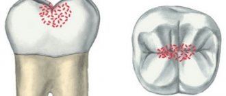
What type of caries is this?
Cervical caries is a clinical form of the carious process localized in the neck of the tooth - a small space between the root and crown, covered by the gum. It is also called basal caries. The disease develops regardless of age. Even an infant can get sick if he is fed haphazardly and often given sweetened water.
Based on the number of lesions, cervical dental caries is divided into:
- single – from 1 to 3;
- multiple – from 3 to 10;
- systemic – 10 or more.
The disease may be:
- primary – develops for the first time;
- secondary – develops again after treatment has already been carried out.
And:
- uncomplicated - if the destruction involves only hard tooth tissues - enamel and dentin;
- complicated - when infection penetrates into the dental cavity, root canals and periodontium with the development of inflammation in these tissues (pulpitis, periodontitis).
Cervical caries
Prevention
Only careful oral hygiene can prevent the occurrence of cervical caries. In addition to brushing your teeth twice a day with toothpastes, it is recommended to use dental floss. To remove plaque from gum pockets and remove hard deposits, you need to visit the dentist twice a year and have your teeth professionally cleaned.
To maintain oral health, it is recommended to include fresh solid vegetables and fruits in your diet: apples, carrots, cabbage, etc. Dairy products as a source of calcium and phosphorus will be beneficial for your teeth.
When the first signs of cervical caries appear, you should make an appointment with a dentist and treat the disease before it becomes complicated by pulpitis.
Other
Stages
Cervical caries: medium, deep and complicated
Cervical caries occurs in stages. They replace each other faster than in the carious process of other localizations and cause complications more quickly.
Spot stage
The main symptom is a white spot on the enamel. The stain is matte - the enamel loses not only calcium, but also its shine. There are no complaints, only occasionally there is a feeling of a sore throat. If the patient does not visit the dentist for preventive purposes during this period, cervical caries may not be noticed.
Surface
The enamel continues to lose minerals and gradually deteriorates, its surface becoming slightly rough. The color may change from white to yellowish. The sore throat intensifies, and it hurts when eating very cold or hot food. At this stage, the patient may already suspect something is wrong if he carefully listens to the signals of his body. But this rarely happens, so this stage in most cases remains without treatment.
Average
At this stage, there is already complete destruction of the enamel and deepening of the carious cavity to the middle of the dentin layer. The defect is black in color and visible in the form of a hole in the tooth. It can only be missed if the cavity is covered by gums. At this stage, it hurts from the effects of temperature and chemical (sour, sweet foods) irritants, but the pain goes away quite quickly. It is at this stage that most patients turn to the dentist.
Deep
The carious cavity increases in width and depth, reaching the dentin border. A thin layer of dentin separates it from the dental cavity with pulp. The pain intensifies, the tooth reacts to all types of irritants, including mechanical ones - pressure when chewing. The tooth can hurt for a long time and quite severely. Food gets stuck in the resulting cavity.
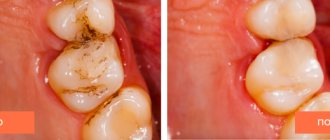
Circulatory
Circulatory caries in a child with a broken tooth
This caries of the cervical region has its own characteristic features. There is a circular damage to the enamel and dentin of the cervical area and the teeth simply break off. The front teeth are mainly affected, the disease proceeds unnoticed and very quickly, reaching deep stages in weeks and even in a matter of days.
It mainly affects children from infants to preschool age, which is why its second name is bottle disease. In children in the first months and years of life, it is associated with improper frequent feeding without subsequent cleansing of the oral cavity. This leads to the accumulation of food debris on the crowns, the proliferation of bacteria and the destruction of several teeth at once.
In adults, circular cervical caries also occurs; its causes are: impaired immunity, smoking and alcohol abuse, poor oral hygiene, crowded teeth, and malocclusion.
Can you take care of your dental health yourself?
Yes, sure. There are a number of rules that, if followed daily, will significantly reduce the risk of developing carious lesions:
- Brushing your teeth twice a day. Allows you to remove soft plaque in which bacteria multiply. To get rid of it reliably, you need to brush your teeth morning and evening with a brush that has medium-hard bristles. Each such procedure should last at least 3-4 minutes. A good solution would be to alternate fluoride-containing toothpaste (1 week) and fluoride-free toothpaste (3-4 weeks). This periodicity is due to the fact that enamel has a saturation limit with fluoride. A week of using fluoride-containing paste will provide the enamel with a sufficient amount of this element, and then fluoride will simply be removed from the body. But after a month, its concentration will decrease and its influx will be required again.
- Supplement the procedure with dental floss at least once a day, since the brush is not able to fully clean the spaces between the teeth.
- Regular brushing after meals. Residues should be removed by normal rinsing. It should be done after every meal. And to get rid of food particles that linger in the interdental space, it is best to use dental floss.
- Be careful with sweets! Bacteria that cause caries multiply much more intensively in an environment rich in sugars and fats. Therefore, instead of another cake, it is better to eat an apple or carrot. On the one hand, you deprive the bacteria of additional nutrition, and on the other, the solid consistency of such products itself cleanses the teeth and removes soft plaque.
The above preventive measures can be carried out independently. However, you cannot do without periodic assistance from a dental specialist. Therefore, we recommend contacting our clinic to determine the risk level of caries for you and your family members. And then, based on these results, SM-Dentistry doctors will develop an individual plan for preventive oral examinations for you. Constant preventative care will ensure your dazzling smile and the health of your entire oral cavity.
And of course, if acute symptoms of the disease appear, you should never self-medicate! This will only worsen the situation and cause complications. If you experience pain, we invite you for a consultation with a specialist at the SM-Dentistry clinic, call tel. or fill out the feedback form on our website.
Forms of deep caries
Deep caries can occur in two forms - acute and chronic. Each has its own characteristics. Acute deep caries is characterized by rapid development and can affect a large area of the tooth in a short time. Upon superficial examination, the cavity in the tooth appears small. But further diagnostics show that this is deep-stage caries, and it has almost reached the tooth root. It is acute caries that is most often accompanied by a complication in the form of pulpitis. Therefore, when treating it, the dentist has to not only prepare the tooth and put a filling on the dental crown, but also remove the nerve and fill the canals.
Chronic deep caries, unlike acute caries, is a slow, sluggish process. Although most of the tooth with this form is destroyed, the carious cavity is smaller in area than with acute caries. The patient may complain of periodic aching pain, which does not depend on external stimuli and occurs on its own.
Depending on the affected area, deep caries can be divided into cervical, fissure or located on the front incisors.
Based on the speed of development of the pathological process, it is divided into compensated (relatively slow development), subcompensated (with an average speed of development) and decompensated (with a high speed of development). Objectively, any form of deep caries can be cured, but it will require varying amounts of time and effort.