Author of the article:
Soldatova Lyudmila Nikolaevna
Candidate of Medical Sciences, Professor of the Department of Clinical Dentistry of the St. Petersburg Medical and Social Institute, Chief Physician of the Alfa-Dent Dental Clinic, St. Petersburg
The human oral cavity has its own microflora and its own specific diseases arise. Let's talk about one of them - fibrous epulis of the gums: what kind of disease it is, what are the causes of its occurrence, how is the treatment carried out.
Growth on the gum: why it appears and how to treat
A growth has appeared on the gum: what is the cause
Infectious diseases
Non-communicable diseases
Types of growth
Gum cancer
Lump after crown installation
Growth on the gum: how to treat
Which doctor should I contact if a growth appears on the gum?
Normally, the gums have a smooth, pale pink texture. Any change, be it a thickening or a growth, signals problems in the body that cannot be ignored, as they pose a health hazard. Why lumps appear on the gums and how to treat them, we will tell you in the article.
Prevention of tartar –
Prevention is, first of all, high-quality and regular oral hygiene, no snacking between meals, and regular use of dental floss. You can read about proper hygiene in the link above. In this article you will find the most detailed description of what proper oral hygiene should be. But we would also like to tell you about what else can help you in the fight against pigmented plaque and dental plaque.
1) Use toothpastes containing pyrophosphates, because... this component prevents the mineralization of dental plaque. Pyrophosphates in toothpastes will not be able to dissolve existing tartar, but they are good at preventing its appearance.
2) Use electric toothbrushes for hygiene. They are at least 2 times more effective at removing plaque compared to regular manual toothbrushes. In addition, the pulsating movements of the Oral-B electric brush heads allow you to loosen tightly attached plaque, and the rotating movements sweep it away from the teeth. But if you have periodontitis, then it is better to use Philips Sonicare sonic brushes rather than Oral-B brushes.
A growth has appeared on the gum: what is the reason?
A lump or growth is a compaction on the gum caused by damage to periodontal tissue, which may appear without any previous signs. Growths appear in people of any age, but mainly in young children, as they unknowingly introduce infection into their mouths. The first thing you need to do when you detect formations in your mouth is to determine the nature of their origin (only a doctor can do this). It comes in two types:
- Infectious. The appearance of cones is caused by the activity of bacteria.
- Non-infectious. Bumps and growths as a result of injury, mechanical or chemical damage.
If a lump has formed in your mouth, you should consult a doctor to rule out possible complications. The doctor will help diagnose the problem and prescribe appropriate treatment.
Fibroma
Description
Benign tumor of epithelial cells. Initially, the bumps on the gums above the tooth are small in size and do not hurt. However, mechanical stress and other negative factors can lead to the transformation of fibroma into a malignant tumor.
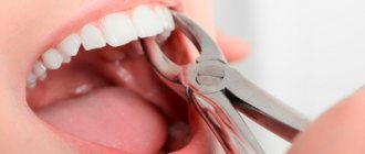
Treatment
Treatment is carried out surgically. The doctor excises the tumor, applies stitches, and prescribes rinses. Further follow-up visits may be required to monitor the recovery process.
Infectious diseases
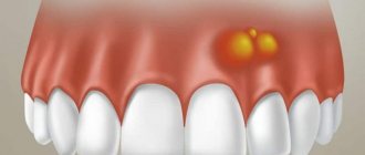
| Gingivitis and periodontitis Gingivitis is the initial stage of gum disease, often accompanied by the appearance of a red ball-shaped growth near the tooth. The next stage is periodontitis. With it, purulent discharge is observed, and the lump acquires a grayish or beige tint. |
| Granuloma or cyst When pulpitis is advanced, the inflammatory process reaches the apex of the root, where a granuloma forms. Often this process goes unnoticed by the patient, so he is in no hurry to see a doctor. Over time, the pathology can develop into a fistula - a white formation on the gum. |
| Flux Purulent inflammation on the gums occurs against the background of advanced caries or pulpitis, a poorly treated root canal. At the first stage, minor pain appears; on the second - swelling and redness; on the third, the temperature may rise and the cheek may swell; on the fourth – the pain becomes sharp and throbbing, swelling increases. |
Diagnostics
The diagnosis is made based on examination by a specialist and histological examination. A differential diagnostic method is used to exclude the presence of hypertrophic gingivitis in the patient. It has similar symptoms to epulis. An x-ray is also used for diagnosis, where, with epulide, rarefaction and destruction of bone tissue due to inflammation are observed.
Non-communicable diseases
| Epulis This is a benign formation. It has a stem and a cap. Most often it occurs due to malocclusion, improper prosthetics, and hormonal changes. In the absence of adequate treatment, the tumor can develop into a malignant one. |
| Exostosis A bone growth that occurs against the background of a jaw abnormality - the bone extends beyond its usual boundaries, forming a lump. Most often, the reason lies in a genetic predisposition, less often - as a result of injury or complex tooth extraction. |
| Hematoma As a rule, it is formed as a result of an impact and goes away on its own over time. If there is damage to the tissue, then surgical intervention is necessary. |
Causes: why does a tumor form?
Epulis occurs under the influence of the following factors:
- Injury to the gums through: an overhanging filling;
- edges of a decayed tooth;
- tartar;
- burn;
- bruise;
- poor quality prosthesis.
Types of growth
New growths on the gums vary in shape, size, and method of formation.
- Angiomatous growth. A soft, bumpy, pink growth that grows very quickly. Often reappears after removal. This growth is diagnosed in children aged 10-12 years; as a rule, it does not occur in adults.
- A fibrous growth is a gum-colored lump. It grows slowly and does not cause pain, so patients are in no hurry to see a doctor.
- Giant cell growth. A rapidly growing neoplasm of red-blue color, from which serous fluid is secreted. The lump is easy to injure.
Classification
There are several types of epulis. Each of them has its own number in ICD-10 (International Classification of Diseases, 10th revision).
Fibrous (K06.82 - benign neoplasms):
Location area: vestibular side of the gum;
- Round or oval shape;
- Slow growth;
- Surface smooth/lumpy surface;
- Doesn't bleed;
- Wide base;
- Thick consistency;
- Light pink (to match the gum color).
Angiomatous:
- Location area: neck of the tooth;
Giant cell (K06.81 - other specified changes in the gums):
- Location area: alveolar part;
Gum cancer
This dangerous neoplasm is worth highlighting separately. Mutation cells begin to divide uncontrollably, which is why a red tumor forms on the gum. The disease progresses quickly and over time can develop into jaw cancer.
What are the symptoms?
- General weakness;
- increased body temperature (37 C°);
- loss of appetite;
- constant drowsiness.
The doctor makes an accurate diagnosis after a detailed examination. As a rule, treatment is long and painstaking. With timely medical intervention, the outcome is favorable.
Types of epulis
Depending on the nature of the pathological process, epulis on the gums is divided into three main types.
- Fibrous. The gum tumor differs in density. Its base is coarse fiber fabric. The tumor grows slowly and asymptomatically.
- Angiomatous. Epulis angiomatous consist of epithelial cells and a large number of blood vessels. The tumor is localized mainly in the molar area. Due to the many blood vessels, even with a slight impact on the tumor, bleeding occurs. On palpation, the lump is soft and painful.
- Giant cell. The tumor formation can be large. Giant cell epulis affects the position of teeth, displacing them. Pain is not typical for this species, but bleeding is possible due to mechanical damage.
The development of giant cell epulis on the gum poses the greatest risk, since the likelihood of the tumor becoming malignant is much higher than with other forms of the disease.
Growth on the gum: how to treat
The treatment plan is developed by the doctor based on the nature of the growth, so in no case should you self-medicate. It is important to understand that if you do not contact a specialist in a timely manner, serious complications can arise.
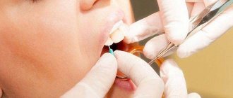
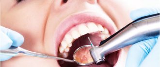
If the cause is an infection, then it must be removed. In case of periodontal inflammation, professional oral hygiene is performed. If the cause is periodontitis and complicated pulpitis, then first the lump is opened and pus is pumped out of it, and then conservative or surgical treatment is carried out.
If there is no pain, for example, with epulis or ecostosis, then the decision about surgical intervention is made together with the patient.
Tartar: removal
When you brush your teeth, you can only remove soft plaque from the surface of your teeth. Partially mineralized plaque, as well as hard dental deposits, can no longer be removed by regular teeth cleaning at home. And in this case, only a dentist can clean the tartar.
1) Cleaning the stone at the dentist’s appointment –
The traditional way to remove hard plaque is ultrasonic teeth cleaning. In this case, stone removal is carried out using an ultrasonic scaler. Removing supragingival calculus is a very simple procedure, and 1 hour is enough to remove calculus from all teeth. But to effectively remove subgingival stones, you need several visits and a highly qualified doctor. Therefore, it is best to remove stones not from ordinary dental therapists, but from periodontists (these are dentists who specialize in the treatment of gum inflammation).
Ultrasonic tartar removal –
There is also the “AirFlow” procedure, with which you can remove small dental deposits, pigment plaque, and polish your teeth. In this case, cleaning and polishing of teeth occurs due to a water-air mixture containing granules of an abrasive substance, sprayed under high pressure from a special tip. But AirFlow will not be effective against massive hard dental plaque, which in any case will first have to be removed with ultrasound.
Tartars: photos before and after their removal
On average, the price for tartar removal is about 100-150 rubles per 1 tooth, including polishing.
2) How to remove tartar at home –
Some patients use recipes for removing plaque, which are replete with many unprofessional websites, promising complete relief from tartar. We'll disappoint you, but these methods not only don't work, but also cause harm to tooth enamel and gums. Just for fun, you can check them out at the link above.
It is impossible to remove well-mineralized tartar and dense pigment plaque with any home remedies. However, not too pronounced pigment plaque and only partially mineralized plaque can still be removed with the help of the following modern dental care products. First of all, we are talking about Oral-B electric toothbrushes - in combination with special highly abrasive toothpastes.
Important: Oral-b electric brushes have a round head with bristles that performs 8,800 reciprocating movements per minute, as well as a minimum of 20,000 pulsating movements. Pulsations help loosen dense plaque (even that which may already be partially mineralized), and rotational movements sweep it away and polish the teeth. This brush is a miniature dentist’s tool and is probably familiar to everyone who has gone to the dentist for professional oral hygiene.
The second component is highly abrasive toothpastes, which can help remove pigmented and partially mineralized plaque. They can only be used once a week. If you are using them in combination with an Oral-b electric brush, it is advisable to use the electric brush only at low speed (in the “whitening/polishing” mode). You can find out how this is done correctly and what toothpastes are used at the link below.
You can also purchase professional teeth polishing products, which the dentist uses to finish polishing teeth after ultrasonic cleaning (they are sold in stores that sell consumables for dental clinics). For example, pastes such as “Detartrine” or “Detartrine – Z” (this one is more effective). They are able to remove even small tartar and not too pronounced smoker’s plaque. Used in combination with an Oral-B electric toothbrush (rotation of the nozzle should be at low speed).
→ Scheme for removing dental plaque at home