Removing a tooth is the last thing a dentist will suggest. Due to a number of subjective and objective reasons, it is impossible to maintain the dentition in ideal condition until old age.
Dental defects are the loss of one or more dental units, leading to a violation of the integrity of the dentition, deviation in the development of physiological occlusion, and an abnormal arrangement of individual teeth.
Reasons contributing to the development of dental defects
Dental defects arise due to:
- untimely treatment of caries;
- inflammatory processes in the oral cavity;
- presence of malocclusion;
- genetic conditioning;
- diseases leading to changes in metabolic processes;
- damage to the jaw apparatus;
- various neoplasms.
Other causes of dental deformities
In addition to deformations of the development of the skull, other reasons can lead to changes in the dentition:
- Poor posture. When posture is impaired, the body begins to adapt. Not only the position of the spine changes, but also other parts of the musculoskeletal system. These adaptive changes affect the dentofacial apparatus. A defect in the dentition develops.
- Bad habits in a child. This may involve prolonged pacifier or finger sucking. Such habits greatly affect the formation of the dentition and can lead to tooth movement and changes in bite.
- Intrauterine pathologies. They may be due to various reasons. For example, diseases suffered by the mother during pregnancy. As a result, deformations of the jaws and tooth buds may develop.
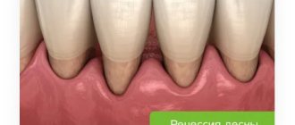
- Periodontal diseases. Weakened tissues cannot hold teeth in their normal position. They begin to shift, the dentition becomes deformed.
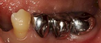
- Partial edentia. When one or more units are lost, the rest begin to shift. As a result, a dental defect develops.
Wedge-shaped defect
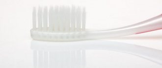
Such defects form in the cervical part of the tooth, near the gingival margin. The defect has a wedge shape. Because of it, the crown becomes thinner at the base. This is a dangerous condition that, without treatment, can lead to destruction and fracture of the crown. It develops gradually: the defect may not be noticeable in the early stages. It is often accompanied by gum disease. The formation of a wedge-shaped defect is associated with improper distribution of the chewing load (in case of malocclusion or the absence of some teeth in a row). Sometimes the problem occurs due to improper brushing of teeth if the enamel is weakened (for example, if a person uses a too hard toothbrush, brushes too vigorously, or immediately after eating acidic foods). A possible cause is also considered a malnutrition of hard tissues (due to problems with metabolism or blood supply). To treat the defect, the defect is filled, the tooth is strengthened and remineralization is carried out.
Types of dental deformities
Teeth can move from their normal position in different ways. Depending on the type of displacement, types of dental deformations are distinguished:
- Rotation of a tooth relative to its axis.
- Tilt towards the palate, cheek, tongue.
- Vertical displacement of teeth, leading to their lengthening.
- The distal displacement of one unit back in relation to the rest of the dentition.
- Mesial displacement is the opposite of distal displacement, since the deformed tooth moves forward.
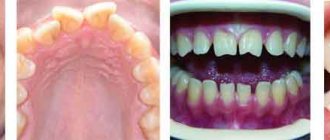
- Crowding is characteristic of an underdeveloped small jaw. There is not enough space for normal development of the dentition. The teeth move in different directions, overlapping each other.
- Gaps and crevices in the dentition develop if the jaw size is large. Visible gaps (diastemas, tremata) form between adjacent dental units.
In complex cases, a combined displacement can be observed, combining different anomalies. Visually, such defects look like uneven teeth.
Pigmentation
If this defect appears, the color of the tooth changes. It may turn brown, gray or almost black, pink or red, yellow. The shade depends on what dye is applied to the crown. For example, if it is tea or coffee, the color will be brown, and if it is nicotine tar, it will be yellow or grayish. If the color of the crown changes unevenly and clearly visible pigmented areas appear, this indicates enamel defects. It absorbs dyes better where there is demineralization and the structure of the fabric is damaged. Pigmentation itself is not dangerous, but it can be associated with other dental health problems. It often appears in the presence of chips, poor hygiene, and some concomitant diseases. To restore pigmented areas, professional teeth cleaning and remineralization are performed. Sometimes whitening, restoration of crowns or installation of veneers may be required.
Diagnosis of dental deformations
Dental deformities are not an independent diagnosis. Treating them only with traditional methods used to correct the bite is not always effective. If the cause that caused the deformation of the dentofacial apparatus is not identified and eliminated, there is a risk of relapse.
To make a diagnosis and select treatment options for dental deformation, doctors at our clinic use:
- Visual examination of the oral cavity. The doctor will definitely ask you when the problem began. This will help determine the causes of the development of malocclusion.
- Paraclinical studies. After a visual examination and medical history, you will be prescribed tests: x-rays of the teeth and alveolar process, x-rays of the temporomandibular joint, tomography, etc.
Examination of the dentition gives the dentist a lot of information about the bite. With its help, you can determine the nature of the deformation, the condition of the periodontium, and roots. Instrumental diagnostic methods are used to diagnose functional and morphological disorders.
With the help of examination and questioning, you can obtain other data on the condition of the dentofacial apparatus:
- Position of teeth in the jaw. The position of the dentition is assessed in relation to the sagittal plane, focusing on the line between the incisors. If this line is displaced, a more precise occlusion study is necessary. The doctor determines the size of the incisal overlap, the position of individual units, and the nature of their occlusal surface.
- Features of the movement of the lower jaw during opening and closing of the mouth. Zigzag movement of the jaw may indicate pathologies of the TMJ and masticatory muscles.
Impaired closure of teeth and their rotation around their axis. Movement of molars and premolars is easily diagnosed during examination.
Instrumental diagnosis of dental anomalies allows an accurate diagnosis.
Fluorosis
A rare disease that occurs when too much fluoride enters the body. It accumulates and can provoke changes in the structure of hard tissues and the appearance of non-carious defects. With fluorosis, colored spots or stripes appear on the enamel. Hard tissues may gradually deteriorate. In this case, deep, extensive defects are formed.
A person can receive an excess amount of fluoride in several cases: with water, when working in hazardous industries and (very rarely) when taking certain medications. During treatment, the cause of excess fluoride is eliminated (for example, replacing water with bottled water), and then teeth are restored, removing defects.
Treatment
Treatment methods for dental deformities are chosen depending on the type of pathology. Most deformities not associated with the loss of antagonist teeth develop in childhood. Therefore, the problem needs to be resolved as early as possible. In orthodontics they use:
- Plates. Can be used immediately after baby teeth are replaced by permanent teeth. The plate puts pressure on the dentition, as a result it is aligned. Installation of plates is indicated for patients aged 7-12 years. At this time, the child’s bones are growing rapidly, so the technique will be effective.
- Trainers. They can be used in early childhood. The trainer is a silicone structure that resembles a mouth guard. They are worn for several hours. The elastic trainer protects the dentition from excessive pressure from the tongue and cheeks and prevents teeth from moving.
- Braces. Can be installed on children over 12 years of age. The orthodontist determines the need to use braces based on the results of the study.
These are the most common options for orthodontic treatment of dental deformities. Specialists at the Zuub.rf dental clinic believe that the approach to eliminating this problem should be comprehensive. It is important not only to visually straighten the dentition and correct the bite, but also to eliminate the cause that led to the development of the anomaly.
Hyperesthesia
This is a condition in which the enamel thins and becomes overly sensitive. Sometimes this can cause the shade of the crown to change, but more often hyperesthesia is manifested by acute aching pain when the teeth come into contact with irritants. The reaction can occur to drinks that are too cold or hot, sour or sweet. Hyperesthesia can have several causes, including too aggressive whitening, poor daily hygiene, regular damage to the enamel when brushing your teeth or due to contact with sour or sweet foods and drinks. It also occurs with calcium deficiency, malocclusion (if occlusion is disturbed) and as a complication of bruxism (teeth grinding during sleep). To treat hyperesthesia, you need to eliminate the cause of thinning enamel (for example, adjusting your daily dental care, correcting your bite, or using mouth guards for bruxism). To remove high sensitivity, remotherapy is performed. To do this, the teeth are coated with a remineralizing composition and fluoride varnish. While the enamel remains highly sensitive, it is better to rinse your mouth with clean water after each meal.
Necrosis
This is the death of hard tissue cells, due to which multiple defects appear on the surface of the crowns, as well as on the tooth root. The structure of the cells changes, and destruction can affect not only dentin, but also the pulp. Necrosis is rare and is usually not directly related to the condition of the teeth. It can be caused by severe toxin poisoning, hormonal imbalance, or severe systemic diseases. Treatment at the dentist is symptomatic: restoration of hard tissues, remineralization, prosthetics for complete tooth destruction. The main therapy must be related to the disease that caused the necrosis, and dental treatment only complements it.
You have questions?
We will call you back within 30 seconds
+7
Injury
The tooth may become damaged due to trauma, and this requires urgent treatment. Usually these are chips, fractures or dislocations that occur due to impacts, accidental falls, or gnawing on hard objects. If the defect as a result of injury is superficial (it is a small chip), it is enough to restore the hard tissues by performing a restoration. If it is a severe bruise, crack, or fracture, more serious recovery will be required. If you quickly consult a dentist, the tooth will most likely be saved (there are such chances even if it was knocked out). In the most severe cases, removal is required, and then implantation and prosthetics are performed.
Symptoms
The specificity of the manifestations depends on the specific type of defect. At the first symptoms indicating the presence of abnormalities, it is recommended to undergo a comprehensive orthodontic diagnosis, the results of which determine the need for dental treatment. Characteristics that deserve attention include:
- Violation of the occlusion of the elements of the upper and lower row.
- Formation of spaces between teeth.
- Curved position of individual units.
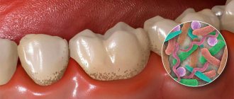
- Intense accumulation of plaque and tartar.
- Complete coverage of the crown surface.
- Bleeding gums during hygienic cleaning.
- Deviation of facial symmetry.
- Speech defects, problems with breathing and posture.
- Extraneous sounds and difficulty opening the mouth.
Various types and varieties of malocclusion are characterized by the presence of common features - in all cases the patient experiences physical and psychological discomfort associated with impaired functionality and aesthetics. Muscle strain provokes increased fatigue and can also cause regular pain, manifested in the occipital and cervical region.