Protrusion and retrusion are types of abnormal position of individual teeth. If such defects develop, it will not be possible to correct the situation on your own. However, the problem can be resolved if you promptly seek help from a specialized specialist. That is why, when you identify the first signs of pathology, you should immediately make an appointment with an orthodontist. We’ll talk about what these anomalies are and how to correct them later in our article.
Protrusion and retrusion - what is it?
Protrusion is a malocclusion in which the front teeth protrude forward at a rather acute angle in relation to the jaw. Retrusion, on the other hand, is a phenomenon in which the front teeth move back.
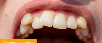
Protrusion
“With the development of such malocclusions, deformation of the dental arch occurs. The consequence of retrusion, for example, is a shortening of the frontal portion of the arch, as a result of which the incisors take a crowded position. Such defects often lead to the development of periodontal diseases and, as a consequence, early tooth loss.”
Smirenko Vnatoly Ivanovich Dentist-orthodontist with more than 18 years of experience
This defect in the dentition does not look aesthetically pleasing, to say the least. In addition, it contributes to the development of corresponding diction disorders. For example, in childhood, such malocclusions can provoke phonemic hearing disorders in a child, which can subsequently cause certain problems with learning at school.
retrusion
Consequences
If jaw protrusion remains untreated, this is fraught with the development of the following complications:
- increased risk of decay in front teeth (which are tilted forward);
- uneven chewing load on the jaws and teeth;
- change in face shape – cosmetic defects;
- deterioration of the jaw joints;
- the presence of large interdental spaces (three and diastemas), which contributes to infectious and inflammatory pathologies of the oral cavity.
To avoid such consequences, doctors recommend going to the clinic as early as possible and undergoing treatment.
Reasons for the development of anomalies
Today, experts identify several common factors that most often cause a lack of space in the dentition, as a result of which protrusion or retrusion may occur:
- the presence of too large neighboring teeth,
- underdevelopment of the dental system,
- supernumerary teeth.
Among other processes that cause abnormal deformation of the position of teeth in a row, the following are distinguished:
- late change of primary teeth,
- incorrect anlage and placement of the rudiments of permanent teeth,
- bad habit of sucking your finger while sleeping or chewing a pencil.
Patients who are accustomed to breathing through their mouth rather than their nose often face a similar problem. In this case, protrusion becomes a consequence of a change in the position of the jaw. As a consequence, corresponding problems arise related to speech function and aesthetics.
Habit of mouth breathing
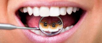
Diagnosis of the disease
Diagnosis of such a defect is not considered difficult. A doctor can make a diagnosis only by external examination of the oral cavity. In addition, the specialist will pay attention to indirect signs, such as mouth breathing or chronic diseases of the respiratory tract.
If tooth protrusion is suspected, the patient is subject to the following types of examinations:
- dental examination;
- patient interview;
- radiography;
- additional research methods (at the discretion of the doctor).
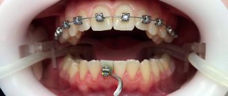
Protrusion after braces
Protrusion can occur during or as a result of orthodontic treatment using a corrective apparatus. Deviation of teeth from a given position after removal of braces is not a rare phenomenon. The thing is that after removing the system, there is no longer any external pressure on the teeth, so the reverse process begins. The teeth strive to return to their original position, which until recently was prevented by orthodontic construction. In a number of situations, such a problem may arise due to incorrect treatment, that is, an elementary mistake by the orthodontist.
To prevent the development of protrusion after braces, it is important to take the retention period responsibly. After removing the corrective device, the orthodontist installs retainers - mouthguards or thin wires that fix the teeth in the desired position.
Retainers
On a note! Experts disagree on how long the retention period should last on average. Most practicing orthodontists are inclined to believe that this period should last at least twice as long as the orthodontic treatment process itself. But everything, of course, is individual.
Before installing an apparatus to correct your bite, it would be a good idea to consult with a specialist in advance about the retention period. Installing retainers allows you to prevent subsequent tooth curvature and consolidate the results after a course of wearing braces.
Classification
Dentists distinguish several types of protrusion of elements of the jaw row:
- Bimaxillary protrusion is characterized by a change in the position of the incisors on both jaw rows simultaneously. This situation may be accompanied by the inability to completely close the jaws and lips.
- Protrusion of the upper jaw is a pathological forward movement of the incisors of the upper dentition.
This pathology is accompanied by insufficient development of the orbicularis oris muscle and shortening of the upper lip. In a relaxed position, the oral cavity remains slightly open. - Mandibular protrusion is defined as excessive forward inclination of the incisors located in the lower dentition.
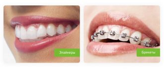
Treatment of protrusion and retrusion
The choice of a specific method for correcting such defects directly depends on the degree of complexity of the anomaly and the consequences caused. As a rule, the standard treatment package includes the following procedures:
- physiotherapy,
- use of corrective devices,
- prosthetics,
- removal of a tooth,
- separation or division of combined teeth.
In mild cases of pathology, experts often advise performing a massage by pressing the tongue on the problematic tooth, pushing it into the correct position. If there is sufficient space in the row, this method is very effective. In more complex cases, an orthodontic system may be required. In order not to trigger the development of an anomaly and subsequently lose a tooth, it is important to promptly seek help from a doctor.
Patient review:
“I am currently undergoing treatment with braces, and recently it turned out that I have protrusion of the front teeth in the upper row. Now the doctor and I are struggling with this problem. The orthodontist gave me elastics, and the traction is very strong. But the question is this: the teeth seem to be stretching back, but there are no gaps between them. I wonder if this makes any sense. Next time I come for an appointment, I want to find out if there are any other treatment options.”
KatiStar92, from correspondence on the forum
The doctor may resort to the removal of individual teeth or separation when the patient has severe crowding. In this case, separation involves the separation of adjacent teeth artificially, usually by removing a thin layer of enamel on the lateral surfaces. This manipulation can be performed during orthodontic treatment.
Patient review:
“I’ve been wearing Dimon braces for over a year. The teeth naturally straightened, but another problem appeared - protrusion of the first fours. The doctor separated the quads and put elastics on the braces. It hurts terribly, but there is nothing to do, you have to endure it. But in my opinion, this is better than removing a tooth. Moreover, my doctor said that in my case, separation would be quite enough.”
JekaUng, from correspondence on the forum
With the development of an advanced form of the anomaly, the patient may encounter such unpleasant external manifestations as large gaps between the teeth and deformation of the dental arch. It is impossible to correct the pathology on your own, so if the first signs of protrusion or retrusion occur, you should immediately contact your dentist.
Why is it necessary to correct tooth protrusion?
The root of the tooth must have a stable position inside the bone socket. This allows the front teeth, which have only one root, to distribute chewing pressure evenly. When protrusion develops, the teeth that are pushed forward are deprived of bone support and find themselves in a dislocated position, subjected to excessive stress from antagonist teeth on the opposite jaw. Therefore, the tilting of the teeth may become worse. At the same time, the aesthetics of the face and the phonetics of speech are increasingly disturbed.
A dental examination is required. The specialist conducts a thorough X-ray and craniometric diagnosis (measurements of facial proportions) in order to identify the cause and severity of the anomaly. Depending on the factor that provokes tooth displacement, treatment is prescribed.
How to diagnose?
- X-ray of children's teeth
Allows you to assess the condition of temporary teeth and rudiments on layer-by-layer sections. Today, this is the most informative type of research, which allows us to eliminate errors in diagnosis.
Recovery after surgery
After a minimally invasive simple session, the patient is activated after 2-3 hours. Most of those operated on notice noticeable improvements within the first hours after minimally invasive surgery. Perhaps, immediately after activation, he will be allowed to go home on his own. But in order to avoid postoperative complications, which are not excluded even after percutaneous (the most gentle) surgery, the following are prescribed:
- antibiotic therapy (against infections and suppurations);
- taking vascular medications (against limb thrombosis, thromboembolism);
- a calm physical regimen, especially gentle on the lower back during the recovery period;
- special physical therapy for good restoration of the operated area, muscle strengthening, and prevention of relapses;
- wearing a support corset during rehabilitation;
- ban on lifting weights, lifelong refusal from heavy sports.
In total, rehabilitation measures take 2 months. But this does not mean that during these 2 months a person will be limited from walking, going to work, doing household chores, etc. No, with a successful intervention, patients even go to work within 3-5 days. However, non-compliance with specific rehabilitation and lifelong regimens, which the doctor thoroughly informs about upon discharge, is associated with high risks of postoperative complications. The first among them is the rapid resumption and more progressive course of protrusion.
Prevention
Fortunately, a herniated disc is treatable and can often be prevented by making certain lifestyle changes and strengthening exercises that can help keep the spine strong and healthy:
- 1. Strengthen the muscle frame. Having strong muscles can help protect the intervertebral discs and prevent back damage and injury. The core consists of the muscles of the trunk, including the abdominal cavity and back. To strengthen your core muscles, you can do a variety of exercises, including Pilates and yoga.
- 2.Increase overall flexibility. Keeping your body mobile and flexible helps reduce spinal injuries. Simple exercises such as bending over can help relax the muscles.
- 3. Maintain good posture. A sedentary lifestyle, for example, sitting at a desk for long periods of time, can lead to back problems and osteochondrosis. Poor posture when lifting heavy objects and strenuous activities that require repetitive movements that place stress on the back can accelerate disc degeneration.
- 4.Maintain a Healthy Weight and Diet A healthy and nutritious diet can help prevent obesity and reduce stress on the spine. Excess weight can significantly contribute to the development of symptoms associated with osteochondrosis, which can lead to protrusion of the intervertebral disc.
Apart from this, vitamins like calcium and vitamin D are also essential for maintaining the strength of bones, including the bones of the spine. A lack of calcium can lead to low bone density and therefore increase the risk of bone diseases such as osteoporosis
Active treatments
In the active portion of physical therapy, the physical therapy therapist will teach the patient various exercises to improve flexibility, strength, stability of the spinal column and restore range of motion. The physical exercise program (physical therapy) is individualized, taking into account health and medical history. Exercises for one person may not be suitable for another person with spondylolisthesis. Thus, the exercise therapy doctor selects an individual exercise program for each patient and the task of the set of exercises is to create a muscle corset that will compensate for biomechanical disorders associated with vertebral displacement.
If necessary, the patient receives information from a physical therapy doctor on how to correct his posture and incorporate ergonomic principles into daily activities. This is all part of the physical self-care or self-medication aspect: through physical therapy, the patient learns good habits and principles to better care for the body.
Incorrect position of incisors
At the stage of primary occlusion, a reverse incisal overlap may form. With normal development, the upper jaw overlaps the lower jaw by 2-3 mm. With reverse incisal overjet, the growth of the upper jaw is delayed and the lower jaw begins to protrude. There may be a delay in the eruption of the canines, especially if the upper incisors are inclined palatally. With such a dental anomaly, orthodontic treatment will be aimed at normalizing the position of the teeth.
Reverse incisal overlap can be caused by incorrect inclination of the upper incisors, as well as incorrect positioning of the lower anterior teeth. This anomaly can be caused by too active growth of the lower jaw. Large gaps (three spaces) between the lower teeth may indicate the presence of this anomaly. Early diagnosis is necessary for successful orthodontic treatment. Treatment will involve moving the lower teeth lingually (inside the mouth) to the desired position.
Surgical treatment of vertebral displacement (spondylolisthesis)
Surgical treatment may be recommended if there is severe displacement of the vertebra or there is no effect of conservative treatment.
The type of surgery depends on the type of spondylolisthesis. Patients with isthmic spondylolisthesis may undergo surgical reconstruction of the defective part of the vertebra. If an MRI scan or PET scan shows that the bone is active at the site of the defect, then it will be restored when the arches are reconstructed. This surgery involves removing any scar tissue from the defect and placing a bone graft in the area, followed by screw fixation.
If there are signs of compression of nerve structures, surgery may include decompression to create more space for the nerve roots to exit the spine. Decompression is often combined with vertebral fusion, which can be performed either with or without screws. In some cases, the vertebrae are returned to their normal position before fusion is performed, and in other cases, only fusion can correct the misalignment of the vertebra. Outcomes and recovery after surgery depend on a comprehensive rehabilitation program.
Clinical manifestations of protrusion
In addition to back pain, additional symptoms may also occur:
1. Pain that radiates down one or both legs. 2. Weakness of the leg muscles, which does not allow the patient to walk normally. 3. Numbness of one or both legs. 4. Difficulty walking even short distances.
Of course, all these problems inevitably greatly interfere with normal life and activities. Pain from a herniated disc usually worsens when a person is active and decreases when they rest. Coughing, sneezing, sitting, driving, and bending forward can increase pain. The pain increases because these movements put more pressure on the nerve. People with a painful bulging disc often try to change positions to relieve pain.
Calculation of diagnostic jaw models
To accurately diagnose dental anomalies, jaw models (casts) are used. This method allows you to copy the shape of the jaws with millimeter precision. Based on the width of the incisors, information is determined about the proper width of the dental arch, which is necessary to create space for all teeth. An orthodontist uses measuring instruments to determine the length and width of the dentition, the degree of displacement of the teeth and their sizes. In all these procedures, the experience of the doctor performing them and carrying out further calculations is important. Any measurement error will lead to unsatisfactory treatment results that will be difficult to correct.
Symptoms
Timely detection of abnormal inclination of incisors ensures faster and more effective correction of the problem.
Since this pathology can be observed both with permanent and during mixed dentition, it is worth paying attention to the following symptoms inherent in protrusion:
- Excessive forward protrusion and outward turning of the lips . This symptom may not be very noticeable externally in people with full lips, while with sufficiently thin lips this pathology will lead to a violation of the aesthetic appearance of the face.
- Inability to close lips completely . A situation in which, in a relaxed position, the gap between the lips reaches 4 mm is considered pathological.
The severity of these symptoms depends on the severity of the pathology. Signs can be either not too noticeable or reduce the aesthetic appearance of a person.
Treatment of vertebral displacement
- Initial treatment for spondylolisthesis (displacement of the vertebrae) is conservative and based on symptoms.
- A short period of rest or avoidance of activities such as heavy lifting, bending, and athletics may help reduce symptoms.
- Physical therapy can help increase range of motion in the lumbar or cervical spine, relieve muscle spasms, and strengthen core muscles.
A physical therapy program is one of the most effective treatments for spondylolisthesis for two main reasons: (1) It can help strengthen the muscles that support the spine, and (2) it can teach the patient how to maintain spinal function and prevent further progression of vertebral misalignment.
Physical therapy includes both passive and active treatment methods.
- Passive treatments help relax the body. They are called passive because the patient does not actively participate. A physical therapy program may begin with passive treatment to allow the body to heal, especially if the patient has pars interarticularis fractures.
- But the main goal of physical therapy is active treatment. These are therapeutic exercises that strengthen the body and help prevent the recurrence of possible pain associated with a misaligned vertebra.
Passive treatment
The doctor may prescribe passive procedures such as:
- Deep tissue massage: This method aims to relieve spasm and chronic muscle tension that occurs due to the body's adaptation to biomechanical disorders associated with vertebral misalignment.
- Hot and cold therapy: alternating hot and cold therapy. By using heat, blood flow can be achieved to a specific area and the increased blood flow brings more oxygen and nutrients to that area. Blood is also needed to remove the byproducts created by muscle spasms and also aids in healing.
- Cold therapy, also called cryotherapy, slows blood circulation and helps reduce inflammation, muscle spasms and pain. Ice packs can be used for cryotherapy, but currently a special physiotherapeutic hardware technique called cryotherapy is used to cool tissues.
- TENS (transcutaneous electrical nerve stimulation): TENS helps reduce muscle spasms and it can also increase the production of endorphins, natural pain relievers.
- Ultrasound: By increasing blood circulation, ultrasound helps reduce muscle spasms, swelling, stiffness and pain. The sound waves penetrate deep into muscle tissue, creating heat that enhances circulation and healing.