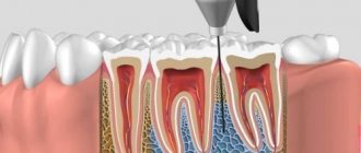
A targeted photograph of the tooth is taken using a radiovisiograph. It is a compact device that operates locally and provides a clear image of an individual tooth for immediate, on-site diagnosis. The image appears on the computer screen immediately after the procedure. This diagnostic technique is used in modern clinics and is considered a mandatory step in dental treatment.
When is it necessary to take a targeted photograph of a tooth?
Radiovisiography is used in the following situations:
- diagnostic search - the image visualizes the carious process and its depth, you can also detect pulpitis or periodontitis;
- the image allows you to assess the condition of the periodontal tissues, investigate the spread of the problem, its clear localization;
- During endodontic treatment (root canal treatment), several targeted images are required to monitor each stage of treatment.
Indications for orthopantomogram
An orthopantomogram (or “OPTG”, “panoramic image of the dental system”) is one of the types of diagnostic radiography. In dentistry, OPTG is of key importance - many types of treatment cannot be started without this diagnostic method.
Method of performing an orthopantomogram:
Technically, it is carried out as follows: the beam source (X-ray tube) and its receiver (film or digital sensor" move around the object under study in opposite directions. As a result, a very limited part of the object of study is in focus, everything else is blurred. Panoramic views are made photographs using orthopantomographs.The volume of radiation is such that panoramic photographs can be taken every day for a month without significant harm to health.
Before the procedure, the patient is put on a special lead apron to protect him from unwanted exposure to X-rays. An orthopantogram is performed from a standing position. A disposable cover is put on a special tube. The tube is clamped by the patient himself with his front teeth. The X-ray tube will rotate around the patient's head for a few seconds. Information from the sensor will be sent to a computer, corrected using special programs, and then this image can be printed on paper or film, and also saved in digital format. No other preparation is required.
Contraindications: first and second trimester of pregnancy, lactation.
Why do modern dentists use radiovisiography?
Compared to classic X-rays, which previously provided images on film, computer radiovisiography has a number of advantages that have forced massive X-ray machines out of dental practice:
- lower radiation exposure to the patient (in order to obtain an image on a computer sensor, less X-ray radiation is needed than to develop an image on film);
- saving time for the doctor and the patient (no need to develop the film);
- a high-quality image that the doctor can enlarge on a computer, examine it more closely, perform color correction, etc.;
- the image file can be stored on the computer for a long time, transferred and reused;
- The image is transferred to the doctor at the dental unit directly during treatment - this significantly shortens the treatment process.
A competent doctor will never begin treatment without confirming his diagnosis and treatment plan with an objective diagnostic method. Sighting image data is an indication for many treatment tactics and explains the need for dental procedures.
Safety of X-rays before implantation
When planning implantation and in the postoperative period, it is necessary to monitor the process of implant healing. Therefore, repeated orthopantomograms and CT scans are necessary. But is this amount of radiation harmful to the body?
| Type of examination | Exposure dose, m3v* |
| Sight shot | minimum radiation exposure - 0.01 |
| OPTG | minimum radiation exposure - 0.01 |
| CT (3D) | with partial CT - 0.02; with full CT - 0.05 |
*The permissible annual dose is 5 m3v. For comparison, the natural background of Moscow is 0.02 m3v.
Features of the technique and differences from standard photography
The difference between this technique is the replacement of conventional X-ray film with an electronic matrix of the visiograph sensor. The sensor connects to a computer and records x-ray radiation. The operating principle is similar to using X-ray film, but has its own differences:
- The radiovisiograph has a sensitive sensor, this reduces the radiation dose to the body, since the device requires less X-ray radiation to obtain an image;
- in one visit, the dentist can take 9–10 photographs without causing harm to the patient’s health;
- control of each step during the procedure, which makes it consistent;
- maximum speed of image acquisition, which is ahead of any other technique;
- the procedure does not cause discomfort to the patient, is easily tolerated and does not cause pain;
- the ability to quickly adjust the parameters of a targeted tooth image on the screen to obtain the most accurate data;
- the doctor can take measurements of the necessary structures automatically;
- storing images in a computer database and quickly searching them;
- Pictures can be transferred via e-mail, within the system and recorded on removable media.
Contraindications for the study
Sight radiography has such a low radiation dose that there are practically no contraindications to its implementation. It is not recommended to conduct radiographic examinations in the first trimester of pregnancy, therefore preparation for bearing a child should begin with sanitation of the oral cavity. Also, the procedure may not be comfortable for patients with an increased gag reflex in cases where examination of large molars is necessary. However, specialists carry out it very carefully, which most often does not cause any discomfort.
How is a targeted tooth image taken?
This technique does not cause pain to the patient. It is carried out in a short time and consists of the following stages:
- protecting the patient with a lead apron;
- installation of the sensor inside the oral cavity;
- the patient should not move for several seconds;
- The healthcare professional takes the photo and it automatically appears on the computer screen.
Some patients may experience a gag reflex if the sensor touches sensitive areas. The employee who performs the procedure will give recommendations on how to avoid unpleasant sensations. If you breathe deeply through your nose during the procedure, you can avoid the reflex. Moreover, you need to be patient for a few seconds. Sometimes, difficulties arise under the influence of anesthesia - this can interfere with holding the sensor. In this case, a clinic employee will also help by fixing the device in the patient’s mouth.
The described technique is a necessary diagnostic step in dentistry, which will make treatment safe and comfortable. Entrust your dental treatment to professionals and forget about toothache.
Thanks to the use of a radiovisiograph installed at the Le Dent clinic, the radiation dose is significantly lower and amounts to about 0.2 mSv. That is, even if the patient needs to take 10–15 targeted photographs of the tooth in one visit, he will receive the same radiation dose as if he had taken one picture on a regular X-ray machine.
To take targeted images of individual teeth, our clinic uses a Kodak 2100 with a visiograph. The radiation dose of this device is 30% lower than that of other similar models of radiovisiographs. Today, this is one of the safest and most convenient installations.
Stages of the procedure
To carry out the procedure, a separate room, equipped accordingly, is required. The procedure itself does not cause virtually any discomfort and consists of several stages.
- The position of the person in the chair, the doctor’s clarification of the location of the problem.
- It is imperative that the patient wears an apron that protects against harmful radiation.
- Fix your head to get a good shot.
- Taking an image with the patient in a stationary position.
The entire procedure from taking a photo to receiving it in electronic or printed form takes a short period of time - about 20 minutes.
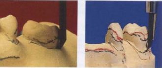
Analysis of the finished image
The dentist himself or a radiologist is responsible for describing the image. If the image quality does not meet the required quality, the procedure is repeated. The specialist examines the patient’s teeth, roots, and jaw bones in detail. If there are light and dark areas, this indicates the appearance of inflammation, cysts and other neoplasms.
In the pictures you can see inhomogeneities, damage to bone tissue, damage to the enamel, cement and root part, damage to the septum and other abnormalities that indicate the presence of a particular disease.
Features of performing a targeted shot for children
The younger generation needs this procedure more than adults, because caries develops more intensively in children. Another reason is to identify deviations and defects at an early stage of tooth growth. If any are detected, the necessary preventive and therapeutic measures are taken.
But it is not advisable to use the targeted photo procedure for children under 2 years of age. It is prescribed only in cases of birth or mechanical injuries, or to monitor the development of teeth.
Advantages and disadvantages
Each research endeavor has its pros and cons.
Advantages:
- Safety.
- High image quality.
- A large amount of information received.
- Convenient storage.
- Speed of execution.
Disadvantages for dentists include:
- It is impossible to detect cracks in the roots.
- Pictures of only one plane.
- Small viewing area.
- Narrow survey area.
- Limitations for use
The procedure is considered harmless and therefore has no visible contraindications for its implementation. Considering the minimum level of radiation (the permissible dose is 1000 microsieverts per year, i.e. about 100 images per year), a targeted image can be taken even during pregnancy. But, nevertheless, it is not recommended to resort to the procedure under the age of 2 years, in the presence of bleeding, reduced immunity, or in poor health.
A radiovisiograph provides a lower dose of radiation, an X-ray machine.
Price of the procedure
| Click to sign up for a FREE consultation |
Imaging services are provided in almost every hospital. Its cost will vary from 150 rubles and above. Sometimes the procedure is included in the cost of treatment.
In public clinics, under the compulsory medical insurance policy, you can take an x-ray and print the image for free.
Thus, a targeted image is a safe and effective procedure that can identify problems with the dental elements in the early stages if the patient seeks qualified help in a timely manner.
Further detailed analysis of the image
A detailed analysis of the image is carried out by a doctor who determines a treatment regimen or prepares for surgery, or by a radiologist. The dentist examines the image and evaluates the color of various areas. He makes a conclusion about the rigidity, density, homogeneity of the bone structures of both jaws, and the placement of parts of the dentition. The analysis makes it possible to identify carious cavities, granulomas, cysts, periodontitis and the consequences of injuries. The image shows areas of inflammation, blood vessels and nerves.
The most common dental diseases on a dental photograph are manifested by the following signs.
- Caries. This is the process of destruction of dental tissue. The picture shows areas of transparency of the enamel and the hard part of the tooth. The clearing zones have uneven, unclear contours.
- Pulpitis. Damage to the bone structure is characterized by heterogeneity of color in the interroot zone against the background of hypertrophy.
- Periodontitis. The progression of granulomatous periodontitis is accompanied by destruction of the hard part of the tooth and cement. Granulomas are observed in the area of tooth root formation, the interdental gap is enlarged, its edges are blurred.
- Periodontitis. The image shows signs of osteoporosis - the density of bone structures decreases, the height of the partitions in the dentition decreases, and pockets form.
To read the photo correctly, the image must be unblurred and in focus.