Author of the article:
Soldatova Lyudmila Nikolaevna
Candidate of Medical Sciences, Professor of the Department of Clinical Dentistry of the St. Petersburg Medical and Social Institute, Chief Physician of the Alfa-Dent Dental Clinic, St. Petersburg
At any age, a person can face such a problem as discoloration of tooth enamel. There are several reasons for this phenomenon, but one of the most common is fluorosis. Uneven coloring of the enamel disrupts the aesthetics of a smile and causes certain psychological discomfort during communication. One of the methods for restoring aesthetics in case of discoloration is microabrasion. We'll talk about what this method is and what indications there are for microabrasion of tooth enamel.
Fluorosis as the main indication for microabrasion
Dental fluorosis is a chronic disease that affects tooth enamel due to excessive accumulation of fluoride in the human body. The main cause of the development of the disease is long-term use of water and toothpaste with a high content of fluoride compounds. With fluorosis, enamel discoloration occurs, which manifests itself in the form of spots. The problem mainly affects children at the stage of formation of permanent teeth.
This problem especially concerns residents of endemic areas. In Russia, these are the Leningrad region, the Republic of Mordovia, the Nizhny Novgorod region, the Urals, and Western Siberia. In these regions, the concentration of fluoride in ordinary water exceeds sanitary standards (>1.5 μg/l). This also includes natural springs and artesian wells. In addition, in many cities, water is fluoridated for prevention. The danger of fluorides is that they accumulate not only in teeth, but also in bones, and if accumulated in excess, they can even lead to bone cancer - osteosarcoma.
The disease occurs in 3 stages:
- mild form of the lesion - small chalky spots and stripes on the surface of individual teeth;
- moderate damage - pigmented spots of yellow or brown color are added, covering several teeth;
- severe form - almost the entire tooth is covered with brown spots, while the crown of the tooth is deformed, the enamel is erased or chipped.
Enamel microabrasion is used only for mild or moderate fluorosis (spotted and chalky-mottled forms). But the indication for the procedure is not only fluorosis.
Microabrasion – gentle polishing of enamel to remove plaque and prevent caries
Every day, the enamel of our teeth is affected by a huge number of external factors.
Excessive consumption of sweets, coffee and tea, smoking - all this leads to gradual demineralization of the protective layer of teeth and, as a result, a change in its color. It is important to understand that this is not only an aesthetic problem. Under bacterial plaque, hard tissues are quickly destroyed, which in the absence of regular hygiene inevitably leads to the onset of carious processes. The situation can be corrected in the early stages using a microabrasion procedure. Microabrasion
is a gentle polishing of teeth to remove pigmented areas and prevent the development of caries. Most often, such changes include white spots, which cause aesthetic discomfort to patients.
Read more about what it is and when the technique is used further in this material.
Contraindications
The procedure is not recommended under certain conditions:
- tetracycline staining;
- moderate enamel underdevelopment;
- age-related staining;
- severely thinned enamel, cracks and erosive defects;
- wedge-shaped defects;
- exposed necks of teeth;
- severe form of fluorosis;
- pathologies of oral tissues;
- pulpless teeth.
Microabrasion is contraindicated for women during pregnancy and lactation. In addition, the procedure is not performed on baby teeth.
What are the pros and cons?
The technique has quite a few advantages, among which specialists and patients often highlight the lack of need for any preparation. In addition, the procedure is completely painless and does not cause any discomfort. If microabrasion is used to prevent further tissue demineralization, then after its successful implementation it is possible to avoid large-scale tooth preparation for its further restoration. In general, the technique saves in cases where other options for removing plaque and pigmentation do not help.
“They performed microabrasion on me. I found white spots on my front teeth, the doctor said this is how caries begins. After the examination, the dentist suggested grinding off a thin layer to ensure that the lesions were removed. I agreed. There was no pain at all during the procedure. Only then did sensitivity to cold and hot appear. Personally, everything went away for me literally in 3-4 days.”
Evgenia S.S., Moscow, from correspondence on the woman.ru forum
Among the disadvantages, one can highlight a fairly impressive list of restrictions on the use of the method. In addition, after the procedure, almost all patients experience increased sensitivity of the enamel layer - hyperesthesia goes away on its own within a few days, but for this you need to strictly follow the instructions of your doctor.
How does the procedure work?
Previously, eliminating cosmetic smile defects was possible only through the use of composite materials and the installation of crowns. Modern technologies make it possible to get rid of uneven staining of teeth without boron preparation; one of these technologies is microabrasion, or micro-abrasive treatment. The essence of the method is to grind off a microscopically thin layer of enamel, followed by polishing. Thus, this is not a therapeutic event, but a cosmetic one.
Due to the fact that the impact is only on the upper layers of enamel - the thickness of the removed layer is usually no more than 0.2 mm - this procedure does not lead to serious irreversible consequences. Microabrasive treatment minimally changes the contours of the enamel surface and is a more conservative method than tooth restoration with composite materials.
Microabrasive treatment does not require anesthesia, as it is painless. Before the procedure, the doctor decides on the need for professional hygiene. The dentist then determines the patient's natural enamel shade using the standard Vita scale and creates a treatment plan. The patient's eyes are protected with glasses.
Algorithm for the procedure:
- isolation of the working field using a rubber dam. Performed to protect the mucous membrane;
- applying an abrasive mixture to the surface of the tooth being treated and rubbing it in using slowly rotating attachments - cups. For microabrasion, the drug Opalustre is used, which contains microparticles of silicon carbide and 6.6% hydrochloric acid;
- rinsing off If the result is positive, move on to the next stage, if not, repeat the previous one;
- polishing the tooth surface with fine fluoride paste to achieve an optimal aesthetic result;
- registration of color changes.
As a rule, one visit to the clinic is enough to eliminate enamel discoloration. The best results are achieved by a combination of microabrasion and clinical bleaching.
Complications after microabrasion
One of the most common complications of the microabrasion procedure is hyperesthesia, caused by a high degree of demineralization of tooth enamel. Treatment of this pathology comes down to rinsing the mouth with a 10% solution of calcium gluconate and excluding sour, spicy foods and too hot or cold dishes from the menu.
An illiterate approach to the selection and application of an abrasive mixture can cause erosion on tooth enamel or lesions on gum tissue. Such complications can be dealt with by regularly applying antiseptic and anti-inflammatory agents to the affected areas, as well as drugs that accelerate regeneration processes.
Efficiency and safety
Microabrasion allows you to get rid of enamel discoloration. As a result, not only the appearance of the tooth improves, but its further destruction is stopped. In addition, micro-abrasive treatment allows you to avoid tooth grinding and restoration using expensive composites. After the procedure, age spots do not return.
Efficiency depends on the degree of damage: microabrasion helps either completely eliminate surface stains on the enamel or mask discolored areas. Provides a natural glossy shine to the enamel surface. The result is noticeable immediately after the procedure.
The main complication after the procedure is increased tooth sensitivity. To eliminate it, it is recommended to use special toothpastes and rinses for sensitive teeth, for example, low-abrasive ASEPTA Sensitive toothpaste. Additionally, it is necessary to use remotherapy products as an opportunity to mineralize damaged areas, for example, ASEPTA Plus Remineralization. This professional toothpaste restores weakened and damaged enamel, saturates it with essential macro- and microelements.
What results can be achieved?
After abrasion of incisors, patients receive the following results:
- the enamel becomes light, while the top layer of tissue is not damaged;
- careful elimination of aesthetic defects;
- prevention of caries due to demineralization of teeth;
- correction of unevenness of hard tissues;
- improving the appearance of the dentition while protecting against decay;
- smoothing the surface after removing braces;
- getting rid of dense pigmented plaque;
Microabrasion is often carried out in childhood after the end of the period of mineralization of hard tissue on the permanent incisors.
Clinical researches
ASEPTA products are clinically proven effective. For example, repeated laboratory tests have proven that regular use of preventive toothpaste ASEPTA SENSITIVE for a month can reduce bleeding gums by 62%, reduce sensitivity of teeth and gums by 48% and reduce inflammation by 66%.
Clinical studies have proven that regular use of professional toothpaste ASEPTA REMINERALIZATION improved the condition of the enamel by 64% and reduced tooth sensitivity by 66% after just 4 weeks.
Sources:
- Report on determining/confirming the preventive properties of toothpaste “ASEPTA PLUS” GENTLE WHITENING” Author: doctor-researcher A.A. Leontyev, head Department of Preventive Dentistry, Doctor of Medical Sciences, Professor S.B. Ulitovsky First St. Petersburg State Medical University named after. acad. I.P. Pavlova, Department of Preventive Dentistry
- Clinical and laboratory assessment of the influence of domestic therapeutic and prophylactic toothpaste based on plant extracts on the condition of the oral cavity in patients with simple marginal gingivitis. Doctor of Medical Sciences, Professor Elovikova T.M.1, Candidate of Chemical Sciences, Associate Professor Ermishina E.Yu. 2, Doctor of Technical Sciences Associate Professor Belokonova N.A. 2 Department of Therapeutic Dentistry USMU1, Department of General Chemistry USMU2
- Clinical studies of antisensitive toothpaste “Asepta Sensitive” (A.A. Leontyev, O.V. Kalinina, S.B. Ulitovsky) A.A. LEONTIEV, dentist O.V. KALININA, dentist S.B. ULITOVSKY, Doctor of Medical Sciences, Prof. Department of Therapeutic Dentistry, St. Petersburg State Medical University named after. acad. I.P. Pavlova
- The role of anti-inflammatory rinse in the treatment of periodontal diseases (L.Yu. Orekhova, A.A. Leontyev, S.B. Ulitovsky) L.Yu. OREKHOVA, Doctor of Medical Sciences, Prof., Head of Department; A.A. LEONTIEV, dentist; S.B. ULITOVSKY, Doctor of Medical Sciences, Prof. Department of Therapeutic Dentistry of St. Petersburg State Medical University named after. acad. I. P. Pavlova
- Report on determining/confirming the preventive properties of toothpaste “ASEPTA PLUS” COFFEE and TOBACCO Author: doctor-researcher A.A. Leontyev, head Department of Preventive Dentistry, Doctor of Medical Sciences, Professor S.B. Ulitovsky. First St. Petersburg State Medical University named after. acad. I.P. Pavlova, Department of Preventive Dentistry
- Report on determining/confirming the preventive properties of commercially produced personal oral hygiene products: Asepta toothpaste used in combination with Asepta mouthwash and Asepta gum balm Head. Department of PFS Doctor of Medical Sciences Professor S.B. Ulitovsky St. Petersburg State Medical University named after Academician I.P. Pavlova. Faculty of Dentistry. Department of Preventive Dentistry.
Description of the process by stages
The microabrasion procedure is carried out in several stages
The patient is asked to take a seat in the dental chair so that he is comfortable. Microabrasion does not require anesthesia, but a positive psychological attitude and human comfort are important. Then he is relaxed, calm and does not interfere with the doctor during work.
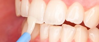
When the patient is prepared to begin the procedure, a special composition in the form of a paste is applied to the surface of the teeth, which contains carborundum, an abrasive gel with silicon and hydrochloric acid. To apply the composition, the doctor uses a special rubber nozzle. After this, keep the paste on the teeth for some time so that it has time to react and soften the enamel layer. Then it is removed with special rubber cups - nozzles.
Now step-by-step microabrasion:
- The dentist uses a special gel (“Axil” or “Rubberdam”) to isolate healthy teeth and mucous membranes from the area that will be treated
- A special retractor is installed
- The paste is carefully applied and rubbed with rotating rubber nozzles into the surface of the tooth.
- The dentist removes excess composition
- Remo-gel is applied to those areas where lesions have appeared, which restores lost minerals and is a stimulant for the restoration of hard tooth tissue. Remineralization is carried out only in cases where there are indications for this
- In conclusion, the doctor examines the teeth that have been treated, gives the patient all the necessary recommendations for caring for the oral cavity and teeth after microabrasion, and gives a special leaflet on these issues.
If necessary, the procedure is repeated after some time. This depends on the depth of penetration of caries and the contamination of the patient’s teeth.
Bleaching for fluorosis
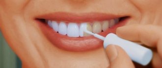
Many people wonder if teeth whitening will help with fluorosis? This method can be applied to the streaked or spotted form, when the spots still have a white tint. Whitening will even out the color of teeth and stains. Minor defects can also be sanded down and the enamel remineralized. If the spots darken, it will be possible to get rid of fluorosis only with the help of prosthetics with orthopedic structures such as veneers, lumineers or crowns.
Diagnosis of dental fluorosis
High-quality differential diagnosis is the key to successful treatment and determination of adequate therapeutic tactics. The professionalism of the dentist is important; based on the results of a visual examination, clinical symptoms and anamnesis, he can determine the form and extent of the disease at the first appointment. A dental examination is carried out according to a standard procedure.
- Mild cloudiness of the enamel is detected by drying the surface, which makes it possible to assess the condition of the protective coating in the area of pigmentation changes.
- The vital staining technique is effective - in case of fluorosis, damaged areas of enamel are not exposed to methylene blue dye.
- Determination of the degree of dental damage is carried out using luminescent diagnostics. A mild form of the disease in ultraviolet rays manifests itself as a weak glow; with moderate and severe degrees, partial quenching of fluorescence is observed.
- To exclude caries, microradiography is prescribed. The study also allows us to determine the depth of the lesion in the middle and deep layers of dentin.
Changes in enamel pigmentation may be associated with other pathologies or a lack of vitamins D and A. In this case, the condition of connective tissues worsens, bone fragility, a tendency to inflammatory processes in the gums and roughness of the surface of the teeth are noted.
