Mechanical damage to a tooth due to injury without violating its integrity is called a bruise. This type of injury is common in children and adults, especially athletes. According to statistics, among all injuries to primary teeth, bruises account for 2.5%; bruises of permanent teeth account for 1.5% of the total number of traumatic injuries in the permanent dentition.
Even if the tooth looks unchanged after a bruise, it is necessary to visit a dentist to prevent the development of complications associated with the injury.
Symptoms
A strong blow to a tooth is accompanied by damage to periodontal tissue, and some fibers and small blood vessels rupture. There are no visible structural damage to the tooth; upon visual inspection, it appears intact. After a bruise, the tooth remains motionless, and minor mobility is rarely observed. The gums in the area of the injured tooth swell.
In the first hours after the impact, patients experience pain in the tooth, which intensifies when biting; the pain is aching in nature. The tooth feels high, and slight bleeding may occur from under the gum near it. When a bruise occurs, the neurovascular bundle of the tooth can be damaged, that is, the pulp is injured, and hemorrhage occurs in the pulp chamber, and the enamel becomes pink in color. A severe bruise can lead to the death of the pulp.
Often, when a bruise occurs, cracks appear on the tooth enamel, which can only be detected with a special examination.
Luxation of a baby tooth
Luxation of a baby tooth is a fairly common dental problem among pediatric patients. In this situation, the upper and lower incisors are most often injured, less often the premolars and molars. In the absence of professional medical assistance, such a condition can cause inflammation of the bone and surrounding soft tissues and lead to deformation of the dentition.
What is a luxated baby tooth?
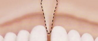
In clinical terminology, luxation of a baby tooth is its persistent pathological displacement in relation to the tooth socket. Depending on the nature of the displacement, the dislocation can be incomplete or complete.
Incomplete dislocation, or traumatic dystopia, is a shift that occurs as a result of rupture or stretching of the structural components of the supporting apparatus of the tooth (periodontal ligaments). In this condition, there is a change in the position of the baby tooth vertically, in the anteroposterior or lateral direction.
Complete dislocation of a baby tooth in a child (extraction) occurs due to a total rupture of the periodontal and circular ligaments and the neurovascular bundle. In this case, the tooth completely loses connection with the socket and falls out of the soft tissues.
Causes of complete and incomplete dislocation of a baby tooth
- Mechanical injury (fall, blow).
- Biting into very hard food.
- Foreign body ingestion.
- Incorrect removal of a nearby tooth.
- Opening bottles with teeth, etc.
Clinical signs of luxation of a baby tooth
With incomplete displacement, the baby tooth changes its position and becomes mobile. If it emerges from the socket, its cutting edge protrudes above the edges of other teeth. During a traumatic rotation, the dystopic tooth may be located at an angle to the longitudinal axis, leading to malocclusion. In this condition, children complain of pain when biting, and minor bleeding from the periodontal gap may also develop.
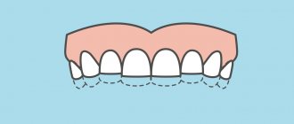
Intrusion, or impacted dislocation of a baby tooth (immersion of the crown into the bone tissue of the alveolar process), is accompanied by bleeding, swelling of the lips and gums (a consequence of stretching of the neurovascular bundle). With a large impact force, the injured tooth can be found in the jawbone or soft tissues.
With complete dislocation (traumatic extraction) of a baby tooth, moderate bleeding from the socket, swelling of the gums and lips, and damage to the soft tissue of the alveolar process are observed.
Treatment
Partially avulsed baby teeth are secured with a metal splint or a plastic tray, and those that cannot be repositioned are usually removed. In case of impacted dislocation, the tooth is left in the socket (within a certain time its growth can be restored). It is secured with a metal splint or a plastic mouthguard. If post-traumatic inflammation develops, removal is performed.
If a baby tooth is completely dislocated and falls out of its socket, treatment is prescribed purely individually, taking into account the condition of the bone tissue at the tooth root and the viability of the pulp. In this situation, with unchanged periodontal tissues, tooth replantation is possible with subsequent anti-inflammatory and restorative drug treatment.
Diagnostics
- Patients with a tooth injury are referred for x-ray diagnostics to exclude a root fracture. To obtain information using targeted radiography, it is sometimes necessary to take several images from different angles, which is undesirable for the patient.
The most accurate information about the condition of the roots is provided by computed tomography - an x-ray examination method that allows you to obtain a three-dimensional image of the tooth. The examination results are displayed on the computer screen and must be transferred to a CD or USB flash drive.
An X-ray examination of a tooth bruise reveals a slight widening of the periodontal fissure.
- The condition of the pulp after injury is monitored using EDI - electroodontodiagnosis. The method consists in determining the reaction of the nerve endings of the pulp to the influence of electric current. The level of electrical excitability of the pulp depends not only on its condition, but also on the degree of formation of the tooth root.
The examination is carried out 2 or 3 days after the injury, since on the first day the pulpal response may be reduced due to traumatic neuritis. Be sure to perform an electrical test on adjacent healthy teeth to compare sensitivity levels. 3-4 weeks after the injury, EDI is repeated.
- Another method of examination for a tooth bruise is transillumination, the essence of which is to pass a beam of cold light through the tooth and evaluate shadow formation. If there are cracks in the enamel after an impact, they will be clearly visible in the stream of light; The technique also helps to detect pulpitis. In modern clinics, all dental units are equipped with light guides for transillumination examination.
Tooth pain after a blow to the face: causes and cures
Go back Despite their strong structure, our teeth are susceptible to damage due to mechanical stress. A hit to the face can immediately provoke a number of problems with them. Perhaps the most unpleasant of them is tooth pain - a symptom indicating the seriousness of damage to the injured area. How can you quickly get rid of this problem?
Causes
The blow to the face itself may occur on some other part of the head, but pain in the tooth will still most likely occur. By all indicators, this injury is a typical bruise - a closed injury without violating the integrity of the damaged object in anatomical terms. With it, the tooth remains in place, without changing its position relative to the jaw and other teeth, but some parts of it are affected. The mechanism of a bruise is simple: damage occurs to the dental ligaments, which fix the tooth in place and attach it to the jaw bone. With a strong impact, there is even a serious risk of their complete rupture. A severe tooth bruise can lead to tangible consequences. First, there is the possibility of damage to the dental nerve, which can lead to loss of sensitivity. Secondly, a blow to the face can also cause destruction of the dental pulp, which can lead to tooth loss. In this case, pain becomes a signal that dental problems require an immediate solution.
Main symptoms of tooth bruise
Knowledge of the symptoms of a tooth bruise will help you in correctly recognizing the nature of the damage. As a rule, experts identify four main characteristics of a bruised tooth:
- Severe pain (especially in the first few hours after a blow to the face), problems with a correct and painless bite.
- A feeling that the tooth has changed its position relative to the jawbone and other teeth (the victim begins to feel as if the tooth is located a little higher or is bent to the side).
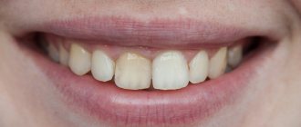
- When blood vessels and the dental nerve rupture, the tooth itself may change color to pink (due to the release of blood inside it). After a few days, the color may change from pink to dark brown (after blood clotting).
- A good indicator of a bruised tooth is the gum near it: if there is damage, it will be inflamed, and the tooth itself may be slightly mobile.
How to cure a bruised tooth?
First aid for a bruised tooth after a blow to the face is to reduce pain. The victim should apply a cold compress to the injury site to help relieve swelling. You should avoid eating solid food for three to five days after the injury. If the pain is too acute, then it is recommended to use such powerful painkillers as analgin, paracetamol or tempalgin. We remind you that in any case you will have to see a specialized doctor: as mentioned above, after a blow to the face there is a risk of rupture of the dental nerve or destruction of the pulp, and therefore in order to avoid complications you will definitely need the help of a professional.
Complications after injury
With bruises, the prognosis is usually favorable, but in some cases the injury can lead to the following complications:
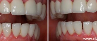
- Darkening of the enamel. After a bruise, the cause of darkening is hemorrhage into the pulp chamber: the pink color of the tooth darkens over time, the enamel acquires a brownish or gray tint. Depulped teeth tend to darken due to the fact that metabolic processes in them stop, the teeth become “dead”, and the enamel becomes dull.
- The death of the pulp leads to the development of pulpitis: the pulp decomposes, inflammation develops in the tooth, which without treatment turns into periodontitis.
- Periodontitis is inflammation of periodontal tissue. It can be: post-traumatic, occurring a short time after the injury; chronic, develop as a consequence of pulpitis. When a purulent infection occurs, there is a high risk of tooth loss and purulent blood poisoning.
- The appearance of a post-traumatic odontogenic cyst at the root apex during the development of post-traumatic periodontitis.
- Stopping the development of roots of permanent teeth in children.
- In case of injury to milk teeth, the following are possible: disruption of the formation of the rudiments of permanent teeth, their death.
Dental injuries in children. First aid.
Dental injuries in children. First aid.
Injuries and damage to teeth are very common in childhood. In the primary dentition, injuries occur in children from 2-3 years of age; this period is associated with the beginning of the development of coordination and the first attempts at independent movement and exploration of space; in the mixed and permanent dentition, the peak of injuries occurs at 8-12 years of age (in most cases, injuries recorded in boys at this age), the predominant causes of injuries at this age are falls, road traffic accidents and active sports. The upper and lower incisors are most often injured. The fate of the tooth and the amount of treatment in the future depend on the correctness and speed of actions of parents and relatives in the first minutes after the incident.
It is very important not to panic and try to assess the extent of the damage.
Complete dislocation (Avulsion)
If a complete rupture of the tooth ligaments occurs during an impact, it may completely leave the socket, and bleeding from the socket is also observed.
The situation is an emergency, requiring urgent contact with a dentist, you have 20 minutes to save the tooth. First of all, we look for the lost tooth, we try to hold only the crown part, without touching the root, if it is dirty, we quickly rinse it with water for a second, and the best option is to immediately return the tooth back into the socket on your own, with the convex side outward, focusing on the neighboring one tooth. To maintain the position, you need to clench your teeth or bite down on a gauze pad. Then go to the clinic to fix the damaged tooth (splinting). If it is impossible to straighten the tooth yourself, put it in milk and contact the clinic as soon as possible, minutes count.
After replantation and splinting, tetanus prevention and careful hygiene are necessary, as well as dynamic observation with X-ray control, and timely filling of the canal in order to avoid complications.
Dislocation, displacement
There is a change in tooth position and mobility. The gums may be damaged, ruptured, or bleed. A misaligned tooth can make it difficult to close your mouth.
Do not remove the tooth from its socket, touch it, or try to close the teeth together. Try to contact the clinic as soon as possible
If it is impossible to contact a doctor quickly, try to return the tooth to its place with light pressure, focusing on a symmetrical tooth, fix the position by closing your teeth, and seek help from the clinic as soon as possible.
In some cases, root canal treatment may be required (50% of cases)
Impacted dislocation (intrusion)
The tooth is motionless, but has become shorter in appearance or is not visible at all; swelling and bleeding from the gums may be observed
Contact the clinic immediately for reduction and splinting, or orthodontic rehabilitation may be prescribed.
In 90% of impacted dislocations, root canal treatment is required.
Fracture
A fragment of the crown of the tooth is missing.
Try to find the fragments and put them in a saline solution or milk, the dentist will fix them back. If the fragments are lost, try to get an appointment with a dentist during the day or the next day to have a composite restoration performed. If the pulp is exposed, it is not always necessary to depulp the tooth; direct coating with a therapeutic dental material based on MTA can be performed. In this case, dynamic monitoring is necessary every 3-6-12 months
In many cases, such consequences can be avoided; take care of protection for your children during outdoor activities, roller skating, scootering, and also playing sports - protective helmets, sports mouth guards.
Treatment
- For mild bruises, treatment consists of resting the tooth for 3-4 weeks by reducing the load during chewing: the menu includes soft and semi-liquid foods, and a blender is used to grind hard foods.
- To ensure rest for baby teeth, temporary bite separation with the help of mouth guards is used; if a permanent tooth is bruised, splinting is performed. The splint allows you to immobilize an injured tooth and redistribute the load during chewing onto healthy teeth.
- If the pulp dies due to an impact, the tooth cavity is opened and the pulp is removed, after which the root canals are filled and a permanent filling is installed. If the crown of a tooth darkens, it can be whitened.
- When a baby tooth is bruised, grinding of the cutting edge of the crown of the antagonist tooth is used to prevent tooth contact and reduce pain. This method is not used for permanent teeth.
- To relieve pain, it is recommended to take an anesthetic tablet (ibuprofen, ketorolac, nimesulide) and apply an anesthetic gel (Dentol, Kamistad) to the gums around the tooth.
- When swelling of the soft tissues of the face accompanies a bruise, cold compresses are applied: a plastic bottle with cold water (not lower than +4°C) is wrapped in a cloth and held on the area of swelling for 15-20 minutes.
- A course of magnetic laser therapy is carried out - a combined effect on injured tissue of a magnetic field and low-intensity laser radiation. The method helps improve healing processes, relieves swelling and inflammation. The course consists of 10 daily procedures lasting 5 minutes.
- UHF therapy is indicated to accelerate tissue regeneration.
Prevention
To prevent dental injuries, you must:
- Observe safety precautions at work, arrange the workplace and carry out potentially hazardous work in accordance with established rules.
- Properly equip playrooms for children and supervise children while playing.
- Follow traffic rules and use seat belts when traveling, as even minor emergency braking of the car carries a high risk of injury.
- Avoid conflict situations that lead to fights.
- Ensure the safety of sports games using special equipment (helmets, masks, dental guards).
- Carefully select sports grounds: they must comply with safety requirements.