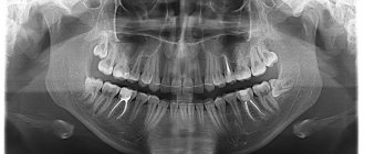
An x-ray is the dentist’s main tool in making the correct diagnosis. However, a conventional orthopantomogram or targeted photograph has limited diagnostic potential and does not provide complete data on the condition of the teeth and maxillofacial area. But technologies are constantly being modernized and today, more informative technology has come to the aid of conventional radiography - dental computed tomography (CT).
What does a 3D dental x-ray show?
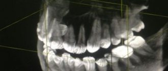
3D dental tomography is a highly accurate diagnostic method that makes it possible to obtain a three-dimensional image of the dental system in different projections. Volumetric images obtained with CT allow the specialist to enlarge, rotate and examine the area of interest from all sides and at different depths:
- The entire maxillofacial apparatus.
- A dentition or an individual tooth.
- Paranasal sinuses.
- Bone and periodontal tissues.
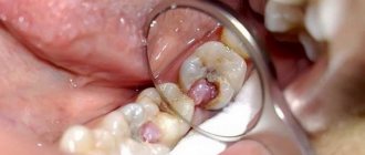
Dental CT allows you to detect inflammation, assess the homogeneity of the filling material and check the quality of installation of a filling, crown or implant, see the number of dental roots and their fragments, identify neoplasms, assess the degree of curvature of teeth, determine the exact parameters of bone tissue (height, width, density, etc.). d.). The information obtained allows the doctor to optimize treatment measures and predict the result.
Dental treatment during pregnancy: possibilities, features, contraindications
Features of dental treatment during pregnancy
The treatment approaches in this case differ from the methods of providing care to an ordinary patient. There are some nuances to this process:
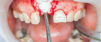
- Dental treatment during pregnancy is necessary if caries, periodontitis, pulpitis have developed, or inflammatory processes occur in the gums (stomatitis, periodontitis, gingivitis);
- Both chemically curing materials and compositions, the curing of which is carried out through the use of special lamps that are not dangerous for the unborn baby, can be used as a filling material;
- There are procedures that are not recommended during pregnancy, and among them are chemical teeth whitening and implantation. The latter is not used due to the fact that implantation is a process that requires a lot of body resources, and they are spent on bearing a child. In addition, often after such a procedure there is a need to use antibiotics, which are contraindicated for expectant mothers. In this regard, the implants most likely will not take root;
- It is necessary to carry out treatment during pregnancy with anesthesia, since the fear and pain that can arise without its use is a serious stress for the woman’s body. Not every drug is suitable for expectant mothers, so it must be selected taking into account the individual characteristics of the patient - and only from the list of approved drugs. Therefore, before starting treatment, it is necessary to first discuss all the nuances with a doctor, including a gynecologist;
- it is permissible to carry out procedures such as installation of prostheses (crowns, bridges);
- General anesthesia should not be used when carrying a child.
One of the most difficult dental procedures is tooth extraction, and it can be done, but only in some cases, since it requires the use of strong anesthetics and, after surgery, antibiotics. The procedure is indicated in situations such as the presence of a large cyst, a purulent inflammatory process, a fracture of the tooth root or crown, severe pain that cannot be treated with other methods. If the situation allows you to postpone surgery, it is usually recommended to perform it after birth.
Dental treatment in different trimesters of pregnancy
At each stage of gestation, medical care has its own characteristics, so the patient should inform the doctor about the due date.
The following periods are distinguished:
- I trimester. Speaking about when you can treat your teeth during pregnancy, doctors assure that this can be done at any time. But at this stage, the specialist needs to be as careful as possible in choosing methods, means and determining acceptable manipulations. At this time, the tissues of the organs and systems of the fetus are intensively formed, as a result of which some medications can negatively affect this process.
- II trimester. It is considered as safe as possible for dental procedures. It is at this time that you should visit a doctor for prophylaxis in order to prevent problems with gums, which can be susceptible to various diseases. This is due to increased blood supply during this period, which can cause bleeding, provoking the development of pathogenic microflora. As for treatment during pregnancy at this stage, it is carried out in case of acute conditions of the patient that threaten the loss of teeth. Manipulations aimed at achieving a cosmetic effect are not performed.
- III trimester. During this period, medical actions are carried out in cases of extreme necessity: in case of acute pain, inflammatory processes, situations in which there is a risk of tooth loss.
Attention! Each patient’s case is individual, so the doctor needs to develop a treatment strategy during pregnancy with an appropriate approach. In addition, it is advisable to consult a gynecologist. Expectant mothers are recommended to visit the dentist once a month.
Features of diagnosis during pregnancy
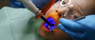
In addition to an in-person examination by a doctor, one of the main examination methods before treatment is an x-ray. It is necessary for a doctor to study tissues that are not visible to the eye. There are often cases when it is impossible to start the process without this procedure, including during pregnancy, since it is impossible to determine the volume and location of the affected areas. Many expectant mothers are afraid of x-rays, believing that the radiation can harm the baby. But it is minimal, so it does not have a negative impact on the child.
Safe pain relief during dental treatment during pregnancy
This is another point that expectant mothers beware of when going to the dentist. Local anesthetics are used for pregnant women. They do not penetrate the bloodstream, affecting only the area in which medical manipulations are performed, so they cannot have a negative effect on the fetus.
Not all painkillers are recommended for use in such cases; the most commonly used drugs are:
- "Ubistezin";
- "Artifrin";
- "Ultracaine";
- "Alfacaine".
Attention! Only a doctor should choose a drug, based on the problem the patient presented with, the general condition of the woman herself and other important factors.
The answer to the question of whether it is necessary to treat teeth during pregnancy is obvious. This must be done if the disease strikes. But it is important to contact a reliable, qualified, experienced specialist for this, who will take all measures for high-quality and safe treatment for the patient and her baby.
Why are dental x-rays prescribed?
A 3D photograph of teeth is performed if the following indications exist:
- Injuries of the maxillofacial area.
- Preparation for endodontic treatment (structure of root canals, pathological processes in the periodontium, degree of pulp damage, etc.).
- Diagnosis of neoplasms (cysts, abscesses, granulomas, tumors).
- Anomalies of development and deformation of the maxillofacial apparatus.
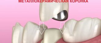
- Quality control of filling and implant installation.
- Planning of orthodontic treatment (identification of impacted and dystopic teeth, analysis of the condition of the tissues around each tooth, etc.).
- Detection of hidden periodontal cavities and pockets.
- Implantation planning (assessment of jaw bone parameters, indications for sinus lift or osteoplasty, modeling the result of implantation).
- Endogenous pathologies of the maxillary sinuses.
Three-dimensional x-ray examination is the gold standard when planning any complex dental procedure or surgery. CT allows you to quickly make an accurate diagnosis, competently plan treatment or dental prosthetics, and monitor the results.
Initial examination
Before dental implantation, you need to visit the dentist's office for an initial consultation. During this procedure, the doctor will carefully examine the oral cavity, assess its condition and give a referral for x-ray diagnostics and tests. Already at this stage, the dentist can:
- determine indications and contraindications for implantation;
- inform the patient about the features of the upcoming operation.
In addition, during the initial examination, the patient must fill out a questionnaire (survey card), in which the presence of bad habits, chronic diseases and surgical interventions should be noted.
| We strongly recommend that you fill out the survey card honestly. Then our doctors will know better how your body will behave during implantation. |
What equipment is used
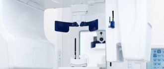
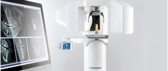
To carry out 3D diagnostics, a three-dimensional computed tomograph SOREDEX Scanora 3D with advanced functionality is used. This is the latest generation equipment, which allows you to obtain three-dimensional images of the anatomical structures of the maxillofacial region in a few seconds, with the least radiation exposure for the patient.
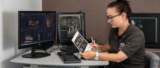
The program analyzes the obtained multiplanar sections and builds them into a 3D model, thanks to which the specialist is able to accurately assess the condition of the dental system, detect all pathological processes occurring in this area and competently plan a treatment regimen.
A virtual 3-dimensional model of the scanned area can be recorded on any digital media (CD, flash drive), which allows the attending physician, if necessary, to view diagnostic data or involve related specialists in the analysis of the received information.
Possible harm
Cone beam dental computed tomography is the safest and fastest diagnostic method. Thanks to the use of a conical X-ray beam, the radiation dose received during the study is 10 times less than when using spiral CT. And the pulsating mode of the X-ray beam further reduces the radiation dose. The three-dimensional computed tomograph SOREDEX Scanora 3D is one of the safest devices in terms of X-ray radiation dose - only 0.035 m3v.
However, despite the safety of the study, CT also has contraindications. If we just talk about dental tomography, it is not performed during pregnancy (in the 1st trimester). 3D dental x-rays with contrast are prohibited for pregnant and lactating women, patients with endocrine disorders (diabetes mellitus, thyroid pathologies), renal failure and intolerance to iodine-containing drugs.
Computed tomography after implantation
In some cases, a CT scan may be required after implant placement. It is prescribed to determine the reasons for the instability of the artificial root, identify complications, and confirm the engraftment of the rod.
If you need to do a CT scan of the brain, you must inform your doctor about the presence of implants. Metal objects can distort the image, but doctors with many years of experience can correctly interpret CT results if a patient has titanium roots or metal pins.
Online consultation with a doctor
If you are concerned about the condition of your teeth. Painful sensations arise, gums bleed for a long time, and seals have appeared on the jaw. You are concerned about previously installed dental implants. And there was a need to take a 3D photo of the teeth. Then, after receiving three-dimensional visualization, it is better to go for an examination or consultation with a dentist to interpret the images and compare the results with your current complaints. It is impossible to independently understand the nuances of a 3D image, much less make a diagnosis. The specialist will explain the situation and give recommendations before the in-person appointment.
Advantages of the method
- The ability to rotate, enlarge, and examine images in any projection and section, which is impossible with conventional 2-dimensional scanning.
- The examination lasts only a few seconds (8-20 seconds).
- Complete diagnostic information.
- Maximum security.
- Digital information format.
- Detection of any pathological processes at an early stage.
- No prior preparation required.
- 3D reconstruction without distortion or artifacts.
- A wide range of purposes - from endodontic dental treatment and implantation to maxillofacial operations.
Cost of dental examination
The cost of an orthopantomogram in clinics in Moscow and the regions starts from 700 rubles. But sometimes patients undergo it for free, for example, during turnkey implantation, when the total price of dental restoration already includes all the necessary diagnostic methods, consultations, implants, dentures and the work of specialists.
Chibisova M.A. Digital and film radiography in outpatient dentistry, 2004.
Author: Bespalov R. D. (Thank you for your help in writing the article and the information provided)
Is there an alternative to CT
There are many other diagnostic imaging methods (x-ray, orthopantomogram, ultrasound, etc.), but only CT provides the possibility of highly accurate, separate images of all types of tissue at different angles and to different depths. Although a panoramic dental photograph remains an equally important diagnostic tool for a dentist today, it can only provide a general overview. In turn, a 3D tomogram allows you to obtain not a single flat image of the jaw, but a whole series of sequential multiplanar images in different projections and without the distortions inherent in a panoramic image.
Example:
due to the different density of bone structures exposed to X-ray radiation, it is impossible to see less dense bone in a 2-dimensional image; accurate information is provided by a 3- D image of the teeth.
Acceptability during pregnancy
Panoramic tomography is extremely undesirable for patients during pregnancy - in such cases it is performed only if there is an urgent need. The procedure poses an increased risk during the first trimester. In later stages of pregnancy, X-ray radiation will not have such a severe effect, but can still cause certain disturbances in the functioning of internal systems. For example, the thyroid gland is especially sensitive to the effects of radiation.
How does the procedure and decoding work?
To take a 3D photograph of teeth, a standing or sitting patient needs to bite a special plate and fix his position in the device using a fixing stand. During the entire scanning time, you must remain absolutely still.
The tomograph sensor makes a series of revolutions around the patient’s head for 8-20 seconds, producing about 200 images in different projections. Processing digital data takes 5-15 minutes, after which the information is written to a disk or flash drive. No preparation is required, you just need to remove all metal jewelry from your neck, ears, and hair before the procedure.