Diagnostics plays an important role in any field of medicine, and dentistry is no exception. It is very important for the doctor to see the full picture of the patient’s condition in order to make an accurate diagnosis and select the appropriate treatment. In dentistry, diagnosing something is not so easy, which is explained by the difficulty of accessing some parts of the oral cavity. In some cases, a 3D image of the teeth helps clarify the picture. What is it and how is it made?
Methods for diagnosing dental condition
Most methods for diagnosing the condition of the teeth and oral cavity fall into the category of visualization, that is, they can clearly show everything that happens in this part of the body. At a minimum, it is now completely impossible to imagine dentistry without radiography. Now this type of examination is included in the standard set of procedures that a patient undergoes when visiting the clinic with any complaint.
On a note! An examination is necessary not only when diagnosing the condition of a specific tooth, but also if the patient is faced with problems with the functioning of the entire maxillofacial apparatus.
Table. Main types of diagnostics in dentistry.
| View | Description |
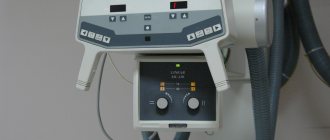
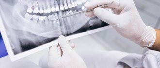
| This is the most common diagnostic option, which can be carried out in any, even the most modest clinic. It provides the opportunity to correctly and accurately assess the condition of the canals in the roots of the tooth and identify developing pathologies in the area of hard tissues. Often used for endotonia. An X-ray image is a small black and white image on a special film or on a CD. | |
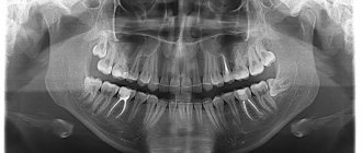
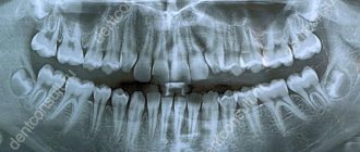
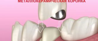
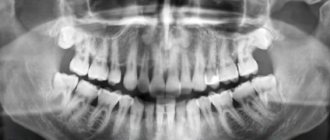
| This is otherwise called a panoramic image of the dental system. Using it, the doctor will be able to identify pathologies in the development of the jaw in general and teeth in particular, detect all carious cavities, understand how much hard tissue is destroyed during periodontal disease, clarify whether endotonic treatment helped the patient, etc. The image will also allow you to see whether there are any or deviations or changes in the condition of the lower sections of the maxillary sinuses. An orthopantomogram is also done in relation to the temporomandibular joints. It is often prescribed before prosthetics. | |
| This is a picture of not just the jaw, but the entire skull in one projection or another. It is necessary for measuring parts of the facial part (cephalometry). Based on this image, you can plan treatment with an orthodontist. | |
| It is this picture that is called 3D, which is discussed in the article. We'll talk about it separately below. |
Where can I take a photo of a tooth?
Computed tomography is becoming an increasingly common procedure offered by dentistry in Moscow. However, unlike a digital orthopantomogram, it can so far only be found in large clinics offering a wide range of services. You can always find out where to take a 3D photograph of a tooth in Moscow from the specialist who referred you for this study. The price for a computed tomography scan of the jaw, or 3D dental image, in Moscow varies from clinic to clinic and starts from approximately 2,500 rubles.
Publisher: Expert magazine about dentistry Startsmile.ru
Author of the material: Ekaterina Gasparova
What is a 3D photo?
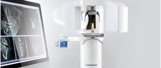
A 3D image or computed tomography of teeth is an x-ray that is performed using a special device - a tomograph (CBCT). The picture is a three-dimensional image that demonstrates the entire dental system in detail. The resolution of the image is high, which means that even the smallest details can be seen in it. The doctor, using a computer program, will be able to thoroughly examine absolutely any part of the patient’s jaw, moreover, at any depth and at any angle. The snapshot is provided on disk, where it is stored. If you need to view the disc, you can insert it into your computer and see everything you need on the monitor. Moreover, sometimes this diagnostic option saves the patient from a number of unpleasant manipulations on the part of the doctor.
On a note! The scanning volumes of such an image vary from 5x5 to 13x15 cm.
An orthodontist, implantologist, maxillofacial surgeon and other doctors can give a referral for such a plan. The technique is very accurate, and often without it, doctors simply will not do most of the manipulations. And the effectiveness of treatment thanks to this image increases significantly.
The advantages of taking a 3D photo are as follows:
- high image accuracy and detail;
- the ability to view all parts of the mouth - the image is three-dimensional;
- errors are excluded when identifying the location of pathology development, since there are no distortions in the image;
- the ability to assess the condition of the jaw from any side;
- the picture is taken quickly - a few seconds are enough to get a detailed image;
- taking a picture is safe for health, since the radiation exposure experienced by the patient’s body is extremely small. Especially if you compare its level with the level with the same x-ray.
What is a 3D dental image?
If you visit an orthodontist, maxillofacial surgeon, implantologist, or even a plastic surgeon for a consultation, most likely one of his first appointments will be to do a computed tomography (CT) scan of the jaw, or a 3D dental scan. This is a modern and very accurate diagnostic technique that allows the doctor to view an image of the patient’s jaw from any angle and in any projection, even when the patient has already left his office. CT, unlike an orthopantomogram, provides a three-dimensional image of the jaw without distortion and allows you to look into any layer of tissue, making a kind of virtual section, without the need to perform unnecessary traumatic procedures on the patient live. The efficiency and safety of diagnosis and treatment are thus significantly increased.
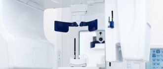
How is a 3D photo taken?
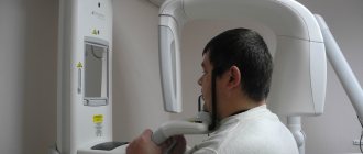
The image is taken using a special tomograph. The procedure is carried out as follows:
- a person takes a sitting or standing static position;
- then a special protective apron is put on his chest - the same as used during radiography;
- there is a special small plate in the mouth that you need to bite with your teeth;
- then you need to rest your forehead against a special support, and grab the handrails with your hands to avoid unnecessary movements;
- then you need to freeze for about 15 seconds at the doctor’s command to get an accurate image;
On a note! The tomograph will make only one full revolution, but will have time to take about 200 mini-images.
- After this, the pictures are processed and compiled into one overall picture - this is how the desired three-dimensional image is obtained. The treatment only takes a few minutes.
At the exit, the patient receives a disk with an image recorded on it, which he brings to the doctor to assess the condition of the jaw system.
How do you take a 3D photo of your teeth?
A dental CT scanner is similar to an orthopantomograph. During the procedure, the patient stands or, in special cases, sits. A protective apron with a lead layer is put on the neck and chest. The patient squeezes a special bite block with his teeth, rests his forehead against the fixing support, and holds the handles with his hands. This allows him to take the correct position and freeze for 14 - 24 seconds, depending on the selected mode and shooting frequency. This is exactly how long it takes for the tomograph’s planar sensor to make one revolution around the patient’s head. During this time, the sensor manages to take about 200 pictures in different projection sections. Processing the data and building a 3D model takes a few more minutes, plus recording the tomogram onto a disk - and after 10 - 15 minutes, the most accurate information about the state of your dental system is in your hands.
Indications for this type of examination
3D tomography has a lot of possibilities, for which it is valued by doctors. Thus, it will allow you to assess the condition of not only the jaw, but also the gums, see the quality of previously installed fillings, and reflect all pathologies and anomalies. Indications for this may include:
- abnormalities in the structure of the jaw and the shape of the teeth;
- suspicions of pathological changes in dental tissues;
- identification of the rudiments of permanent teeth in children, assessment of the condition of milk teeth;
- the presence of cysts, inflammation, cracks in the roots of the tooth;
- suspicion of traumatic injuries to the jaw;
- preparation for implant installation;
- the presence of tumors in the jaw area;
- preparation for plastic surgery;
- preparation for operations in the jaw area.
Thus, a detailed three-dimensional image is necessary in all complex cases that require detail and a thorough assessment of the patient's current condition. It is especially important in assessing abnormally growing teeth, as well as in case of complex fractures and dental pathologies.
On a note! A CT scan is also often performed during preparation for implant placement. Moreover, its efficiency and effectiveness are much higher than that of orthopantomography. It is often necessary when performing a so-called sinus lift.
Table. Areas of application of CT.
| Sphere | Explanation |
| Orthodontics | The ability to assess how distorted the bite is. |
| Surgery | Assessment of the possibility of certain manipulations by the surgeon, as well as the possibility of installing teeth. |
| Maxillofacial Surgery | Assessment of the condition of the jaw and surrounding areas of the skull. |
| Therapy | The ability to detect deep-lying caries and choose the right treatment option. |
Computer X-ray of teeth: indications
Among the indications for computed tomography of the jaw, or, as it is also called, 3D dental imaging, are anomalies in the structure of the jaw, the temporomandibular joint, the presence of impacted (not erupted or not fully erupted) and dystopic (rotated in the wrong direction or grown in the wrong direction) in its place) teeth; complex fractures of the jaws, damage to the dentition; preparation for maxillofacial operations. CT is also significantly superior to orthopantomography in preparing and performing implantation, in diagnosing inflammation in the area of tooth roots, determining the number of roots and root canals of a tooth, diagnosing cracks in the roots and detecting tumors at the initial stage; when assessing the severity of periodontal and periodontal disease. When planning a sinus lift or bone grafting, you will also have to take a 3D photo of your teeth. If bone tissue is clearly visible on a regular x-ray, then CT also allows you to see soft tissues, blood vessels, canals and changes in the mucous membrane. It is believed that a 3D image of teeth is more informative than one panoramic image and a set of targeted images of all teeth together.
Are there any contraindications?
This procedure also has contraindications. Whatever one may say, there is an effect on the body from x-rays. In general, the load is small, especially if you remember the SanPiN standards (1 mSv/year), and is only 0.045-0.06 mSv. However, there is still a load with CT. So the indications for a CT scan must be justified - it is not advisable to just take a picture.
A 3D photo cannot be taken or is permitted with caution under the following conditions:
- pregnancy, especially the first months when the fetus is actively developing;
- breastfeeding period;
- problems related to the thyroid gland;
- diabetes;
- certain forms of allergies;
- chronic renal failure.
Attention! In some cases, a doctor, despite contraindications, may prescribe a 3D image, but only if the risk of the study is completely justified, and doctors have no other choice.
There is also a so-called tomography with contrast agent. It makes it possible to obtain a more detailed picture of soft tissues, so it is used extremely rarely to take photographs of the jaw. But it has many more contraindications than conventional CT. Substances that may cause undesirable effects should not be used as contrast. It is important to inform your doctor or specialist before the examination if you have any allergic reactions.
How to prepare for a 3D photo?
Step 1. The first thing you need to do is visit a dentist to understand whether it really makes sense to spend money on such an expensive examination. In some cases, a dental CT scan may not be strictly necessary.
Step 2. Next, if the doctor has confirmed the need for an examination, it is important to evaluate the market for this service in your city and choose a clinic based on the price-quality ratio.
Step 3. Then it is important to sign up for a convenient day and arrive at the clinic at the appointed time.
Step 4. Before taking the photo, it is recommended to clean your mouth by chewing gum or brushing your teeth.
Step 5. After the picture has been taken, you need to receive the results and take them to an appointment with your doctor.