Hello! Today we will talk about a procedure used in dental practice, x-rays. And the most common question we hear from our clients is how safe is this procedure?
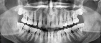
Our clinic performs two types of X-ray examinations using a radiovisiograph and a computed tomograph. Using them, we can take a picture of one or several teeth, as well as a panoramic picture of the entire oral cavity.
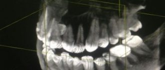
What is the difference between a radiovisiograph and a computed tomograph? On a radiovisiograph we see all teeth in a two-dimensional projection. It's essentially a photograph of a tooth. On a computed tomograph we can see all organs in a three-dimensional projection.
What can a doctor see on an x-ray?
He can see:
- total number of teeth;
- anatomical features of the structure of the maxillofacial region;
- level, bone height;
- possibility of installing implants.
X-rays are also done for the purpose of establishing a diagnosis, when the doctor cannot make one based solely on the patient’s complaints.
X-rays are also done to monitor the doctor’s work. It shows how:
- canals are sealed;
- tooth removed;
- an implant was installed.
17-hydroxyprogesterone is a precursor to cortisol, a hormone produced by the adrenal glands and involved in the breakdown of proteins, glucose and fats, maintaining blood pressure and regulating the immune system.
Synonyms Russian
17-OPG.
English synonyms
17-hydroxyprogesterone, 17-OHP, 17-OH progesterone, progesterone -17-OH.
Research method
Enzyme-linked immunosorbent assay (ELISA).
Units
ng/ml (nanograms per milliliter).
What biomaterial can be used for research?
Venous blood.
How to properly prepare for research?
- Do not eat for 2-3 hours before the test; you can drink clean still water.
- Unless directed by a doctor, women should be tested on days 3-5 of the menstrual cycle.
General information about the study
The test determines the amount of 17-hydroxyprogesterone (17-OPG) in the blood. 17-Hydroxyprogesterone is a precursor to cortisol and is used by the body to produce it.
Cortisol is a hormone produced by the adrenal glands; it is involved in the breakdown of proteins, glucose and fats, maintaining normal blood pressure and regulating the activity of the immune system. The production of cortisol is stimulated by adrenocorticotropic hormone (ACTH), which is produced by the pituitary gland. Cortisol concentrations typically fluctuate throughout the day, with hormone levels peaking at 8 a.m. and then decreasing in the evening. In addition, the level of cortisol in the blood increases during illness and stress.
Several special enzymes are required to produce cortisol. When one or more of these are deficient or dysfunctional, abnormal amounts of cortisol and its precursors are produced, causing 17-hydroxyprogesterone to accumulate in the blood. The adrenal glands use excess amounts of 17-hydroxyprogesterone, producing a lot of androgens. Excess androgens, in turn, can cause masculinization and promote the development of male sexual characteristics not only in men, but also in women.
So, hereditary deficiency of special enzymes and, as a result, excessive amounts of androgens lead to a whole group of functional disorders in the body, collectively known as “adrenal hyperplasia.” Most often it is caused by a deficiency of the enzyme 21-hydroxylase, which is the cause of the disease in 90% of cases. Adrenal hyperplasia is inherited in mild or severe form.
In more serious cases of congenital adrenal hyperplasia, 21-hydroxylase deficiency and excess androgens can result in a female child being born with blurred genitalia, making it difficult to determine the baby's sex. Boys with this disease appear normal at birth, but subsequently their sexual characteristics begin to develop prematurely. In girls, hirsutism is likely, first in childhood and then during puberty, then irregular menstruation occurs and signs of masculinization appear. Almost 75% of patients of both sexes affected by 21-hydroxylase deficiency due to congenital adrenal hyperplasia produce less aldosterone, a hormone that regulates salt retention. In newborns of both sexes, this can lead to a life-threatening “salt loss crisis”: too much fluid accumulates in the body and therefore too much salt is excreted in the urine. In some cases, patients with these symptoms may have low blood sodium levels (hyponatremia), high potassium levels (hyperkalemia), low aldosterone, and increased renin activity. This severe, although less common, form of the disease is often detected during a preventive medical examination of newborns.
In the milder, most common form of the disease, only partial enzyme deficiency may be observed. Sometimes this form is called late, or non-classical, congenital adrenal hyperplasia - its symptoms can appear at any time in childhood, puberty or in adults. However, they may be mild and may develop slowly over time. And although this form of adrenal hyperplasia does not pose a threat to life, it is still dangerous for problems with growth, development of the body, as well as abnormal puberty in children, which can cause further infertility.
75% of newborns with 21-hydroxylase deficiency caused by adrenal hyperplasia also produce less aldosterone, a hormone that regulates salt retention in cells. The loss of too much fluid and salts along with urine threatens acute insufficiency of the adrenal cortex and the so-called salt loss crisis.
What is the research used for?
- For a preventive medical examination of newborn children - in order to find out whether the baby has congenital adrenal hyperplasia or any hereditary disease that is caused by specific gene mutations associated with a deficiency of enzymes involved in the formation of cortisol. Almost 90% of cases of congenital adrenal hyperplasia are caused by mutations in the CYP21A2 gene, which leads to 21-hydroxylase deficiency and accumulation of 17-hydroxyprogesterone in the blood.
- To screen for congenital adrenal insufficiency before symptoms appear, or to confirm adrenal hyperplasia if symptoms of the disease already exist.
- For the diagnosis of congenital adrenal hyperplasia in older children and adults who have a milder form of “late” hyperplasia. If a diagnosis of 21-hydroxylase enzyme deficiency is made, treatment is prescribed that involves suppressing the production of adrenocorticotropic hormone and replacing deficient cortisol with glucocorticoid hormones. However, a 17-hydroxyprogesterone test may be periodically necessary to monitor the effectiveness of treatment.
- Together with tests for other hormones - to exclude adrenal hyperplasia in patients with symptoms such as hirsutism and irregular menstrual cycles.
- To monitor the condition of women suffering from polycystic ovary syndrome, infertility, and occasionally to monitor the condition of patients with suspected adrenal or ovarian cancer.
When is the study scheduled?
- During preventive medical examination of newborns. If increased values are obtained, it is repeated to confirm the initial result. It may be prescribed if the child has symptoms of adrenal insufficiency or disorders associated with the loss of salts from the body. Signs of the disease may include lethargy, weakness, lack of appetite, dehydration, and low blood pressure.
- If congenital adrenal hyperplasia is suspected in a child (indistinct genitalia, signs of masculinization, acne and early growth of pubic hair). Sometimes - if a milder (late) form of congenital adrenal hyperplasia is suspected in older children or adult patients.
- Girls and women with hirsutism, irregular menstrual cycle, masculinization or infertility (these symptoms are very similar to the symptoms of polycystic ovary syndrome).
- Periodically in case of 21-hydroxylase enzyme deficiency (to monitor the effectiveness of treatment).
What do the results mean?
Reference values
| Reference values | ||
| Up to 2 months | 0.96 - 10.46 ng/ml | |
| 2 months - 1 year | 0.66 - 2.81 ng/ml | |
| 1-10 years | 0.2 - 0.8 ng/ml | |
| For 10-18 years old - Tanner stage: | ||
| I | ||
| II | ||
| III | 0.33 - 1.98 ng/ml | |
| IV | 0.33 - 2.31 ng/ml | |
| V | 0.33 - 2.64 ng/ml | |
| Men | 0.2 - 3.1 ng/ml | |
| Women | Folliculin phase | 0.4 - 1.51 ng/ml |
| Luteal phase | 1 - 4.51 ng/ml | |
| Postmenopause | 0.2 - 0.9 ng/ml | |
| Pregnancy | 1-6 weeks | 1.32 - 3.30 ng/ml |
| 7-14 weeks | 1.1 - 2.8 ng/ml | |
| 15-24 weeks | 1.65 - 4.62 ng/ml | |
| 25-33 weeks | 1.98 - 10.2 ng/ml | |
| 34-40 weeks | 2.64 - 13.20 ng/ml |
Reasons for increased 17-OPG levels
Normal levels of 17-hydroxyprogesterone indicate the absence of congenital adrenal hyperplasia due to 21-hydroxylase deficiency.
A significantly increased concentration of 17-hydroxyprogesterone in a newborn most likely indicates the presence of congenital adrenal hyperplasia.
Moderately elevated levels of this enzyme may indicate a less severe form of adrenal hyperplasia, and 11-beta-hydroxylase deficiency (another enzyme defect associated with congenital adrenal hyperplasia) is also likely.
Reasons for decreased 17-OPG levels
Low or decreasing concentration levels in patients diagnosed with hyperplasia indicate a positive effect of treatment.
What can influence the result?
- In prematurely born children, increased levels of 17-hydroxyprogesterone are often observed.
What is an x-ray?
This is one of the types of radiation. If you think that X-ray radiation is present only in the X-ray room, then we can say right away that you are very mistaken. Let's remember physics lessons. We receive small doses of radiation every day:
- outdoors, from a natural source of radiation - the sun;
- at home from household sources located in the apartment: from a TV, various gadgets, a refrigerator, etc.
Radiation is measured in sieverts. What is the rate of radiation that a person can consume? Acceptable is 1000 sieverts per year.
Now let's remember mathematics and calculate how many X-rays can be taken for a person. A targeted image of one tooth is equal to 2-3 microsieverts. You can take 300-500 x-rays per year. A panoramic photograph (all teeth) is equal to 16-18 microsieverts. You can take 60-70 x-rays per year.
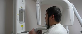
Now let's look at computed tomography. 1 photo taken on this device is equal to 60-80 microsieverts. You can take 16-20 x-rays per year.
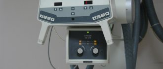
We are now considering the maximum dose limits for X-ray equipment. Our clinic has a radiovisiograph and a computer tomograph of the latest generation made in Germany. The load on the devices is reduced to a minimum.
Previously, when taking an x-ray, the exposure time (you hear it as a beep) was approximately 2-3 seconds. Now this time has been reduced to 0.05 seconds.
Now let's talk about household radiation. If you watch TV for 3 hours at a distance of less than 2.5 meters from the screen, you get approximately 0.5 millisievert, i.e. Watched TV for 5 days, get 1 x-ray. If you sit in front of a computer monitor for more than 3 hours, you get 1 microsievert, 1 x-ray. We flew by plane to Turkey, where the flight is 3-3.5 hours, you get 10 millisieverts or 5 targeted shots. And add radiation from TVs, refrigerators, microwave ovens and you will understand that you will not receive terrible radiation in the X-ray room.
Edematous-proteinuric hypertensive gestosis (OPG-gestosis)
Edematous-proteinuric hypertensive gestosis (OPG-gestosis) develops more often at the 25th week of pregnancy or later, sometimes shortly before birth. Predominant in primiparous women. Risk factors are diabetes mellitus, kidney disease in the prenatal period, Rh incompatibility, the age of the pregnant woman is less than 18 and over 35 years, obesity, polyhydramnios, multiple pregnancies, etc. Characteristic of this pathology is high sensitivity to endogenous pressor peptides and amines.
Hypertension is nephrogenic in nature. Blood pressure level is one of the criteria for the severity of OPG-preeclampsia. So, with the first degree of severity, the blood pressure level reaches 150/90 mm Hg, with the second it reaches 170-180/100-110, and with the third it exceeds 180/110 mm Hg. An increase in diastolic blood pressure with a decrease in pulse pressure often predominates. Sometimes a complication such as eclampsia occurs. Preeclampsia, which develops against the background of previous hypertension, chronic glomerulonephritis and other diseases, has a more severe course.
OPG-gestosis must be distinguished from other variants of increased blood pressure in pregnant women, which are usually transient in nature. These include:
a) transient hypertension during hypercortisolism of pregnant women (physiological Cushing's syndrome) is caused by additional synthesis of ACTH in the placenta. Obesity, moon face, hirsutism, pink stretch marks and hypertension occur. After childbirth, a complete involution of symptoms is observed. No treatment required;
b) functional increase in GH during pregnancy: acromegaloid facial features appear, a moderate increase in blood pressure (it should be remembered that the criterion for hypertension in pregnant women is a blood pressure level > 130/80 mm Hg). Symptoms disappear after childbirth.
Diagnostics.
For OPG-preeclampsia: comprehensive examination of the kidneys. In the blood there is depression of all sterol and protein hormones, except prolactin. Total hyperlipidemia. Strengthening the adhesive properties of platelets, a chronic form of DIC syndrome is possible.
Treatment.
When treating hypertension in pregnant women, the safest first-line drugs are dopegit, long-acting nifedipine, hydralazine, labetolol. (beta-blockers can be used in the third trimester of pregnancy, and in earlier stages they cause fetal growth retardation. ACE inhibitors and AT1 receptor blockers are contraindicated (teratogenic effect, possibility of fetal death). Diuretics are not recommended due to the risk of reducing blood volume and reducing placental perfusion .
Can X-ray examinations be performed on pregnant women?
In the first half of the term, the study is done only according to strict indications. In the second half of the term, you can do as much research as you like.
Another interesting fact. People often ask X-ray technicians to wear more protective lead aprons. Let's say right away that this is useless. You receive the same dose of radiation from covered and exposed parts of your body.
And remember that a doctor will never order an x-ray just out of curiosity. X-ray is one of the types of diagnostics of the area of proposed treatment and the anatomical features of the oral cavity.
Come to our clinic and we will make every effort to make your stay comfortable and enjoyable!