From this article you will learn:
- how much does it cost to do a dental x-ray,
- what you need to know about radiation doses,
- Is it possible to take a dental photograph during pregnancy?
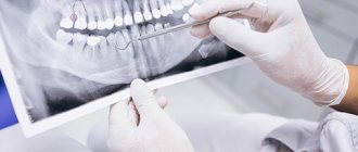
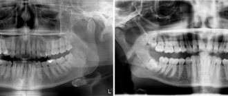
Dental X-ray is a traditional way to determine the quality of root canal filling, as well as to diagnose inflammation at the apex of the tooth root (with apical periodontitis). X-rays of human teeth in dentistry are most often carried out using targeted X-rays. A targeted photograph of a tooth received this name because it is small in size and shows the condition of only a few teeth and the bone tissue around them (Fig. 1-2).
Modern targeted X-rays produce a very low dose of X-ray radiation (compared to what it was 15-20 years ago). This is due both to the advent of ultra-sensitive photographic films, which require a significantly lower dose of radiation, and secondly, to the advent of special intraoral sensors that capture the image digitally and display it on a computer screen. Such digital images require several times less X-ray radiation - even compared to modern films.
Sighting and panoramic radiography in dentistry –
However, targeted images are not suitable for planning bite correction, assessing the condition of bone tissue before implantation, or planning the installation of future implants. They are not very convenient for planning treatment and prosthetics for a large number of teeth, and often do not allow detection of perforations and cracks in the tooth root. Focused photographs of the teeth may not be enough in cases where it is necessary to find problems with root canal filling in order to be able to properly treat the teeth.
Therefore, dentists very often refer patients to more complex options for x-ray examination of teeth and jaw bone tissue -
- dental computed tomography (CT),
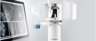
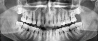
- orthopantomogram (Fig. 3),
- teleroentgenogram (for bite correction).
In this article, we will go into detail about the pros and cons of targeted dental x-rays, and what you need to pay attention to if you need to have a dental x-ray (for your own safety). For detailed reviews on the remaining specified methods of radiography in dentistry, read the links above.
Sight image of a tooth –
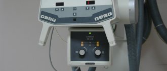
A targeted photograph of a tooth can be recorded either on photographic film or using a special intraoral sensor that detects X-ray radiation and transmits the image to a computer screen (such a device is called a radiovisiograph or simply a visiograph - Fig. 5). In both cases, an X-ray machine is used as a radiation source (Fig. 4), i.e. the only difference is in the way the image is captured - either on X-ray film or using a digital sensor.
Taking an image using a visiograph -
Digital vs Film Photography: Pros and Cons
Targeted dental x-rays using film were once the only examination option in clinics. It must be said that film photographs have a number of disadvantages that have significantly reduced their use. They require expensive consumables (film, reagents), time to develop photographs, there are difficulties with storing photographs, and over time they fade and are lost. There are also differences in patient safety.
Even modern X-ray films require 4-8 times the radiation dose compared to digital X-ray sensors. For example, the radiation dose to a patient for 1 film image is 10-15 μSv (microsieverts), and for a picture on a visiograph it is on average 1-3 μSv (this dose corresponds to the background natural radiation received by each person in 1 day).
The patient's exposure time when using film X-ray is 0.5-1.2 seconds, and using a digital visiograph sensor - 0.05-0.3 seconds. It is precisely by reducing the required exposure time when using a radiovisiograph that the radiation dose is significantly reduced. Thus, in one day of treatment at the dentist you can take no more than 3 film photographs and 5-6 digital photographs. And as you will see below, the use of a visiograph in some cases even allows you to take an X-ray of a tooth during pregnancy, but in compliance with all safety rules and for urgent indications.
How to take a picture of a tooth using a visiograph: video
Important: always try to take digital photos and inform them in advance that you want them saved to a flash drive. Firstly, then you will always have the pictures at hand, and you can always show them to another doctor. Secondly, the photographs taken for control after treatment will be your guarantee that if you received poor-quality treatment, you will always be able to prove it (the clinic will no longer be able to lose your photographs and rewrite the medical record).
Thirdly, if a digital image is printed on a printer, then the quality of the image will depend not only on the quality of the digital image, but on the resolution of the printer (rare clinics have printers that print in high resolution). Therefore, a photo in digital format will have better quality than a photo printed on paper.
Intraoral
Intraoral x-rays are used using a dental apparatus. Now equipment is being produced for this purpose, which can take both classic photographs using special film and digital ones.
Interesting! A portable version of this equipment can be purchased for 70,000-100,000 rubles.
Intraoral diagnostics
The patient must be completely still during dental x-rays, otherwise the image will be unclear. In order not to move, the patient sits in a special comfortable chair.
There are several radiography methods
Intraoral radiography is the main tool for identifying most dental diseases. Here are the modern intraoral radiography methods that are used today:
- intraoral contact;
- occlusal (intraoral bite);
- interproximal;
- telephoto
Interproximal radiography
For many years, the main way to detect dental diseases was contact x-rays, performed according to the bisector rule or isometric projection. This technique was created by the Polish dentist Cieszynski back in 1907. The main expectation of this technique is to obtain a clear image of the periapical tissues. To do this, the beam is centered at a point that is a projection of the apex of the root of the molar being examined. Another goal of the method is to obtain life-size images of molars. To keep distortion to a minimum, the isometric rule is applied. Simply put, the beam goes to the top of the tooth root, and always at a right angle to the bisector of the angle formed by the axis of the tooth and the plane of the film.
Important! If the beam goes any other way, the displayed tooth length will certainly be distorted.
Principles of "half angle" and "parallel radiography"
The image taken in this way should not be shorter than the actual dimensions of the tooth, and its length should not exceed the natural one by more than one tenth. On the other hand, it is impossible to follow the rules of isometry exactly. This is because it is very difficult to clearly identify in each patient the bisector of the angle created by the dental axis and the plane of the film. Therefore, they use the angle of inclination of the tube, calculated by observation and comparison for each dental group.
Here are the angles at which the X-ray tube should be tilted to the horizontal plane for each group:
- How to store urine for analysis before submitting it to the laboratory for a child and an adult
- molars - 25-30 degrees;
- premolars - 35;
- fangs - 45;
- incisors - 55.
Zonografiya
If you are shooting a bite, the angle of inclination should be 20 degrees greater. It is important to follow the orthoradial rule here. That is, the beam at the moment of the picture goes at a right angle to the tangent that was drawn to the dental arch in the area of the tooth being examined. Then his image is not distorted by overlapping images of molars in the neighborhood.
Interesting! For contact photography, a 2x3 or, alternatively, 3x4 cm film is used. And for bite-shot photography, a 5x6 or 6x8 cm film is used.
X-ray film showing a section of the jaw
In order to prevent injuries to the mucous membrane, the corners of the film must first be cut off. After that, it is placed in two small envelopes: first in one made of special paper that does not allow light to pass through, and this in a waxed envelope. The bag thus obtained is placed in the mouth, and the patient must press it against the hard palate, as well as against the alveolar process of the area being examined.
Important! For dental x-rays, the patient must be in a chair. His head must be fixed on the headrest exactly as required for the procedure.
If the upper jaw is being examined, it is important that the head is in a position where the wings of the nose and the external auditory canal are vertical. The doctor inserts the film exactly so that the edge does not intersect with the occlusal plane and protrudes from behind the teeth by half a centimeter.
X-ray film showing molars
If the lower jaw is examined, the patient’s head must be fixed so that the line that connects the angle of the open mouth and the tragus of the ear is strictly vertical. He needs to lift his head a little.
Sometimes it is necessary to specially adjust the beam so that separate images of the roots of multi-rooted teeth are received, or to determine how the roots interact with neoplasms. Then oblique intraoral projections are already used.
X-rays can be used to obtain a life-size image of a tooth.
Important! This method has a significant disadvantage - the inability to find out in what condition the marginal sections of the interalveolar ridges are. This is because it shoots with a slanted beam, making the image shorter. So this is useless for diagnosing the pathology of the gums and other tissues surrounding and fixing the tooth.
Interproximal
This method is used when it is necessary to obtain a high-quality image of the marginal sections of the alveolar jaw processes. In this way, bone resorption can be determined without embellishment over time. This method also represents the best opportunity to detect the carious process - approximal and cervical.
Cervical caries
The doctor here uses special film holders to place the film in the patient’s mouth so that it does not intersect with the dental crowns, but is at a certain distance from them. This way you can get a picture of symmetrical places on both jaws. The film is fixed with a piece of thick paper: it is attached to its wrapper and clamped between closed teeth. The beam goes at right angles to the film and the tops of the teeth.
Important! In the photographs you can immediately see the crowns of the teeth and the marginal sections of the alveolar sections of both jaws.
Interproximal radiography
Interesting! To study all the teeth at once, three to four images are required.
Occlusal
This method is popular and simple. They resort to it in the following cases:
- it is necessary to examine large areas of the alveolar process;
- you need to find impacted and dystopic teeth;
- when diagnosing illnesses in representatives of the younger generation;
- you need to examine the hard palate;
- it is necessary to obtain an image of the oral cavity for examination for calculi of the salivary glands - submandibular and sublingual;
- intraoral contact x-rays cannot be performed.
Occlusal radiography
Contraindications for intraoral contact x-rays are:
- 6 types of eye lenses and 6 solutions for their care: choosing the best
- jaw injuries;
- stiffness of the temporomandibular joint;
- tendency to reflex vomiting.
X-ray bite
X-rays can be used to find out what the cortical jaw plates are like in cysts, neoplasms, and also to find out the reaction of the periosteum. When taking bitewing x-rays, the doctor must follow the rules of bisector and tangent. The patient holds a 5x6 or 6x8 cm film with his jaws. During the x-ray procedure of the upper jaw, the film is not only clamped between the jaws, it is also first inserted deeply into the patient’s mouth. He sits in a special chair in such a position that the midsagittal cranial plane is clearly vertical, and the line between the tragus of the ear and the wing of the nose is horizontal.
Interesting! The beam goes to the root of the nose at an angle of 80 degrees.
With the help of such diagnostics, you can find out whether a person has any neoplasms
Long focal length
This is a technique, the essence of which is to shoot with non-intersecting rays. It was proposed in 1960. It is gaining popularity around the world, gradually replacing contact intraoral x-rays. Using the long-focus method, you can forget about the disadvantages of the contact method, taking advantage of all its advantages:
- vertical coverage of a fairly large area of the alveolar process;
- image of the entire tooth being examined;
- clear bone structure.
Long focal radiography
A rather tangible advantage of this method is that the image of the marginal sections of the alveolar processes is produced exactly in the form that completely corresponds to the real one. This allows this method to be used very widely in periodontology. For this purpose, the X-ray film is placed in the patient’s mouth so that it in no case intersects with the long axis of the teeth. To achieve this goal, the doctor uses special film holders or clamps to stop the bleeding. In principle, you can even use cotton or gauze rollers here.
X-ray film is placed in the patient's mouth
For this technique, a device with a more powerful tube is used, as well as a long, 360 mm, localizer tube. There must be a distance of 15 to 30 mm between the object and the film. The central beam always hits the film at an angle of either 90 or 15 degrees.
Interesting! The image is almost life-size.
Extraoral
Extraoral, also known as extraoral, technique is used by various equipment, not just dental equipment. Here you definitely need 13x18 or 18x24 film, as well as special cassettes with intensifying screens.
Extraoral radiography
It is used for sialography and fistulography.
Here are the possible indications of an extraoral x-ray:
- inflammation;
- tumors;
- injuries;
- extensive cysts;
- such periodontal diseases when it is impossible to take an intraoral x-ray.
Fistulography
Important! To compare joints, be sure to perform the procedure on the right and left.
Overview
This technique can be used in the following projections:
- straight;
- lateral;
- anterior semi-axial.
Survey radiography
This way you can completely photograph the skull. To perform a direct projection, nasofrontal or, alternatively, nasomental adherence to the cassette is required. X-rays in the nasofrontal projection should be done for cranial injuries and illnesses. It is also useful for sialography and fistulography.
- Radial artery. Topography. Deep palmar arch. Branches of the radial artery.
X-ray in nasofrontal projection
Important! Frontal X-rays are not designed to evaluate dental health.
Direct shots must be complemented by side ones. But the summative effect of both sides makes it too difficult to study the facial bones.
Sialography
Most often, only extensive changes in the bones can be noticed. Lateral x-rays reveal where foreign bodies are located.
X-ray of teeth in semi-axial projection
Axial, like anterior semi-axial x-ray, is done when it is necessary to examine all the structures of the cranial base and bones of the middle facial zone. For example, these:
- eye sockets;
- maxillary sinuses;
- zygomatic bones.
Dental X-ray – price for 2022
The cost of one digital X-ray in Moscow will average from 250 to 350 rubles in different clinics. In addition, this price may only apply to a diagnostic initial image, while all other images taken during the treatment phase may cost less (about 150 rubles per image). Therefore, you should carefully read the clinic’s price list.
It should also be noted that there are a large number of clinics in which the cost of dental treatment is indicated on an all-inclusive basis. Accordingly, the cost of treating your tooth will already include the required number of x-rays (usually 2-4 pictures), for which you will no longer pay anything extra.
What else you need to pay attention to is that the price list of some clinics may state that the cost of a targeted x-ray indicated on the clinic’s website applies only if you are a patient of this clinic. Therefore, if an image is taken to be submitted to another clinic, its cost may be 100 rubles higher).
In addition, if you need a printout of a digital image, some clinics may also charge you about 50 rubles for this. The same applies to the description of the x-ray image: if you want to receive a written description of the image taken by a radiologist, then in some clinics they may additionally ask you for about 100-150 rubles.
Radiation doses and safety –
A patient's radiation exposure is measured in either microsieverts (µSv) or millisieverts (mSv). The recommended radiation dose for the population received as a result of X-ray studies (according to the recommendations of SanPiN 2.6.1.1192-03) should not be more than 1000 μSv per year (= 1 mSv per year).
Below we will give examples of different types of images in dentistry and the corresponding radiation exposure to the patient (data from the Ministry of Health of Russia dated July 22, 2011 and December 21, 2012)…
- Targeted images on a digital radiovisiograph – → lower jaw in adults – 2 μSv, → lower jaw in children under 15 years old – 1 μSv, → upper jaw in adults – 5 μSv, → upper jaw in children under 15 years old – 3 μSv.
- Sight shots using film – 10-15 µSv.
- Digital panoramic image – 55 µSv, but if the patient is less than 15 years old – 24 µSv.
- Digital teleroentgenogram – 7 µSv.
Conclusions: thus, targeted images using a radiovisiograph provide the lowest radiation dose compared to other types of x-ray examination in dentistry. During one visit to the dentist, you can take 5-6 pictures on a digital radiovisiograph without risk to health, but no more than 100 such pictures during the year. A digital orthopantomogram (panoramic x-ray of the jaw) can be done 1-2 times a month, but no more than 10 times during the year. Panoramic films on film provide a greater radiation dose to the patient, and they can be taken less frequently than digital ones.
Is it possible to take a dental x-ray during pregnancy?
Is it possible to take a photograph of a tooth during pregnancy? This issue is regulated by the recommendations of SanPiN 2.6.1.1192-03. These recommendations do not prohibit taking dental x-rays during pregnancy, but it is strongly recommended to use x-rays only in truly necessary cases, for example, in case of acute pain and the provision of appropriate emergency care.
It should be noted that over the past 20 years, the radiation doses received by patients with 1 x-ray of the teeth have become tens of times less, thanks to the advent of radiovisiographs and ultra-sensitive photographic films, which require significantly lower x-ray radiation (24stoma.ru). Therefore, the risks of fetal pathologies have decreased significantly in recent years.
Of course, if possible, X-ray examination of teeth should be avoided, but there is nothing terrible about this today, because The radiation dose of 1 image on a radiovisiograph is approximately equal to the radiation dose of any person to natural background radiation in 1 day. In addition, the irradiation time on the visiograph will be only 0.05-0.3 seconds, which, if protective measures are observed (lead apron), can significantly increase the safety of the procedure.
Additionally, it is worth considering the following recommendations –
It is better not to do a dental x-ray during pregnancy in the early stages, because at this most important time for the laying of organs and tissues of the fetus. And if x-rays are taken, then it is in the second half of pregnancy, because the risks to the fetus during this period are significantly reduced. Please note that you can only take pictures using a modern digital radiovisiograph of the latest generation, because Their radiation dosages are significantly lower than those of outdated digital radiovisiographs and, even more so, film devices.
Are there any contraindications to X-ray examination?
Dental X-rays are not performed on patients with severe bleeding in the mouth, or those who are unconscious or in critical condition.
Pregnancy is considered a relative contraindication. In this condition, women should not undergo dental procedures involving radiation until 4-5 months. But in each individual case, the expectant mother needs to personally consult a doctor, since situations are different.
A breastfeeding woman can undergo an X-ray examination. But it is advisable to resort to digital methods and skip one or two feedings after the session.
Analysis of targeted dental images –
Analyzing X-rays is not difficult if they are of good quality.
Almost any patient will be able to see signs of periodontitis or cysts in the image, and will also be able to determine how well his root canals are filled. All you need here is skill. But it is worth remembering that absolutely everything cannot be diagnosed using X-rays, for example, inflammation of the nerve of a tooth. You can diagnose from the image -
- caries (but not in full),
- periodontitis (granulomas, cysts),
- quality of root canal filling after treatment of pulpitis and periodontitis,
- perforations and root cracks,
- fragments of instruments in root canals,
- presence of periodontal pockets...
1) A group of images indicating the development of inflammation (periodontitis) at the apexes of the roots of previously untreated teeth. In this case, you will always see a clear or vague darkening at the apex of the tooth root, which can be of different sizes and shapes.
2) A group of photographs taken after root canal filling. The first 2 pictures (Fig. 14-15) show what well-filled root canals look like. The following images show poor quality treatment and complications that arose (read the descriptions in each image).
What types of X-ray diagnostics are used in dentistry?
The main types of images obtained during x-ray photography of the dental system:
- Sighting. Provides information about the condition of one or more teeth and adjacent tissues. This helps the specialist assess the condition of dentin, root canals, periodontium, and blood vessels.
- Panoramic (OPTG, orthopantomogram). It is a detailed flat image of the jaws, teeth, joints, and sinuses. This image helps to detect carious areas, defects in fillings, impacted teeth, and cysts. And also draw up a correct plan for implantation, prosthetics, and treatment.
- Occlusal. This is the so-called bite radiography, which is often used when examining children, as well as when it is impossible to obtain clear results using targeted radiography.
In addition, photographs can be film or digital, when the resulting image is transmitted using a special sensor to a computer monitor. In the second case, the study is called radiovisiography.
Summary: important points
As a practicing dentist who knows the system from the inside, I want to draw your attention to the following points that are important for your safety. If the clinic has an X-ray machine, then a license must be obtained for it, the issuance of which presupposes the mandatory presence of a certified radiologist on the staff of the dental clinic. However, in reality, even in large clinics and public clinics, it is not always the case that x-rays will be taken by a trained specialist.
Even if he is, he may go on vacation or get sick, and a regular nurse (dental assistant) will take the pictures instead. This is a gross violation that leads to both the production of low-quality images and an increase in the radiation dose. In small clinics, the risks of receiving a poor-quality x-ray examination are much higher, and the first thing that makes you suspect a forgery is if the picture is taken not by a special employee, but by a nurse from the dentist you came to see.
The second very important point: if you see that the x-ray is not done in a special room, but the x-ray machine is located right next to the dentist’s chair, then you should change the clinic and the doctor. The fact is that all objects in this office (including the dental chair and doctor’s instruments) will have an increased background radiation, and this is no longer safe for health. We hope that our article on the topic: X-ray of the jaw and teeth was useful to you!
Sources:
1. Dental education of the author of the article, 2. Based on personal experience in therapeutic and surgical dentistry, 3. National Library of Medicine (USA), 4. “Digital and film radiography in outpatient dentistry” (Chibisova), 5. “X-ray diagnostics in dentistry" (Lutskaya I.K.).
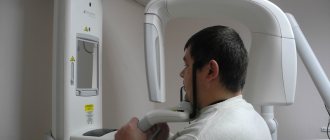
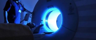
How is dental x-ray examination performed?
The photo is taken in a special room with lead-protected walls and floors. The doctor asks the patient to remove metal jewelry and accessories, as they may distort the results of the study. The vital organs of the body are protected by an apron with lead inserts. Features of the study depend on the method used.
When receiving a targeted image, the doctor directs an x-ray beam to the area of the jaw being examined from the side of the face or to an area of the oral cavity. An orthopantomogram is obtained in a different way. The patient fixes his head in a special device, grabs the plate in a disposable cover with his teeth and remains motionless while the mobile part of the device makes a full revolution around the head.