Harm of X-rays for pregnant women
Sometimes, while expecting a child, women need to sanitize their oral cavity. And in order to properly plan therapy, an orthopantomogram is often required. “Is it possible to take dental x-rays during pregnancy? What are the consequences? - expectant mothers often worry.Indeed, irradiation is undesirable during this crucial period. But in reality, not everything is as scary as you might think at first glance.
How dangerous is x-ray?
In many cases, dental treatment is simply unthinkable without X-ray examination. When you can't do without it:
- when filling thin and curved tooth canals;
- to detect hidden carious cavities, caries under fillings;
- when the question arises whether wisdom teeth need to be removed (for example, if they are incorrectly positioned);
- to diagnose a tooth root fracture, cyst, determine the degree of inflammation of the tissues around the diseased tooth;
- when the issue of removing or restoring a tooth is being decided.
Analysis of targeted dental images –
Analyzing X-rays is not difficult if they are of good quality.
Almost any patient will be able to see signs of periodontitis or cysts in the image, and will also be able to determine how well his root canals are filled. All you need here is skill. But it is worth remembering that absolutely everything cannot be diagnosed using X-rays, for example, inflammation of the nerve of a tooth. You can diagnose from the image -
- caries (but not in full),
- periodontitis (granulomas, cysts),
- quality of root canal filling after treatment of pulpitis and periodontitis,
- perforations and root cracks,
- fragments of instruments in root canals,
- presence of periodontal pockets...
1) A group of images indicating the development of inflammation (periodontitis) at the apexes of the roots of previously untreated teeth. In this case, you will always see a clear or vague darkening at the apex of the tooth root, which can be of different sizes and shapes.
2) A group of photographs taken after root canal filling. The first 2 pictures (Fig. 14-15) show what well-filled root canals look like. The following images show poor quality treatment and complications that arose (read the descriptions in each image).
Cost of services
Registration online
1200
Primary appointment (examination, consultation) with a pediatric dentist
1200
Registration online
Service code B01.064.003
Registration online
for free
Repeated appointment (examination, consultation) with a pediatric dentist
for free
Registration online
Service code B01.064.004
see all prices
In the first three months, it is not advisable to take dental x-rays for pregnant women, since the fetus has not yet formed. Radiation can have an adverse effect on dividing cells, cause chromosome abnormalities and provoke intrauterine developmental defects. By the second trimester, at 16–20 weeks, the fetus is already fully formed - a tiny dose of radiation will not harm the baby in any way, so pictures can be taken without fear.
Dental X-rays during pregnancy are much safer than examinations of the pelvis or chest, because the mother’s belly is reliably protected by a lead apron, and dental examinations are always carried out using special devices with reduced radiation exposure. This applies to both old models and modern visiographs.
Pregnancy is normal
Many people perceive pregnancy as an almost pathological condition, when a woman needs to be locked in a sterile room with airlocks and continuously fed with some kind of vitamins.
In fact, vitamins and so on are not bad, but only in a situation where pregnancy occurred suddenly and the expectant mother has not been eating properly for a long time. That is, we have the task of correcting an already existing deficiency, for example, in iron or folic acid. If the mother eats a nutritious and varied diet, eats fish, vegetables, meat and dairy products, then she is unlikely to need to take anything additional. For example, the same Cochrane provides a meta-analysis that indicates a weak evidence base in favor of the use of additional doses of folic acid. Therefore, there is no need to perceive a normal pregnancy without pathologies as something special. The child is reliably protected by the hematoplacental barrier and many membranes from harmful external factors. The mother's immune system is quite successful in protecting him from most dangers. The most dangerous viral diseases with a teratogenic effect, such as rubella, are no longer scary for vaccinated people. Therefore, we just walk, breathe fresh air, eat well and rejoice at the imminent addition to the family.
Periods of pregnancy.
The trimesters of pregnancy vary in their risks and characteristics. It is important for doctors to take this into account when prescribing medications and choosing treatment tactics.
The first trimester is the most critical for a child's development. It is during this period that the embryonic petals, internal organs are laid and the main systems of the body are formed. Any serious violations during this period usually lead to the fact that the embryo development process does not pass the “tests” and the pregnancy is terminated. This mechanism is important from an evolutionary point of view for culling genetically defective embryos at an early stage. It is estimated that 15–20% of pregnancies end in spontaneous abortion in the early stages. This number is probably even higher, since many women are unaware of their pregnancy and mistake its natural termination for a missed period.
Therefore, as a rule, any really harmful effects on the child in the first trimester simply end it. Very often mothers are frightened because, for example, they took antibiotics without knowing that they were pregnant. If the child successfully continued further development and moved into the second trimester, then most likely everything is fine. Although, of course, if we are talking about taking any medications, then we must definitely consult with an obstetrician about the potential consequences. During this period, it is important to avoid drugs that are potentially toxic to the embryo as much as possible, except for vital indications.
In the second trimester, the fetus is already more or less formed, internal organs and systems have taken shape. The child begins to actively use facial muscles and may already smile. At this stage, there is active development of the skeletal and muscular systems. Harmful effects can no longer lead to termination of pregnancy, but to disruptions in the development of systems that were formed at the time of the influence of the factor. For example, if the mother took tetracycline before six months of pregnancy, then the baby teeth will form with enamel defects and will be colored orange-brown. After the sixth month, the staining will already be on the permanent teeth.
In the third trimester, the main organs and systems completed their formation. Intensive weight gain and size growth are underway. At this stage, the risks of using any drugs are much lower than before. The most important risks that the doctor considers are provoking premature labor and infectious problems.
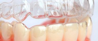
Visiograph - an alternative to x-ray
A visiograph is a modern analogue of an x-ray machine. Its advantages:
- Allows you to take an image instantly, high-quality data is immediately displayed on your computer monitor.
- A narrow beam of rays is directed exclusively at the diseased tooth, does not affect adjacent tissues, and certainly cannot “touch” the abdomen.
- The duration of exposure (irradiation) is 10 times lower than when using old-style X-ray machines and is only 0.05–0.3 seconds. Radiation exposure is only 2 microsieverts (when using old devices - from 7 to 80 microsieverts).
For comparison, just sitting in front of the TV for three hours and enjoying your favorite series, a mother receives a radiation dose of 5 microsieverts. The safe radiation dose per year is 1,000 microsieverts (that’s 500 shots). Of course, you shouldn’t take the procedure lightly, but you shouldn’t panic and overestimate the danger to the baby either.
Radiation doses and safety –
A patient's radiation exposure is measured in either microsieverts (µSv) or millisieverts (mSv). The recommended radiation dose for the population received as a result of X-ray studies (according to the recommendations of SanPiN 2.6.1.1192-03) should not be more than 1000 μSv per year (= 1 mSv per year).
Below we will give examples of different types of images in dentistry and the corresponding radiation exposure to the patient (data from the Ministry of Health of Russia dated July 22, 2011 and December 21, 2012)…
- Targeted images on a digital radiovisiograph – → lower jaw in adults – 2 μSv, → lower jaw in children under 15 years old – 1 μSv, → upper jaw in adults – 5 μSv, → upper jaw in children under 15 years old – 3 μSv.
- Sight shots using film – 10-15 µSv.
- Digital panoramic image – 55 µSv, but if the patient is less than 15 years old – 24 µSv.
- Digital teleroentgenogram – 7 µSv.
Conclusions: thus, targeted images using a radiovisiograph provide the lowest radiation dose compared to other types of x-ray examination in dentistry. During one visit to the dentist, you can take 5-6 pictures on a digital radiovisiograph without risk to health, but no more than 100 such pictures during the year. A digital orthopantomogram (panoramic x-ray of the jaw) can be done 1-2 times a month, but no more than 10 times during the year. Panoramic films on film provide a greater radiation dose to the patient, and they can be taken less frequently than digital ones.
How to minimize risks
What should an expectant mother do so as not to worry about the baby’s health?
- Not all clinics, especially municipal ones, have visiographs. It is better to inquire about this in advance.
- Be sure to use a protective apron and collar.
- You need to inform your doctor about the exact timing of your pregnancy. In case of a delay, even if the test shows a negative result, you must inform the specialist. In unclear situations, diagnosis (and treatment) are carried out as if pregnancy was definitely established.
- Severe pain, swelling, inflammation are good reasons not to delay a visit to the dentist, even in the first trimester. Unless absolutely necessary, the doctor will never prescribe dental x-rays during pregnancy and will try to do everything to postpone treatment to the 2nd, safe trimester or to the postpartum period. However, some acute conditions may require emergency intervention, such as purulent pulpitis or acute periodontitis. In this case, the mother needs to remember that a rapidly developing infection in the oral cavity will cause much more serious damage to the baby than 1-2 pictures on a visiograph.
- If there are concerns that the X-ray has somehow damaged the baby, you should consult with a geneticist and do an ultrasound. This will reassure those mothers who may have taken the photo without knowing that they were expecting a baby.
Since dental treatment during pregnancy is still associated with certain difficulties, the best thing an expectant mother can do is to go to the dentist and have all her teeth treated at least a month before the date of planned conception. And already in pregnancy, mommy should brush her teeth thoroughly and eat foods high in calcium.
Dental X-ray – price for 2022
The cost of one digital X-ray in Moscow will average from 250 to 350 rubles in different clinics. In addition, this price may only apply to a diagnostic initial image, while all other images taken during the treatment phase may cost less (about 150 rubles per image). Therefore, you should carefully read the clinic’s price list.
It should also be noted that there are a large number of clinics in which the cost of dental treatment is indicated on an all-inclusive basis. Accordingly, the cost of treating your tooth will already include the required number of x-rays (usually 2-4 pictures), for which you will no longer pay anything extra.
What else you need to pay attention to is that the price list of some clinics may state that the cost of a targeted x-ray indicated on the clinic’s website applies only if you are a patient of this clinic. Therefore, if an image is taken to be submitted to another clinic, its cost may be 100 rubles higher).
In addition, if you need a printout of a digital image, some clinics may also charge you about 50 rubles for this. The same applies to the description of the x-ray image: if you want to receive a written description of the image taken by a radiologist, then in some clinics they may additionally ask you for about 100-150 rubles.
Risks for mom
You can lose a tooth
We try not to use antibiotics, especially in the early stages of pregnancy.
This greatly changes the treatment tactics if we discover a chronic or aggravated purulent focus that has arisen due to an affected tooth. In a normal situation, we can try to save the patient's tooth under the additional cover of antibiotics that accumulate in the bone tissue. For example, this is a fairly common fluoroquinolone - ciprofloxacin. Everything is great, but it is strictly prohibited for use by pregnant women and children under three years of age due to the fact that it can cause defects in the formation of joints and cartilage. In a situation where the prospects for a tooth without antibiotic therapy in a pregnant woman are questionable, and the risks of developing an infection are high, we are usually inclined to remove such a tooth. It is much better to remove a potentially problematic tooth than to risk the health of the baby or the expectant mother. You cannot leave a chronic purulent lesion: it is dangerous for the child and for herself if it eventually worsens. If periostitis or, especially, phlegmon develops, the mother’s life will have to be saved. Then you will definitely have to prescribe antibiotics and other drugs.
Orthodontics
We stop the orthodontic treatment process if the patient reports pregnancy. First of all, we do this due to the impossibility of X-ray monitoring of the treatment process. Working with a bite blindly is a very bad idea and can end in even bigger problems. Most often, the patient remains at the stage when she found out about pregnancy. Either we continue to use the same aligner, or we simply fix the current condition in the case of braces.
Periodontal problems
The pregnant woman's body directs all efforts to pregnancy.
At the same time, he may ignore some of his own needs and problems. For example, large doses of sex hormones ensure good blood supply to the uterus and the desired condition of the mucous membranes. But in parallel, these same mechanisms lead to gum hypertrophy in the oral cavity. This is especially pronounced in the last trimester, when it becomes noticeable. According to various authors, gingivitis in pregnant women occurs in 25–100% of women, usually from the second to the eighth months of pregnancy. Moreover, in the region, 80% of women experience bleeding gums and have at least chronic catarrhal gingivitis. For the most part, all these symptoms will go away after childbirth and normalization of hormonal levels. However, without high-quality professional hygiene, there is a risk of developing more severe forms of periodontitis and damage to its structures. This also creates additional risks of accumulation of subgingival dental plaque and the development of multiple cervical caries.
To prevent such problems, we always recommend professional hygiene and give recommendations on what to do at home.
What's the result?
Pregnant women are very complex, fragile and emotionally vulnerable patients.
We always try to be especially attentive to their treatment. If a difficult situation arises that requires the prescription of some potent drug, we often arrange a consultation with the obstetrician who is caring for the patient’s pregnancy in order to jointly make the least risky decision for her and the baby. Pregnant women have other specific difficulties. For example, we cannot keep them in a chair in one position for a long time. Expectant mothers should not lie on their backs for a long time, as this position compresses the blood vessels supplying the uterus. After some time, the child usually begins to push indignantly and demand to change the position. It is necessary to give the opportunity to turn on one side or even stand up and walk around, if the treatment protocol allows.
In pregnant women, the volume of the bladder is greatly reduced, which is compressed by the growing uterus. This also has to be taken into account when planning long-term manipulations. In short, there are a lot of nuances, and they are all important. The most important thing is to remember that refusing dental treatment and support often carries greater risks than using local anesthesia or even x-rays.
Carrying out procedures
All dental procedures are carried out after a preliminary consultation with a gynecologist and a general assessment of the need for dental intervention. A thorough examination and high-quality anesthesia are the key to effective treatment of diseases of the hard and soft tissues of the oral cavity.
Regular examination by a dentist and timely prevention of oral diseases are integral components of the health of the mother and fetus. You should not put off visiting a specialist and hope that the problem will solve itself. Modern dentistry has made a big leap in treatment, pain relief and treatment procedures for pregnant women, so if you doubt whether it is worth treating your teeth during pregnancy, then yes, it is definitely worth it.
Anesthesia, painkillers and antibiotics
Anesthetics during pregnancy are dangerous because they can penetrate the placenta and also increase blood pressure due to the high content of adrenaline. However, its concentration in a ratio of 1:200,000 has been proven to be safe for both mother and child. It is recommended to use ultracaine and ubistezin as pain-relieving injections during pregnancy. And it is better not to use lidocaine, which is more popular in dentistry, as a “freeze”: it causes cramps and muscle weakness. Also, during pregnancy, anesthesia is strictly prohibited. If complex treatment or tooth extraction cannot be avoided, the doctor may prescribe painkillers to the patient. Ibuprofen and paracetamol are considered the safest during pregnancy, but you need to keep in mind that taking ibuprofen is not recommended in the 3rd trimester. Antibiotics should also be avoided by expectant mothers, but if there is a risk of complications and the development of bacterial infections, drugs from the group of penicillins and cephalosporins are acceptable.
Risks of dental caries
A child is a key task for a pregnant woman’s body.
If he needs something, the body would rather give up resources to its detriment than allow a shortage for the future baby. A huge amount of calcium is required for the formation of bone and muscle structures of the fetus. Normally, if mom continuously chews cottage cheese pancakes, washing them down with whole milk, calcium will not be a problem. In the case of pathology of calcium metabolism or poor nutrition of the mother, the concentration of calcium in saliva may decrease. This leads to a decrease in its remineralizing properties, since normally saliva replenishes calcium ions washed out under the influence of bacterial acids. As a result, if nothing is done, during nine months of pregnancy you can not only get new foci of acute caries, but also have time to lose several teeth, where the carious process from chronic will turn into a rapid acute course.
In the first trimester, we try not to touch small carious lesions and deal only with remineralization. But in the future, caries must be treated.
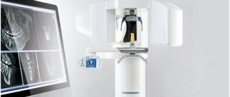
Benefits of CT
CT scanning of the jaw has many advantages. That is why this diagnostic method has found wide application in dental practice.
Advantages:
- Safety. Radiation exposure during the study is minimal.
- Information content.
- Image clarity.
- The ability to zoom in on a computer monitor and examine the desired area in detail.
- Fast execution (less than one minute).
- Comprehensive examination of the entire dentofacial apparatus.
- Possibility of saving received data on electronic media.
On the left is a panoramic image of the jaws, on the right is a CT image
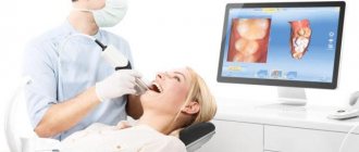
Is an x-ray always necessary before treatment? What are the indications for it?
The doctor will see little with the naked eye. Dental diseases are insidious and make themselves felt when they go deep enough, but before that they “sit quietly.” Therefore, a doctor can completely cure inflammation and remove infection only with a complete picture in hand. Of course, if there is no urgent need for an X-ray, the doctor will do without diagnostics - that is, the final decision, of course, rests with a specialist.
The best way to avoid serious treatment during pregnancy is to visit the dentist at the planning stage of the baby and eliminate dental problems in advance