- home
- Dental news
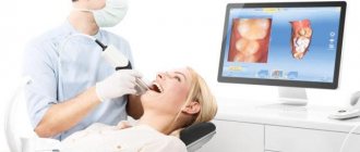
- 3D dental x-ray vs panoramic photo: innovation wins
|
|
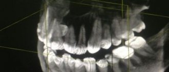
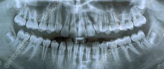
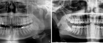
Panoramic image – an x-ray image of the jaw apparatus in a panoramic projection.
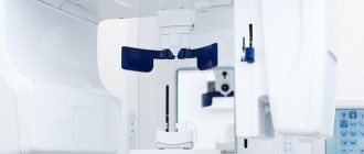
It shows a two-dimensional image of all the teeth of the upper and lower jaw and the adjacent bone tissue. 3D dental x-ray is a three-dimensional model of the jaw system obtained using computed tomography. Allows you to obtain detailed information about the hard and soft tissues of the tooth and jaw, and make a “slice” of any tissue in a three-dimensional projection.
When X-rays, OPTG and CT may be required
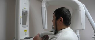
An x-ray is a targeted photograph of one or more teeth. OPTG or orthopantomogram is a panoramic image that captures both jaws. CT is a computer tomogram. It allows you to obtain three-dimensional or volumetric images of the entire human jaw system. Each of these types of diagnostics is used for certain indications:
- X-rays are performed when treating a specific tooth: in the presence of caries, pulpitis, periodontitis, suspected cyst or granuloma. Allows you to determine the extent of tooth damage, as well as the condition of the tissues around the root,
- An orthopantomogram is indispensable in the presence of inflammatory processes in periodontal tissues and jaw bone. It is carried out both during multiple dental treatment and in preparation for orthopedic, orthodontic treatment or dental implantation. Allows you to assess the condition and volume of bone tissue, clarify the position of the maxillary (nasal) sinuses of the upper jaw, nerves and other anatomically important elements,
- CT scan is performed for certain indications. Most often in the presence of tumors of the jaw system (to determine their volume in all dimensions), as well as before dental implantation, especially in complex cases.
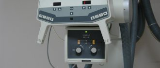
Today in dentistry (and in medicine in general) digital devices are used instead of film ones. They have a much lighter load, and they also use shorter shutter speeds to create a photo. For comparison: a modern digital visiograph takes up to 0.3 seconds to take an image, a film X-ray machine requires up to 1.5 seconds.
The Smile-at-Once clinic uses the latest generation CT-Scan tomograph. It allows you to get both a targeted or panoramic, and a three-dimensional digital image. Thanks to minimal radiation exposure, the equipment is completely safe and allows repeated diagnostics without harm to the human body.
Dentistry "Eurodent" - the best 3D dental x-ray in Almaty
The Eurodent dental clinic is equipped with the most modern diagnostic equipment, which makes it possible to accurately and timely diagnose a wide variety of dental diseases. Our clinic has the latest generation computed tomography device, GALILEOS Comfort PLUS, from the German company SIRONA. The improved characteristics of the device allow you to obtain a huge amount of information that is not available with other types of diagnostics (including a panoramic image).
The data provided by 3D dental examination using GALILEOS Comfort PLUS can be used:
- In endodontics - for preoperative diagnostics, to determine deep root damage, when diagnosing injuries to the dental system, to determine the degree of external and internal resorption of root canals, when performing apictoectomy;
- In implantology - to obtain a complete picture when assessing the implantological and orthopedic parameters of prosthetics, to install implants in the least traumatic way;
- In surgery - in the diagnosis of injuries, fractures, displacements of teeth, their roots and jaw bones, in the diagnosis of neoplasms, diagnosis of sinuses, orthodontic operations;
- In orthodontics - when diagnosing pathologies of the palate, jaw, diagnosis of impacted teeth, root resorption, during orthodontic analysis;
- In therapeutic dentistry - for complex diagnosis of clinical conditions, when planning conservative treatment of periodontal diseases, to clarify the results of X-ray studies, etc.
If you have a question: “Where can I get a dental x-ray done?” and “How much does it cost?”, then feel free to contact our clinic. Eurodent dentistry guarantees its patients the European level of diagnosis and treatment, while the cost of our services is quite affordable for all categories of the population.
We are waiting for you for a free initial consultation!
Based on: 9 votes
Is it dangerous to take pictures?
According to SanPiN1, when performing X-ray procedures for preventive purposes, radiation exposure should not exceed 1000 microsieverts (µSv) per year. We are talking specifically about prevention, since for medicinal purposes the indicator can be much higher.
In terms of the number of shots it looks like this:
- 500 targeted shots (1-3 µSv),
- 80 OPTG (13-17 µSv),
- 20 digital CT scans (50-60 µSv).
For comparison, here is a table that shows the radiation doses that a person receives during diagnostic procedures in dentistry, in other areas of medicine and in life in general. The last table shows parameters that are truly dangerous to the body and can be fatal.
| In dentistry | In other areas of medicine | In life | Dangerous indicators |
| 1-3 μSv – one targeted shot | 30-60 µSv – one digital fluorography, 150-250 µSv – old type film FOG | 5 µSv – 3 hours in front of a computer or TV | 750 thousand µSv – minor changes in blood composition |
| 13-17 μSv – one panoramic image (OPTG) | 500-700 µSv – one mammography procedure | 20-30 µSv – one 2-3 hour flight | 1 million µSv – mild radiation sickness |
| 50-60 μSv – one CT procedure in dentistry | 2000 μSv – one head CT procedure | 2000-3000 μSv – natural dose of radiation per person per year (food, solar radiation, air) | 7 million µSv – lethal dose of radiation |
As can be seen from the table, irradiation can cause harm (minor and short-lived) only when a dosage of 750 thousand µ3V is reached, while only one thousand µ3V is allowed for diagnosis. Therefore, 20 CT images or 80 panoramic images will not cause any harm to the body.
Is it possible to carry out diagnostics during pregnancy?
According to SanPiN, X-ray examinations are allowed in the second half of pregnancy using protective equipment, provided that the radiation dose does not exceed the same 1000 μSv. However, it is recommended to refrain from taking x-rays in the first and last 12 weeks, i.e. in the first and last trimesters.
Do not be afraid of undergoing diagnostic procedures during pregnancy. Even ordinary caries is an infection that, if not properly treated, can spread throughout the body and lead to infection of the fetus. Therefore, it is better to receive a small and completely safe dose of radiation than to carry out complex dental treatment blindly, not knowing how deep the inflammatory process is.
After the baby is born, i.e. During breastfeeding, dental x-rays can be taken, even more than once (within reasonable limits). Radiation doses are minimal, so radiation does not accumulate in breast milk, and there will be absolutely no harm to the baby. There is also no need to pump or skip feedings.
Three-dimensional x-ray (3D): indications, harmfulness, photo
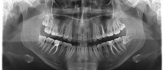
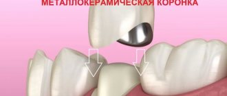
Radiography is a modern method that allows you to assess the condition of the roots and obtain an incredibly accurate image. In modern dentistry, an image gives a complete picture of the entire row or a separate area. Such an examination greatly simplified the dentist’s tasks. By taking a 3D image, doctors get a clear picture of all changes in the structure. The root on X-ray film is clearly visible in three dimensions, which allows you to correctly assess its condition.
The panoramic image fully lives up to its name. Within a few minutes you can get the exact status of the entire row. Why do a dental x-ray if you have a computed tomography? The photo has clear quality and high resolution, which allows you to get a very accurate picture. Our experienced and qualified doctors will be able to correctly identify all pathologies, eliminate any shortcomings and defects without dental errors.
If you need a computer X-ray of your teeth: the indications for the procedure should definitely be taken into account - 3 d x-ray is prescribed for:
surgical intervention;
· jaw fracture;
· prosthetics;
· for preliminary analysis before treatment.
This is a new step in solving any dental problems. Such diagnostics do not have a harmful effect on the human body and can be used for the prevention and treatment of various ailments. In Europe, this diagnostic method has been used for a very long time. It helps avoid conducting therapy “blindly” and allows the doctor to avoid any inaccuracies. A dental photograph can be stored for decades, printed or stored on electronic media. This is the main advantage of modern technology.
Where can I take a photo? In our center you can perform a panoramic x-ray, which will allow you to obtain a three-dimensional image. Pre-registration for diagnostics can be done by phone or with the administrator.
There are comprehensive answers to the question of what 3D radiography shows - it demonstrates:
· condition of periodontal tissues;
· beginning dental diseases;
· condition of tooth roots;
· inflammatory process.
Situations when taking photographs is strictly prohibited
Such situations practically never occur. On an individual basis, X-rays are considered in cases where the patient receives radiation in other areas of life: for example, in hazardous work, while undergoing chemotherapy or radiation therapy. But again, when X-raying the state of the jaw system, the radiation is so small that it will not affect the overall picture.
Thus, you should not be afraid to take photographs - in single quantities they are quite acceptable and will not affect your health at all.
It is important to understand that such diagnostic procedures allow for better treatment, especially with dental implantation, the results of which will last not a couple of years, but for many years of life. 1 Sanitary rules and regulations (SanPiN) 2.6. 2.6.1.1192-03 for the design and operation of X-ray rooms, devices and the conduct of X-ray examinations.
Panoramic shot or 3D x-ray of the jaw: which is better?
Modern adult and pediatric dentistry is impossible without accurate diagnostics. During a visual examination, the doctor can only obtain about 50-60% of all information, because information about the roots of the teeth and the condition of the jaw is simply not available to him. Naturally, to obtain the necessary data, an X-ray examination of the teeth is prescribed, the results of which allow one to obtain a complete picture of the condition of the patient’s dentition. X-ray examination of the jaws allows the dentist to consider all the nuances. Until recently, the most accurate type of diagnosis was considered to be a panoramic photograph of the teeth, which gives a general understanding of the condition of the teeth, their roots and the bones of both jaws. Using a panoramic image, you can accurately diagnose most diseases of the teeth and periodontal tissues.
But it’s not for nothing that dentistry is called one of the most dynamically developing areas of medicine - most advanced technologies are used both in diagnosing and in treating dental diseases.
An innovative solution was the use of computed tomography when examining patients. Scanning the patient’s oral cavity using a tomograph, in contrast to a panoramic X-ray, makes it possible to obtain information in the smallest detail. Compared to a “flat” panoramic image, a 3D X-ray of the jaw allows you to obtain a three-dimensional model of the area under study, while it is possible to “examine” each layer separately. Computer diagnostics gives a clear picture of bone density, the presence of hidden inflammatory processes, neoplasms, etc.
The table below shows comparative characteristics of the capabilities of different types of diagnostics:
| Panoramic shot | 3d x-ray |
| General picture of the condition of the jaws and periodontal tissues | Accurate image of all tissues of the area under study, the ability to obtain a lateral projection, integration with a facial scanner |
| Static two-dimensional (flat) image | 3D volumetric imaging |
| Low degree of detail - you can distinguish between hard and soft tissues, inflammation and neoplasms are visible | High degree of detail - the smallest structural changes are visible in both hard and soft tissues |
| More powerful radiation during the photo | Minimal radiation exposure during diagnostics (2 times less than with panoramic) |
| There are restrictions on diagnosing diseases | High accuracy of diagnosis of any pathological changes |