According to statistics, every third person suffers from pathologies of bones and joints. One of the first places is occupied by diseases of the jaw joints. The problem is that in the early stages of the development of the disease, symptoms may be absent or mildly expressed, and increased tissue wear is characteristic. However, therapy must be started as early as possible, which will speed up recovery and also prevent complications.
X-rays of the jaw allow timely detection of many diseases of the temporomandibular joint. Therefore, when the first symptoms of such ailments appear, you must contact a medical center for an x-ray.
When is a jaw x-ray taken?
Tell the patient exactly whether he needs and can have x-rays of his jaws
, should his attending physician. General indications for such a diagnostic procedure are:
- Suspicion of jaw development abnormalities (in childhood or adulthood). These may be congenital clefts of the hard/soft palate, impaired growth and proper development, or malocclusion.
- Suspicion of infectious-inflammatory and specific pathologies. These include osteomyelitis, periostitis, syphilis, actinomycosis, tuberculosis or necrosis of the jaw bone.
- To identify various acquired defects resulting from injury or removal of oncological formations.
- Suspicion of the presence of a false joint.
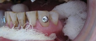
- Suspicion of a benign or malignant neoplasm.
What can you see in the photographs?
The following can be seen on x-rays of the upper and lower jaws:
- defects - cracks, fractures, presence or absence of bone fragments;
- pathological changes in bone tissue - areas of tissue thinning, compaction, thickening or thinning of the cortical plate;
- areas of necrosis - sequestra;
- sclerotic process;
- various periosteal layers, osteophytes (bone growths);
- tumor-like neoplasms.
Is it possible to take dental x-rays during pregnancy?
The only contraindication for X-ray diagnostics is pregnancy. X-rays do not harm the mother's body, but can harm the fetus. Radiation is dangerous for cells that are in the process of dividing. In the body of an adult, these processes are not so pronounced, while in the fetus the formation of tissues and organs occurs. X-rays entering a dividing cell can damage parts of the DNA chain, as a result the new cell will be different from the old one, which can cause pathology. Therefore, women during pregnancy should avoid any X-ray diagnostics.
How is the procedure performed?
X-ray of the jaw of an adult or child is a simple procedure. It can be done in different projections in order to better examine the structure of the bone tissue and make an accurate diagnosis.
X-ray of the lower jaw
To obtain general information about the condition of the lower jaw, an X-ray is taken in a direct projection. It allows for primary diagnosis of inflammatory, oncological and traumatic diseases. The patient is positioned as follows: lying on his stomach, face down, resting the tip of his nose and forehead on the cassette. The X-ray machine sensor is installed at the occipital protuberance.
A lateral view of the lower jaw is taken to assess the condition of the body, ramus and teeth of the desired side. The patient lies on his side, with his cheek on the cassette located at a slight angle.
To diagnose diseases of the lower jaw, images are also taken in the axial projection. The patient takes a position lying on his stomach, stretching his head forward with his chin as much as possible. At the same time, it is pressed against the cassette by the front surface of the neck and lower jaw.
X-ray of the upper jaw
To assess the condition of the bone tissue of the upper jaw and the chin area of the lower jaw, a nasomental placement is performed. To do this, the patient lies on his stomach, face down, resting the tip of his nose and chin on the cassette. The sensor is installed perpendicular to the cassette, 2 pictures are taken - with the mouth open and closed.
Diagnosis of pathologies using x-rays of the jaw
If you have symptoms indicating dysfunction of the temporomandibular joint, you must contact a diagnostic center for research. You can make an appointment for an x-ray in our clinic, where modern equipment is used to ensure the safety of the procedure for patients.
Pathologies can be diagnosed using x-rays:
- traumatic arthritis;
- neuralgia;
- purulent arthritis;
- rheumatoid arthritis;
- osteoarthritis;
- osteomyelitis;
- temporomandibular joint dysfunction syndrome;
- injuries of bone and joint tissues.
The technique provides high information content, which allows the doctor to make a correct diagnosis in a timely manner, prevent further development of pathologies and the occurrence of complications. In addition to X-rays, the doctor may prescribe a computed tomography scan, which will allow you to most accurately diagnose the disease and identify the causes of disorders in the temporomandibular joint.
Panoramic shot of the jaw
A panoramic X-ray, or orthopantomogram, displays the entire upper and lower jaw in a direct projection. In such a picture you can see almost all the anatomical features of the patient’s dental system, all kinds of neoplasms, defects, fractures of tooth roots, and so on. An orthopantomogram allows you to identify the following pathologies:
- damage to tooth roots by caries;
- hidden carious cavities;
- cysts, granulomas, tumors;
- pathological changes in the roots and peri-root space.
When X-rays, OPTG and CT may be required
An x-ray is a targeted photograph of one or more teeth. OPTG or orthopantomogram is a panoramic image that captures both jaws. CT is a computer tomogram. It allows you to obtain three-dimensional or volumetric images of the entire human jaw system. Each of these types of diagnostics is used for certain indications:
- X-rays are performed when treating a specific tooth: in the presence of caries, pulpitis, periodontitis, suspected cyst or granuloma. Allows you to determine the extent of tooth damage, as well as the condition of the tissues around the root,
- An orthopantomogram is indispensable in the presence of inflammatory processes in periodontal tissues and jaw bone. It is carried out both during multiple dental treatment and in preparation for orthopedic, orthodontic treatment or dental implantation. Allows you to assess the condition and volume of bone tissue, clarify the position of the maxillary (nasal) sinuses of the upper jaw, nerves and other anatomically important elements,
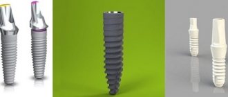
- CT scan is performed for certain indications. Most often in the presence of tumors of the jaw system (to determine their volume in all dimensions), as well as before dental implantation, especially in complex cases.
Today in dentistry (and in medicine in general) digital devices are used instead of film ones. They have a much lighter load, and they also use shorter shutter speeds to create a photo. For comparison: a modern digital visiograph takes up to 0.3 seconds to take an image, a film X-ray machine requires up to 1.5 seconds.
The Smile-at-Once clinic uses the latest generation CT-Scan tomograph. It allows you to get both a targeted or panoramic, and a three-dimensional digital image. Thanks to minimal radiation exposure, the equipment is completely safe and allows repeated diagnostics without harm to the human body.
Decoding the results
An x-ray of an adult's jaw is usually interpreted by a radiologist.
Other specialists may also be involved in this work: dentist, facial surgeon, otolaryngologist. Specific pathologies of the upper and lower jaws have certain radiological features:
- Chronic osteomyelitis. Depending on the type of disease, the images may show foci of resorption of various shapes with a shadow of necrotic masses inside. In more severe cases, communication of the sequestra with the oral cavity is visualized.
- Acute osteomyelitis. When examining the image, you can see areas of bone tissue resorption that do not have clear boundaries.
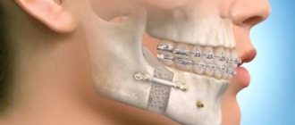
- Fractures or cracks in the jaw bones. This pathology reveals itself by the fact that thin, elongated shadows are quite clearly visible in the photographs. In such cases, it is important not only to identify the fracture, but also to see the bone fragments, if any, and to understand how much they are displaced.
- Chronic periostitis. In the image with such a pathology, periosteal thickenings will be visible. If the disease has become severe, areas of ossification of the periosteum and new bone tissue along the edge of the jaw are visualized.
How are dental x-rays done?
The procedure does not require preparation, it is done quickly and painlessly. All manipulations take place in the X-ray room. The shooting process will vary slightly for different types of x-rays.
Bitewing x-rays and spotting x-rays are done in a similar way. A matrix or x-ray film wrapped in light-proof paper is applied to the problem tooth. A radiation tube is brought to the tooth and an image is taken. Information is transferred from the matrix to the computer. The photo can be printed or saved digitally. If film was used for x-rays, it is first developed in a dark room. This takes a little longer.
For panoramic and 3D images, special devices are used - orthopantomograph and tomograph. The procedure is done standing or sitting, it depends on the design of the device. You will have to remove metal jewelry, dentures, and piercings before the x-ray. You will be wearing a lead apron to protect you from x-rays.
Next, the patient's head is fixed in the desired position, and then filming begins. It lasts 20-30 seconds. The emitter and receiver will move around you, they will make noise. Don't pay attention to this, your task is to sit still. Any movement can affect the accuracy of the photo. At the end of the procedure, the film is developed, and the digital data is processed by a computer.
Why does a dentist refer a patient for dental x-rays?
An x-ray gives the doctor the opportunity to most accurately assess the condition of the problem unit or all organs of the oral cavity. And also study the clinical picture in detail. In particular:
- detect foci of inflammation that have not yet manifested themselves;
- identify neoplasms in the bone and soft tissues of the dental system;
- establish the signs and stage of development of periodontitis or periodontal disease;
- establish linear dimensions, volume, structure of bone tissue before implantation;
- more accurately assess the correctness of occlusion (tight closure of the dentition);
- detect anomalies of the dental system organs;
- determine the position of erupting wisdom teeth.
As a rule, radiography is prescribed before the start of orthodontic treatment, when planning implantation and prosthetics, before and after filling the tooth canals. This study is also used to evaluate the results of conservative and surgical treatment.
Free consultation
30-40 minutes
inspection and diagnostics
treatment plan and cost
Make an appointment
Advantages and disadvantages of x-rays
Dental X-ray has become a leading diagnostic method due to its advantages:
- Information content. X-rays allow you to study the internal structure of the entire dentition. The accuracy of the data obtained can reach tenths of a millimeter.
- Easy to carry out. X-ray does not require preparation from the patient. The procedure is prescribed after the initial examination, it is immediately carried out, a diagnosis is made and treatment is prescribed.
- Availability. There are x-ray rooms in almost every dental clinic.
- Efficiency of the procedure. The picture is taken instantly; CT and orthopantomography will take 20-30 seconds.
- It has virtually no contraindications. Dental X-rays are performed for both children and adults. Contraindicated only for pregnant women.
- Relatively low cost. The price of the service depends on the region, clinic and equipment it uses. Overall, this is a cheap diagnostic method.
The only disadvantage of dental x-rays is the certain dose of radiation that the patient receives. However, it is not that big. If you do not abuse x-rays, it will not have any effect on your body.
How many times can a dental x-ray be taken?
According to legal regulations, the annual radiation dose from X-ray diagnostics should not exceed 1000 μSv (microsievert). If you divide this figure by the dose received after each procedure, it turns out that in a year you can do:
- up to 500 targeted images with a radiovisiograph,
- up to 100 targeted shots per film,
- up to 80 panoramic digital images,
- up to 40 film orthopantomograms,
- up to 20 computer images.
How dangerous is a dental x-ray?
When X-ray diagnostics are performed, the patient receives a certain dose of radiation. It depends on the type of x-ray and the equipment used to take the picture. For a targeted film image it is up to 15 μSv, radiovisiography - up to 3 μSv; digital orthopantomography - up to 17 µSv, film - up to 30 µSv; computed tomography - up to 60 μSv.
We receive about 4000 μSv per year from natural background radiation, that is, every day we receive the same radiation dose as during visiography. When making a flight, we rise to a certain altitude, where the background radiation is higher. In one plane trip you can get up to 20 µSv, it’s like doing an orthopantomogram. If you are reading this text on an old cathode ray monitor, in a few hours it will emit the same amount of radiation as a radiovisiograph in an x-ray room during a targeted shot.
X-rays are safe for the patient if they are not exceeded. To prevent this from happening, always undergo x-ray diagnostics in one place. This will make it easier to calculate how much radiation you received in a year.