The main task of a dental surgeon during the treatment of a fracture of the upper or lower jaw is to restore the anatomical structure of the broken bone and the correct relationship of the dentition. Many techniques help to achieve this, but the effectiveness of treatment also depends on how correctly and quickly first aid was provided.
Before hospitalization
First aid to the victim includes:
- stopping bleeding (pressing or packing the wound, applying cold);
- if necessary, cardiopulmonary resuscitation;
- pain relief (analgin, revalgin intramuscularly);
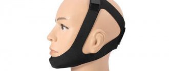
- immobilization of the jaw with the help of fixing bandages (contraindicated if the victim is unconscious, since this increases the risk of suffocation from the retraction of the tongue or vomit entering the respiratory tract).
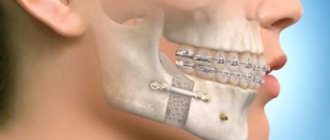
Osteosynthesis using ultrasound
With the help of metal fasteners, the jaw bones can be fixed quite firmly. But during surgery, it is most often necessary to cut through facial tissue and the salivary glands or branches of the facial nerve can be damaged.
It is less traumatic to perform osteosynthesis using ultrasound. In this case, the bone-fixing devices can be inserted shallowly into the bone and a small amount of scars remains on the patient’s face.
The doctor uses low-frequency ultrasound on a titanium plate with spikes. A plate with holes for the dental bur is placed at the fracture site and adapted to the shape of the jaw. An instrument is then used to make shallow holes in the bone through the plate. After which low-frequency ultrasound vibrations are directed to the base of the spines. In this way, the spikes gradually sink into the bone tissue, reliably fixing bone fragments. In this case, an antiseptic solution is supplied through the instrument, which treats the wound.
The bone tissue around the spines becomes denser under the influence of ultrasound. This occurs due to the large contact area, reducing pressure on the bone due to the use of a spike and the internal compression force of the bone tissue.
Ultrasonic osteosynthesis can reduce the time of surgery and reduce the amount of postoperative trauma. The method gives fewer complications and provides a good cosmetic effect.
Osteosynthesis
Indispensable for complex, comminuted and multiple fractures with displacement, loose teeth and complete absence of teeth, for periodontal disease and other inflammatory diseases of the gums in the area of injury. Osteosynthesis is also effective in cases of fracture of the condylar process complicated by dislocation of the articular head of the lower jaw.
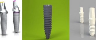
Fastening materials can be steel knitting needles and rods, pins, nitride-tinan wire with shape memory, quick-hardening plastics, polyamide thread, special glue.
However, osteosynthesis with metal miniplates is considered the most convenient and safe method today. They allow you to cut through the skin and muscles on only one side, which simplifies the operation itself and shortens the recovery period. Another undeniable advantage is the ability to reliably fix fragments in areas with significant dynamic loads.
International standards
A real revolution in maxillofacial surgery occurred in 1958, when M. Muller, M. Allgower, R. Schneider, H. Willenegger organized the International Association for the Study of Internal Fixation (AO/ASIF - Ardeitsgemeinschaft fur Osteosynthesefragen/Association for the Study of Internal Fixation – working association for the study of osteosynthesis/association for the study of internal fixation).
According to the postulates of AO/ASIF, the osteosynthesis technique implies that:
1. the structures used must be made of bioinert metal alloys;
2. bone fragments must be anatomically accurately compared and fixed;
3. the use of gentle surgical techniques ensures the preservation of blood supply to bone fragments and surrounding soft tissues;
4. stable fixation of fragments is ensured by interfragmentary compression;
5. early application of functional load is indicated;
6. restoration of contractile activity of muscles and movement in the joint.
AO/ASIF staff also developed and implemented metal plate systems for mandibular osteosynthesis:
· dynamic compression plates;
· reconstructive (blocking) plates (Locking reconstruction plates);
· blocking (locking) plates (Locking plates 2.0 mm);
· universal plates (Universal fracture plates);
· mandibular plates (Mandible (Mandible plates 2.0 mm).
They developed dynamic compression plates (DCP), through which it was possible to create compression between fragments for their primary fusion. The design of these plates includes oval holes with beveled walls, which allows the fragments to be brought closer together when the screws are tightened. The use of dynamic compression plates made it possible to achieve stable internal immobilization, reduced the number of cases of delayed fusion of fragments, and eliminated the need for additional fixation. But their use still does not eliminate the risk of microcracks in the area of the fracture line and the development of osteoporosis in the bone at the site of contact with the plate.
The locking plate/screw system, with threaded plate holes and locking screw heads, was developed to prevent bone necrosis under the plate. The system provides rigid fixation of bone fragments using a plate and a plate and screws to each other - this helps prevent the screws from unscrewing and avoid possible displacement of the fragments while tightening the screws in the hole of the plate. The plate itself is located at a certain distance from the surface of the bone, which prevents the development of lysis.
Foreign studies have not revealed significant differences in the effectiveness and possible development of postoperative complications during osteosynthesis with locking plates with screws and non-locking plates.
Another system developed by AO/ASIF experts is the LCP (locking compression plate system with angular stability) is a design of multi-cell plates with numerous holes and consists of two parts: a threaded one for fixing the head of a locking screw and a hole for creating dynamic compression by eccentric insertion of standard cortical or cancellous screws.
Installation of the plate requires special tools and is carried out according to clearly established technology.
If the adjustment of the LCP to the shape and relief of the outer surface of the lower jaw bone is carried out in accordance with the established requirements, then this creates ideal conditions for the fusion of fragments during osteosynthesis of multiple comminuted fractures of the lower jaw of various locations, in case of suppuration of a bone wound, traumatic osteomyelitis, in case of a fracture with the occurrence of a bone defect tissue, fracture of toothless jaws. Limited contact of LCP with bone helps prevent the development of bone necrosis under the plate.
Splinting the jaw
This is the immobilization (fixation) of bone fragments using a special plastic or wire structure.
The technique, created by military doctors at the beginning of the 20th century, is successfully used by dentists today. The materials used to make the splint have changed, and the methods of applying it have been improved.
Today, a specialist has many types of tires in his arsenal:
- from standard Vasiliev tape splints, the simplest and cheapest method of treatment;
- to aluminum Tigerschdedt splints, which are made individually for each patient, due to which they are more effective. In addition, they evenly distribute the load and minimally injure the teeth.
The type of splinting depends on the type of injury and can be unilateral (when one jaw is fractured) or bilateral (when both are damaged).
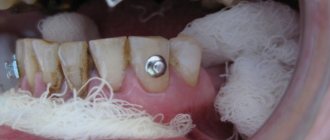
If the teeth are preserved, it is not difficult to apply a bent dental wire splint. It is bent to the shape of the dental arch and fixed with bronze-aluminum wire ligatures, which, like a hairpin, cover the tooth on both sides. Manipulations are performed under local anesthesia.
When both jaws are fractured, a structure with a more rigid base is installed; in addition to wire, hooks and rings are also used that immobilize the lower jaw.
Metal Kirschner spokes
This type of wire was first used to treat fractures of the lower jaw in 1933. Intraosseous insertion of these wires can be carried out both percutaneously (without incisions) and with soft tissue incisions.
In 1975 V.V. Donskoy used an original technique, with which he inserted a wire into the branch of the lower jaw through the mucosa without an incision, then carried out a reposition, and fixed it like a splint to the teeth or to a splint. Later, in 1988, Deryabin E.I. and Osipov V.Yu., and Yu.G. Kononenko and G.P. Ruzin proposed modifications of this method in 1991. Today there are many techniques where the wire suture is combined with knitting needles, staples, surrounding wire ligatures, etc.
Is it possible to do without splinting?
Even if the case is not severe - the fracture is one-sided, closed and without displacement - it is imperative to take measures to prevent the development of such unpleasant complications as:
- accidental displacement of fragments,
- re-injury
- development of inflammation of soft tissues,
- infection of the fracture site.
To do this, it is necessary to immobilize the jaw by any available method. This can be a sling bandage, but it is much more convenient and effective to use a splint. In case of a complicated fracture, splinting is absolutely indispensable, regardless of the location of the injury.
Indications for osteosynthesis of the jaw with plates
The main indication for installing metal plates for osteosynthesis is the prevention of complications that cannot be effectively prevented by applying a traditional splint. In the case of osteosynthesis, an important task for the surgeon is to compare the fragments and fix them in the correct position. If successful, fusion will take up to 3–4 weeks.
Indications:
- fracture behind a row of teeth;
- inflammation that provoked injury;
- significant displacement of bone elements;
- high mobility of teeth at the fracture site;
- incorrect position of the jaws relative to each other.
Treatment tactics for displaced fractures
In such cases, before applying a splint, it is necessary to compare the jaw fragments, for which reduction orthopedic devices are used. A broken upper jaw requires traction using special dental splints.
Such injuries are very dangerous because they can cause asphyxia. But correctly provided first aid will prevent suffocation. Clear the oral cavity of foreign bodies or blood, lay the victim face down, placing a cushion rolled up from clothes, blankets, etc. up to the chest.
Discussion
Despite the development of medicine in general and maxillofacial surgery in particular, the problem of providing emergency medical care for fractures of the facial skull remains not fully resolved. Due to the increase in the number of road traffic accidents and domestic conflicts, and the passion of young people for traumatic sports, there has been an increase in the number of victims with injuries to the facial skull, among which fractures of the lower jaw occupy the first place [12].
The difficulty of early diagnosis of fractures of the angle of the mandible is associated with the insufficient information content of routine methods of radiation examination (radiography of the skull in frontal or lateral projection), late referral of victims to specialized maxillofacial hospitals and, as a consequence, the choice of irrational methods of treatment, which in turn leads to development of various kinds of complications, reduction in the quality of treatment and life of patients.
Rehabilitation after a jaw fracture
For successful treatment of a jaw fracture, anti-inflammatory and restorative therapy, physiotherapy, mechanotherapy and special oral hygiene are also important.
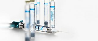
- Within 3-4 days after the injury, antibiotics must be prescribed to prevent inflammation, which are injected directly into the area of injury.
- General strengthening therapy is taking vitamins C, P, D and group B, drugs that stimulate tissue regeneration and restore the level of leukocytes in the blood.
- Among the effective physical procedures, we note UHF therapy, general ultraviolet irradiation, and magnetic therapy. After the third procedure, swelling and pain are noticeably reduced, swelling subsides. For better healing of fragments, 2 weeks after a jaw fracture, electrophoresis is performed using a two to five percent solution of calcium chloride.
- Mechanotherapy, or physical therapy, accelerates the restoration of jaw function and helps if, after an injury, the mouth opens poorly or does not open at all. It can also be practiced at home, starting 4-5 weeks after the fracture, when the splints are removed and a callus has formed.
- Special hygiene involves irrigation at least 8-10 times a day. For unconscious victims, their teeth and mucous membranes are treated with a special solution at least twice a day.
Fracture of the lower jaw - symptoms and treatment
Diagnosis of patients with fractures of the lower jaw is carried out in order to establish the location, number of fractures, as well as to determine injuries to nearby tissues, vessels and nerves.
Diagnostic methods are basic and additional (instrumental).
Basic diagnostic methods are carried out with a standard set of instruments in the emergency room, dental office or dressing room. The doctor finds out the patient’s complaints, learns about previous diseases and previous operations. At this stage, it is very important to establish the time of injury, since the duration of the injury plays a big role in further treatment tactics.
During the investigation, the doctor will find out whether the injury was sustained at home, at work, or during a fight, and whether it is of a criminal nature. In case of injury during violent acts, the doctor is obliged to report the incident to the police. After that, an internal affairs officer talks with the victim, finding out the details of what happened. In this regard, there is often a concealment of the real reasons for the injury, and the patient sometimes invents very ridiculous and implausible stories.
Sometimes collecting complaints and medical history is difficult or impossible due to the patient’s serious condition or alcohol/drug intoxication.
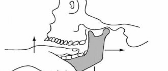
A clinical study begins with an external examination; post-traumatic swelling, changes in skin color, the presence of abrasions, hematomas, and bleeding are detected. Next, palpation is carried out, during which the nature of the swelling is revealed, inflammatory infiltrate, hematoma, and subcutaneous emphysema are excluded. Palpation of the lower jaw is carried out symmetrically, starting from the branch of the lower jaw and ending in the chin. The presence of a bony step indicates a fracture of the mandible. The degree of mobility of the fragments and the direction of their displacement are also determined.
When examining the oral cavity, special attention is paid to the mucous membrane of the alveolar part of the lower jaw, identifying violations of integrity and bleeding. The relationship of the dentition and the displacement of the central line of the lower jaw when opening and closing the mouth are determined. The landmarks are the labial frenulum, the line between the central incisors of the upper and lower jaw.
Often, the opening of the mouth with jaw fractures is limited. When rocking the lower jaw, relying on the chewing teeth and the basal edge, you can determine the degree of pathological mobility of the lower jaw and the location of the fracture.
Additional diagnostic methods:
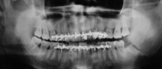
- X-ray : an X-ray of the skull is taken in a direct projection in order to exclude fractures of other parts of the face and an X-ray of the lower jaw in lateral projections, the number of fractures, the presence or absence of displacement, diastasis (gap) and the presence of teeth in the fracture line are analyzed.
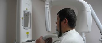
- An orthopantomogram is a more modern method, with functionality similar to an x-ray. One detailed image is taken, and it is possible to view it on the monitor screen using various programs.
- Computed tomography allows you to most accurately identify fractures of the lower jaw, measure the distance of displacement of fragments, diastasis, determine the size and number of fragments, and the presence of foreign bodies. Using certain setting modes, you can determine the violation of soft tissues (muscles, blood vessels, nerves) and the presence of hematomas.
- Assessment of the general condition of the body - examination of organs and systems of the body, identification of viral and chronic diseases. For this purpose, a physical examination, digital chest x-ray, ultrasound examination, and laboratory diagnostic methods are used. If necessary, consultations with related specialists are carried out. All this helps to select the most effective therapy for the patient, taking into account the characteristics of a particular clinical case.
How to eat?
Since during intensive therapy and during the recovery period the jaws are rigidly fixed and habitual chewing of food is out of the question, correction of the diet is necessary during this period.
Food should have the consistency of low-fat sour cream. These are broths, pureed soups, carefully chopped vegetables and fruits, milk drinks, liquid cereals. Spices are excluded, salt consumption is limited. The temperature of the dish should not be higher than 45-50 °C. The most convenient way to eat food is through a straw.
You need to gradually switch to your usual diet after removing the splint. This is important not only for restoring chewing functions, but also for preventing disorders in the gastrointestinal tract.
Purpose of the operation
When performing an operation, the surgeon sets himself the following goals:
- Combine the fragments;
- Fix the bone with special splints until its integrity is restored;
- Secure the fragments with metal plates and knitting needles;
- Fix the dentition with special structures;
- Create conditions for favorable bone healing.
If such goals are achieved using conservative methods, then there is no need for surgery. Disturbances in the bone structure also cannot be ignored. Otherwise, unpleasant changes develop that lead to impaired chewing and articulation, as well as chronic pain in the jaw.
How much does it cost to treat a broken jaw?
The price depends on the nature of the injury, whether osteosynthesis was performed, what splints were used, and whether the patient attended physical therapy procedures. But let's say for sure that the service is not cheap. Osteosynthesis alone will cost from 14,000 to 55,000 rubles.
It is also necessary to consider the cost of subsequent dental treatment to restore lost teeth or damaged teeth after splinting. Our service will help you choose a competent specialist and not waste your money. Compare prices and services of different clinics, read reviews from real patients.
Types of osteosynthesis
There are several methods of osteosynthesis; the doctor decides what type of operation the patient needs. Most often, surgeons combine several methods with each other to achieve better results.
Osteosynthesis of the jaw can be:
- Open. It is usually used for severe fractures. During the operation, soft tissue is cut and bone fragments are exposed. They are connected to each other and non-functional small fragments are removed, compressed soft tissues and fascia are released. However, with such an operation, there is still the possibility of tissue peeling off from the bone, then the callus at the fracture site will not be formed correctly. And this can affect the patient’s quality of life. In addition, stitches remain on the skin and paresis (decreased activity) of facial muscles is even possible. Depending on the type of fastening device, it is possible that the incision on the face will have to be made again to remove the fastener.
- Closed. The doctor combines bone fragments without cutting facial tissue;
- Focal. The fixing fastener is applied directly to the fracture site;
- Extrafocal. Fasteners are placed on top of the skin, above the fracture site.