An isolated fracture of the alveolar process occurs due to bending or displacement at the site of application of force. Anatomical structure. The alveolar process of the maxilla is a curved bony structure, has a vestibular, palatal surface and a margin on which eight alveoli are located. It consists of a spongy substance enclosed in compact plates. The outer compact plate is thinner than the inner one, especially in the area of the front teeth. The alveolar part of the lower jaw contains eight holes on each side. The incisor sockets are compressed from the sides, and their bottom is located closer to the labial compact plate. Therefore, in the area of these teeth, as well as canines and premolars, the lingual wall of the sockets is thicker than the labial wall. The total thickness of the compact plates and spongy substance in the area of the base of the body of the lower jaw is less than in the alveolar part.
Causes
In addition to mechanical trauma from an impact, the cause of a fracture of the alveolar bone may be some disease that disrupts the normal state of bone tissue:
- osteomyelitis - inflammation of bone tissue;
- fibrous osteitis, characterized by bone dystrophy, thinning of the bone structure;
- various cysts and neoplasms that cause bone degeneration.
If the patient has the above diseases, even a slight impact on the bone can lead to a fracture of the alveolar process.
In children aged 5 to 7 years, that is, at the stage of growth of permanent teeth, trauma to the alveolar process can also be diagnosed due to the presence of follicles of permanent teeth in the bone, which subsequently disappear.
Treatment options
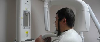
First of all, patients are prescribed an odontometric and x-ray examination. The information obtained helps determine the degree of fracture, the presence or absence of fragments, the condition of the pulp, as well as the number of fractures and their direction. If there is minimal damage, for example, a fracture of a front tooth with a small chip, its natural shape is restored using appropriate composite materials.
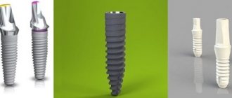
If oblique, comminuted or vertical fractures are found, in most cases the tooth must be removed, since its root is not capable of becoming a full-fledged basis for restoration with a pin. Usually the problem is solved by tooth extraction and further use of implantation.
If a tooth is broken at the root, it can also be restored. Otherwise, a metal-ceramic crown is used, the appearance of which is completely identical to the shape and color of the natural tooth.
If the pulp is damaged, it is usually removed. Then the canal is filled and the tooth is restored using a filling. But you must also make sure that its shape looks natural. Otherwise, closing the jaw may cause subsequent damage to the restored tooth. Another possible problem is the formation of an incorrect bite.
If a dead tooth is fractured at the root, implantation is used or a crown is installed. When a wisdom tooth is fractured, specialists usually remove them immediately. Limited access to these teeth makes them extremely difficult to fully restore.
Symptoms of alveolar bone fracture
An alveolar ridge fracture can be diagnosed if:
- sharp, spasm-like pain in the jaw when chewing or swallowing saliva,
- swelling of the mucous membrane,
- lacerations of the gums (with or without bleeding),
- painful closing of the jaws,
During palpation, the damaged part may move or a separated area may be palpated. Externally, the fracture is characterized by contusions and bruises on the face in the area of the fracture. External examination and x-ray show a crack in the alveolar process or complete separation of the bone from the main cranial bone.
Depending on the angle at which the blow occurred, the broken part may move in different directions, inward or deep into the mouth. If the blow falls on the lower jaw, in this case the impact occurs with the help of the lower jaw on the upper jaw, and the broken part moves upward.
Root fracture
In the event of a tooth root fracture, a specialist analyzes its position to select the appropriate treatment. The fracture can occur near the neck, at the apex, in the middle part, or at the border of the upper and middle parts. In most cases, transverse fractures occur.
Diagnosis of such damage does not cause any difficulties. First of all, a person feels pain while chewing food and biting it. During palpation, mobility of the injured element of the dentition is observed. When percussing and closing the jaw, the patient experiences discomfort and pain. And if the fracture is located near the crown, the enamel may take on a pink tint. Radiography helps to clearly establish the number of cracks, as well as their position.
Diagnostics
The fracture is diagnosed by an oral and maxillofacial surgeon. An x-ray may show different degrees of damage:
- partial damage - damage to part of the bone, not complete detachment;
- non-displaced fracture – damage to all parts of the bone;
- complete fracture - the x-ray shows a gap between the separated part and the skull;
- fracture in different places - damage to the alveolar process in different places, bone fragmentation;
- fracture with deformation - a completely torn off part, displaced at a different angle.
Clinical manifestations
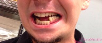
The first sign of a fracture of the alveolar process is a sharp, severe pain. Usually its occurrence is associated with mechanical trauma. The patient cannot completely close his mouth, swallow and chew. The bite is broken.
Upon examination, a hematoma or rupture of soft tissue at the site of injury is usually detected. If the fracture is partial, the deformity may be mild. In complete displaced fractures, bone fragments can come out, breaking through the soft tissue.
The teeth in the area of injury are usually mobile and dislocated. In most cases, severe bleeding is observed.
If a child is injured, the consequences may be adverse. When the alveolar process, which contained the rudiments of permanent teeth, is fractured, they die, so the only solution in the future will be prosthetics.
Treatment
The treatment method is selected depending on the established severity of the fracture. Before any manipulation, the doctor performs anesthesia. Torn tissues are treated with antiseptics to prevent infection. Then reconstruction is carried out (if there are fragments) and fixation of the jaw.
If there is displacement, the doctor opens the gums in the area of the fracture (revision) to eliminate the sharp edges of the fragments, after which he manually puts the displaced part in place. This must be done in such a way that it corresponds to the correct bite. After this, the gum is sutured and an iodoform dressing is applied to the wound.
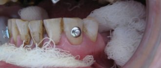
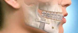
To fix the alveolar process, a smooth splint-bracket is used, which is attached to three healthy teeth on both sides of the chipped part. If this is not possible, that is, if the teeth on one side are not stable or are completely absent, then I use five additional teeth for fixation. For greater immobilization of the dentition, wearing a chin sling may be indicated.
If the injury occurs in the anterior part of the upper molars, then fixation occurs through the installation of a single-jaw bracket, which is fixed on the damaged area (attached with ligatures to healthy teeth).
In case of complete absence of teeth, a splint made of quickly hardening plastic is installed.
After all surgical procedures, the patient is prescribed antibacterial therapy and medications to accelerate healing and prevent the development of inflammation in the injured area.
Fracture of the upper jaw - symptoms and treatment
When providing first aid to the patient, it is necessary to stop the bleeding and prevent aspiration (penetration into the respiratory tract) of blood and vomit. If the lower jaw is not damaged and there are a sufficient number of teeth on both jaws, it is necessary to apply a sling-like bandage, pressing the lower jaw to the upper jaw, or perform immobilization (immobilization) with a rigid chin sling [4].
If there is a risk of respiratory failure, immediate insertion of an airway is required to maintain the conductivity of the airways [1]. In addition, it is necessary to provide pain relief and quickly transport the patient to specialized medical institutions. The most important thing at this stage is to preserve the life and health of the patient.
There are many methods of non-surgical treatment of fractures of the upper jaw, for example, various types of bandages and external fixations, which are currently practically not used.
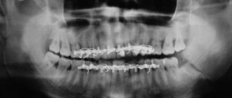
The most common method of orthopedic treatment of fractures is bimaxillary splinting - the application of splints and brackets to the dentition with reposition of fragments and fixation of the bite in the patient’s usual position. This method is conservative and low-traumatic, but in some cases it does not allow obtaining good fixation of fragments of the upper jaw, especially in high and complex fractures. On average, fractures of the upper jaw require immobilization and limitation of chewing load for a period of 4-5 weeks.
The most modern and adequate treatment method at the moment is osteosynthesis (fixation with titanium bone structures) of fractures of the upper jaw. This is a surgical procedure performed through intraoral incisions. With this treatment option, it is possible to accurately compare and fix the fragments to create conditions for their fusion [7].
In the treatment of high fractures, a coronal approach is also used, which allows for cosmetic and wide access to the bones of the entire midface and orbits [5]. Timely implementation of osteosynthesis allows you to prevent late postoperative complications, facilitate the patient’s rehabilitation and speed up recovery.
Fractures with gross violations of the integrity of the upper jaw and significant displacement of fragments towards the pharynx are recommended to be treated surgically. There is no clear opinion regarding other types of fractures - tactics are dictated by the patient’s condition and the specific clinical situation.
It is worth noting that it is very important to constantly wear intermaxillary fixation for tight contact of fragments and to prevent their mobility, especially under the influence of chewing load [9]. High-quality oral hygiene and patient observation by an oral and maxillofacial surgeon are also necessary.
Recovery from fractures takes from four to six weeks, depending on the nature of the fracture, the characteristics of the patient’s body and the method of treatment.
Patients with fractures of the upper jaw should eat liquid food in the early stages, and soft food in the later stages. Eating hard foods and active chewing should be limited. Other recommendations are given based on general somatic and neurological disorders (bed rest, etc.).
Rehabilitation period
If the trauma of the alveolar process did not damage the roots of the teeth, then with proper reconstruction and fixation, after about 8 weeks, a bone callus will form, and the process will grow back to the jaw. The treatment prognosis is favorable.
If the roots of the teeth are damaged, the broken parts may no longer take root. As a rule, it is not possible to achieve consolidation even with successful treatment.
Rehabilitation after a fracture is aimed at full or maximum restoration of the functions of the injured part of the body. It may consist of a set of procedures, which, depending on the characteristics of the injury, may include:
- physical therapy,
- physiotherapy,
- massage.
In order for further rehabilitation to have a positive effect, each of the components of the complex must be selected by a qualified specialist, taking into account all the individual characteristics of the patient.
Crown fracture
This is one of the types of transverse fractures, it is quite easy to identify. The choice of treatment depends on the size of the chipped segment and the condition of the pulp. If the dentin is not damaged, but only part of the enamel is affected, a composite filling is the solution. In case of a fracture of the front tooth, extensions or veneers can be used.
If dentin is affected, doctors use special insulating pads. In cases where a tooth breaks in half and the pulp is damaged, it is removed and the canal is filled. Next, a pin can be installed followed by the use of a filling composite in order to restore the original shape.
A fracture of the neck of the tooth is also common. There are cases when the tooth wall breaks completely, but the pulp sac is not damaged. Typically, in such situations, specialists are in no hurry to get rid of the root, since it can become a good basis for a pin. In this case, in any case, the pulp is removed.