Addresses where you can take a panoramic photo of your teeth in dental clinics in Izhevsk
- Central
- Lenina street, building 5
- +7 3412 222 707 show all
- Mon-Fri 8:00-20:00, Sat 8:00-14:00, Sun
4.0 ratings: 10
- VIP-dent
3.7 ratings: 7
- Sky
4.2 ratings: 5
- Maestro
4.0 ratings: 4
- Kazmaska
4.2 ratings: 6
- Your dentist Nemirova
3.8 ratings: 7
- Lada-esthete
4.4 ratings: 9
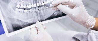
Dental X-ray is the basis for making an accurate diagnosis
Dental X-ray is one of the mandatory stages of diagnosis and treatment. Often, only the results of dental photographs clarify the situation; it allows making a final diagnosis and, accordingly, prescribing appropriate therapy or performing the necessary surgical manipulation. Moreover, the doctor often prescribes x-rays not only before, but also directly during the treatment process in order to assess its correctness and adjust as necessary. This research helps make treatment high-quality, with long-term results. There are no special contraindications, only during pregnancy you should warn your doctor about this.
The work of a radiologist in regional dentistry in Astrakhan is well established, the queue moves quickly. The study is carried out using modern equipment - we strictly comply with the standards of radiological control and patient safety. Sensitive devices have low radiation levels.
You can take a dental x-ray in Astrakhan in our clinic not only to see a local doctor, but also to confirm the opinion of another specialist. We take pictures of baby teeth, molars, filled canals, after resection and other types of surgical intervention. If wisdom teeth are not erupting correctly, an x-ray is indispensable. The image reveals cysts, cracks in teeth, and clarifies the degree of inflammation during periodontitis. It shows how deeply the teeth are affected by caries, and reveals jaw anomalies invisible to the eye.
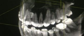
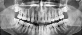
Panoramic photograph of teeth: importance and necessity
It is convenient for us to take an X-ray of a tooth in Astrakhan - both a targeted image and a panoramic one, when a three-dimensional image is obtained. A panoramic photograph of the teeth includes the upper and lower jaw, sinuses, maxillary joint, etc. Orthopantomography (as specialists call the picture) is needed for the work of an orthodontist, orthopedist, when installing dentures, crowns, implants and braces. It makes it possible to assess the condition of the periodontium, impacted teeth, roots and adjacent tissues.
Cost of dental photographs in dentistry
Making dental x-rays in Astrakhan as accessible as possible for dental patients is our task. The price of images is truly affordable for all categories, this also applies to panoramic x-rays. The exact price of the images is indicated on the website of the clinical dental center - each category has its own price.
View prices for paid radiology services (code 300) >>
Make an appointment with a dentist >>
Fresh questions about the service panoramic dental imaging in Izhevsk
- E
Ekaterina asks a question to the Avangard Baby Clinic, October 27, 2022, 20:59 Hello, is it possible for you to treat all of a child’s teeth under general anesthesia and how long will it cost? The child is 2.6 years old. - D
Dmitry asks a question to the Lada-esthete clinic, March 12, 2022, 18:40 Hello. I need to remove two teeth under general anesthesia, since the last time local anesthesia did not take me and they removed me alive. The teeth are level with the gums. Tooth 8 is the upper jaw and 5 is the lower jaw. What do I need for this...
- D
Dmitry asks a question to the Lada-esthete clinic, March 12, 2022, 18:14 Hello. I need to remove two teeth under general anesthesia, since the last time local anesthesia did not take me and they removed me alive. The teeth are level with the gums. What do I need to provide you with for this and how much will it cost?
- D
Denis asks a question to the Avangard Baby Clinic, March 08, 2021, 04:32 The gumboil needs to be removed and two teeth cured. I’m afraid to treat it in the usual way, but in a dream I want to try it. Health allows no chronic diseases. How much will it cost and when can I come, preferably this morning.
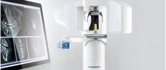
Panoramic shot
Panoramic images directly at the Medfodent Plus clinic
An orthopantomogram (OPTG) is a diagnostic panoramic image of the lower and upper jaws, which allows you to assess the condition of the teeth, temporomandibular joints, maxillary sinuses and other areas of the temporomandibular region. Important: Due to low radiation doses, orthopantomography is used in pediatric dentistry and in other cases where it is necessary to minimize exposure to X-rays.
Description of the procedure
You can undergo first-class diagnostics at a preliminary stage during treatment planning or already during treatment to obtain more accurate results. Using an orthopantomogram, a thorough examination is carried out so as not to miss the slightest pathology that interferes with effective treatment. X-ray examination of the dental system using this modern diagnostic equipment allows for gentle dental treatment.
Orthopantomogram is used:
- to identify all areas affected by caries;
- when identifying periodontitis;
- to identify features of teething, various anomalies in the development of jaws, teeth, etc.;
- in preparation for prosthetics or dental implantation;
- in periodontal and orthodontic treatment.
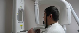
How is the procedure performed?
During filming, the patient's head is held with special clamps to prevent displacement. This is necessary to obtain more accurate images. As a rule, a high-quality photo is obtained the first time. The process takes just minutes.
A comprehensive digital examination is most complete and visual, promotes high-quality and effective treatment, and virtually eliminates erroneous diagnoses and procedures.
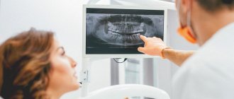
Patients see existing problems “from the outside”, it is easier for the doctor to explain possible treatment options, and at the end of the consultation they receive: examination results: two treatment plans - recommended and alternative.
Treatment plan
After passing all stages of a comprehensive examination, the patient receives 2 options for a treatment plan with a clear explanation: recommended and alternative.
- The plan clearly reflects all stages and their costs.
- The patient receives the results (plan) and has the opportunity to make the right decision.
- The patient can agree on the date and time of the appointment to undergo the recommended procedures (based on the accepted version of the treatment plan).
ATTENTION!!! The cost fixed in the plan does not change during the treatment process.
Features of the event
The doctor instructs the patient to remain still. Then it is necessary to remove jewelry, clothing items with metal parts, and the radiologist proceeds directly to the examination.
Cone beam computed tomography of teeth is performed standing or sitting. The patient is offered a lead apron or vest; he places his head on a special stand so that his forehead rests against the bracket. You must stand still for 20-25 seconds, during which time the equipment records panoramic images. Next, the program produces a 3D design, with the help of which the specialist draws conclusions about the state of the dental system.
The examination is completely painless.
Preparation measures
In order to properly prepare for the examination, you need to clarify whether it will be carried out with contrast. This depends on the indications for the procedure; contrast is necessary to identify neoplasms in bone or soft tissues, or suspected tumor processes. If contrast is not needed, the procedure does not require preparation.
When you schedule a dental cone beam computed tomography scan with contrast, you must come to the clinic on an empty stomach. You will have to refrain from eating 6 hours before the scheduled procedure, so most often it is carried out in the morning.
Before the examination, you must remove jewelry, metal parts and objects.
What can the new tomograph do?
- excellent quality of 2D panorama images
- 3D scanning with the lowest possible radiation dose - Low Dose Technology - ensures high safety, which is very important when examining children, for postoperative monitoring, and when planning the installation of implants
- 5 scanning area formats in 3D mode will allow you to most reliably plan treatment for any task in the maxillofacial area: 5 x 5 cm - optimal when planning the installation of a single implant or for local diagnostics, the lowest possible level of radiation for the patient
- 6 x 8 cm – covers the entire dental arch when planning multiple implant placements and allows the use of surgical implant guides
- 8 x 8 cm – covers the entire dentition, including the lower and upper jaws, as well as part of the maxillary sinuses. Used for implantation on the upper and lower jaws; for the use of surgical templates in implantology; for differential diagnosis of sinusitis in ENT practice
- 8 x 15 cm – covers the upper and lower jaws, including the upper respiratory tract, cervical spine or sinus area. Both maxillary joints and sinuses can be examined. It is used both in dentistry for a 3D panorama, and in the diagnosis of the cervical spine and respiratory tract, in the ENT diagnosis of diseases of the paranasal sinuses (sinuses)
- 13 x 15 cm – covers the entire maxillofacial area: both jaws, both joints, cervical spine, respiratory tract and sinuses can be examined simultaneously. Application: maxillofacial surgery; gnathic surgery; diagnosis of TMJ (temporomandibular joint), diagnosis of injuries to the maxillofacial area; ENT diagnostics.
Sign up for a consultation
to clinic specialists Mon-Fri: from
09
to
20
, Sat-Sun: from
10
to
16
(4852)
230-631
Make an appointment
What does a 3D dental x-ray show?
3D dental tomography is a highly accurate diagnostic method that makes it possible to obtain a three-dimensional image of the dental system in different projections. Volumetric images obtained with CT allow the specialist to enlarge, rotate and examine the area of interest from all sides and at different depths:
- The entire maxillofacial apparatus.
- A dentition or an individual tooth.
- Paranasal sinuses.
- Bone and periodontal tissues.
Dental CT allows you to detect inflammation, assess the homogeneity of the filling material and check the quality of installation of a filling, crown or implant, see the number of dental roots and their fragments, identify neoplasms, assess the degree of curvature of teeth, determine the exact parameters of bone tissue (height, width, density, etc.). d.). The information obtained allows the doctor to optimize treatment measures and predict the result.
Indications and contraindications
The image obtained as a result of three-dimensional computed tomography allows you to obtain accurate information about the structure of the nose and the dental system: bone tissue, teeth, root canals. Such a study allows the specialist to determine the following:
- caries, the depth of damage to teeth;
- periodontitis;
- cysts, granulomas of tooth roots;
- education;
- diseases of the temporomandibular joint;
- anomalies of development, teething, growth of teeth;
- location of injury and/or consequences;
- curvature of the nasal septum and the condition of the paranasal sinuses.
The price depends on how extensive the study is planned: you can study only a separate area, one or both jaws.
Cone beam computed tomography of teeth is recommended for previous injuries and traumatic tooth extractions, planned interventions (prosthetics, implantation, surgery, orthodontic measures).
This diagnostic method is contraindicated during pregnancy (carried out only in cases of urgent need), young children with a height of up to 120 cm, hyperkinesis, and allergies to iodine during a procedure with contrast. Mental disorders in the form of claustrophobia are a relative contraindication: a procedure that does not involve the patient being inside the tomograph can, in some cases, be carried out using another type of equipment.
3D image of teeth in dentistry
Diagnosis in dentistry is often complicated by the fact that the doctor is not able to see the oral cavity or an individual tooth in the most detailed angles. Therefore, to obtain an accurate picture of the condition of the dentition, hardware examination methods are used, which allow you to see what is inaccessible with a normal visual examination.
Technologies are constantly improving, and a 3D image of the teeth has been added to the traditional X-ray and orthopantomogram. As a rule, it is computed tomography that is prescribed by most plastic and maxillofacial surgeons, orthodontists and implantologists.
This is a modern diagnostic method that allows a specialist to see a detailed image of the jaw in different projections and from any angle. The examination in 3D format visualizes the oral cavity for a thorough examination of the dentition for the presence of pathologies and assessment of the general condition.
Content
- What does a 3D CT scan of teeth show?
- Indications for the procedure
- Main contraindications
- Can this be done for children?
- Main types of examination
- How do they take 3D photographs of teeth in specialized clinics?
- What are the benefits?
- Where can you take a 3D photo in St. Petersburg?
What does a 3D CT scan of teeth show?
This type of research is considered one of the most informative diagnostic methods in dentistry. With its help, it is possible to obtain the most reliable information about the condition of bone tissue, the circulatory system of the face and soft tissues.
Speaking about what a 3D photograph of teeth shows, we can highlight the following:
- Identification of foci of pathologies in the studied areas using a large-scale image.
- Visualization of the condition of the jaw in three-dimensional format.
- Diagnosis of cysts, tumors and other neoplasms.
- Evaluation of the effectiveness of previous treatment.
- Position of teeth, including unerupted ones.
- Separate areas of the oral cavity at the required angle.
3D dental x-ray allows you to visualize the condition of not only bone, but also soft tissue. This greatly simplifies the process of diagnosing many pathologies and inflammatory processes.
In addition, the resulting images will show the beginnings of teeth, the location of nerves and blood vessels. The popularity of this diagnostic method is due to the fact that without its use, correct installation of orthodontic structures, implantation and planned surgical interventions in the maxillofacial area are impossible.
Indications for the procedure
The use of dental computed tomography is necessary for most orthodontic and surgical procedures. This procedure is also indicated in the following cases:
- Pathologies of the temporomandibular joint.
- Anomalies in the structure of the jaw.
- Fractures of the maxillofacial region (to determine the severity of the condition).
- Preparation for upcoming dental prosthetics.
- Periodontitis and periodontal disease.
A 3D photograph of teeth is taken if there are suspicions of inflammatory processes that cannot be diagnosed by other methods.
Main contraindications
3D computed tomography of teeth is based on x-rays, so this procedure cannot be called completely safe. The radiation exposure during this examination ranges from 0.045 to 0.06 mSv. This is not a very high figure, given that the annual exposure limit is 5 mSv (according to the Russian Ministry of Health).
As for contraindications, they are standard for all types of x-ray examinations. The main limitation is the period of pregnancy (especially in the first trimester). In this case, the situation is considered individually, based on the ratio of harm to the child and benefit to the mother.
If a contrast agent is used (it is not used so often in CT scans of the teeth and jaw), then the following can be added to the contraindications:
- Patients suffering from thyroid diseases.
- Kidney failure.
- Allergy to drugs containing iodine.
The lactation period is not a contraindication, but after the procedure at least 48 hours must pass before the next breastfeeding.
Can this be done for children?
Many parents think that the use of 3D computed tomography is impossible in childhood due to the negative impact on the child’s body. This is not entirely true, because special attention to this study is given specifically in pediatrics.
If you follow all safety rules and do not exceed the frequency of procedures (for children – once a year), then the examination will not lead to any pathological changes in the child’s body.
3D CT in pediatric dentistry allows you to assess the condition of the sinuses, gums and see the rudiments of baby teeth. This method also allows you to identify malocclusion pathologies in a child.
Main types of examination
Three-dimensional images are taken using various types of tomographs. It is extremely important to know which device the examination will be used on, because the dose of radioactive radiation received and the degree of information content depend on this.
There are several main methods:
- Step-by-step computed tomography. It was carried out on old tomographs that produced images of low resolution and detail. Now it is no longer used, because the minimum radiation dose was 1500 μSv (with a standard of 1000 μSv).
- Multislice CT (MSCT). Body scanning is carried out using sensors located on a rotating tube. The table moves slowly through the ring, allowing you to take pictures in several planes at once. This method is characterized by the fact that the detectors are arranged in several rows. Such equipment is better suited for examining lymph nodes, muscles and other soft tissues. The radiation dose ranges from 300 to 400 μSv.
- Cone beam CT (CBCT). This technique is considered the most modern and informative in the field of dentistry. The main advantage is the maximum accurate image of teeth and bone tissue, which cannot be achieved using MSCT. CBCT is also considered the safest type of computed tomography (on average, the radiation dose ranges from 40 to 120 μSv).
How do they take 3D photographs of teeth in specialized clinics?
The average time of the procedure is no more than 1 minute. During this short period of time, the device takes about 200 pictures in various projections.
Before the procedure itself, you need to remove all metal objects in close proximity to the area being examined (chains, piercings, etc.). The examination itself looks like this:
- The patient's head is securely fixed using immobilization devices. This is very important, otherwise the pictures will turn out blurry.
- A protective apron is put on the chest.
- The rotating tube of the tomograph rotates around the head, taking a series of images.
- All information is immediately displayed on the computer monitor connected to the device.
- Further interpretation of the images is carried out by a specialist.
What are the benefits?
Computed tomography is a completely painless procedure that does not cause any discomfort to the patient. The only thing is that he must remain motionless for a short amount of time.
The use of 3D images is due to the following reasons:
- Minimum radiation dose.
- The examination procedure lasts no more than a few minutes.
- Get results quickly.
- Three-dimensional images have a high diagnostic value.
- 3D images eliminate errors in prosthetics and dental surgery.
Where can you take a 3D photo in St. Petersburg?
Diagnostic X-ray - invites you to undergo a 3D CT scan of the jaw if your physician has prescribed this procedure for you. We have no queues, so you can quickly receive images for further delivery to a medical institution.
For the convenience of our clients, we have organized courier delivery within St. Petersburg. You also have access to recording results on digital media, extended consultation and other additional services. You can find out more detailed information on the website.
To make an appointment, you need to call the specified phone number, or leave an online application.
Sign up for a study by phone
+7 (812) 332-52-54