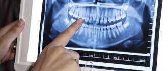
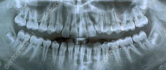
An x-ray is the dentist’s main tool in making the correct diagnosis. However, a conventional orthopantomogram or targeted photograph has limited diagnostic potential and does not provide complete data on the condition of the teeth and maxillofacial area. But technologies are constantly being modernized and today, more informative technology has come to the aid of conventional radiography - dental computed tomography (CT).
What does a 3D dental x-ray show?
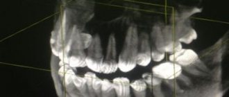
3D dental tomography is a highly accurate diagnostic method that makes it possible to obtain a three-dimensional image of the dental system in different projections. Volumetric images obtained with CT allow the specialist to enlarge, rotate and examine the area of interest from all sides and at different depths:
- The entire maxillofacial apparatus.
- A dentition or an individual tooth.
- Paranasal sinuses.
- Bone and periodontal tissues.
Dental CT allows you to detect inflammation, assess the homogeneity of the filling material and check the quality of installation of a filling, crown or implant, see the number of dental roots and their fragments, identify neoplasms, assess the degree of curvature of teeth, determine the exact parameters of bone tissue (height, width, density, etc.). d.). The information obtained allows the doctor to optimize treatment measures and predict the result.
Types of dental crowns for chewing teeth
When choosing a material for creating a microprosthesis, you should take into account the functional purpose of the teeth.
The molars of the posterior zone of the dentofacial apparatus are subjected to strong mechanical and chemical stress when chewing food. Eating food that differs in taste, composition and hardness can quickly damage the nozzle, which is made of aesthetic material, but vulnerable to the abrasive properties of some products. In addition, the material should not cause allergies to the soft tissues of the oral cavity. Therefore, it is better to entrust the choice of material to a specialist. I am an orthopedic dentist, Sergey Samsakov, taking into account the results of the research, I recommend the optimal material option for creating a crown in each specific case:
- Metal-ceramic crowns
- a successful combination of strength, cost and beauty have made this type of onlay the most popular for installation on a tooth. Metal-ceramic crowns are installed with mandatory circular grinding of the tooth being restored.
- Metal crowns
are reliable products with a budget price. These are long-term onlays with good biological compatibility and do not require preparation of healthy tooth tissue. They can be made in combination with precious alloys. The only negative is the possibility of developing an allergic reaction. - Ceramic prostheses made of zirconium dioxide
are an ideal option for placing crowns in patients with contraindications to the use of metal structures. The high cost of the ZrO2 crown is fully compensated by the durability and aesthetics of the dentition. - Metal-plastic
dental prostheses are very fragile and are used by the doctor as temporary crowns on a tooth with the subsequent installation of a permanent crown made of durable material.
The structural reliability of the prosthesis allows you to protect the living tooth from further abrasive processes and maintain the ability to fulfill its purpose - thoroughly chewing food.
Why are dental x-rays prescribed?
A 3D photograph of teeth is performed if the following indications exist:
- Injuries of the maxillofacial area.
- Preparation for endodontic treatment (structure of root canals, pathological processes in the periodontium, degree of pulp damage, etc.).
- Diagnosis of neoplasms (cysts, abscesses, granulomas, tumors).
- Anomalies of development and deformation of the maxillofacial apparatus.
- Quality control of filling and implant installation.
- Planning of orthodontic treatment (identification of impacted and dystopic teeth, analysis of the condition of the tissues around each tooth, etc.).
- Detection of hidden periodontal cavities and pockets.
- Implantation planning (assessment of jaw bone parameters, indications for sinus lift or osteoplasty, modeling the result of implantation).
- Endogenous pathologies of the maxillary sinuses.
Three-dimensional x-ray examination is the gold standard when planning any complex dental procedure or surgery. CT allows you to quickly make an accurate diagnosis, competently plan treatment or dental prosthetics, and monitor the results.
What is the cost of a dental crown?
The cost of restoring a lost or defective tooth depends on many factors, the main ones:
1. The status of the dental clinic in general and the treating orthopedist in particular - the higher the doctor’s rating, the more expensive the cost and quality of the services provided.
2. Material for making a crown: the lowest price of metal products, ceramic crowns and products made of zirconium dioxide cost from 27,000 thousand rubles per unit. A crown containing precious metal is the most expensive.
All stages of preparation and treatment: consultation, hardware diagnostics, filling and other procedures that ensure oral health also require certain financial costs. The services of a dental technician are included in the final cost of installing a crown on a tooth.
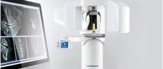
What equipment is used
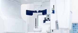
To carry out 3D diagnostics, a three-dimensional computed tomograph SOREDEX Scanora 3D with advanced functionality is used. This is the latest generation equipment, which allows you to obtain three-dimensional images of the anatomical structures of the maxillofacial region in a few seconds, with the least radiation exposure for the patient.
The program analyzes the obtained multiplanar sections and builds them into a 3D model, thanks to which the specialist is able to accurately assess the condition of the dental system, detect all pathological processes occurring in this area and competently plan a treatment regimen.
A virtual 3-dimensional model of the scanned area can be recorded on any digital media (CD, flash drive), which allows the attending physician, if necessary, to view diagnostic data or involve related specialists in the analysis of the received information.
Indications for prosthetics
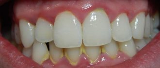
- Chipped crown (when the defect cannot be corrected by installing a filling);
- multiple cracks in the enamel;
- pathological abrasion of enamel;
- fluorosis;
- tetracycline teeth;
- color change (darkening, yellowing);
- destruction of the supragingival part of the tooth more than 70% with preservation of the root;
- thin, weakened enamel;
- replacement of an old restoration.
Possible harm
Cone beam dental computed tomography is the safest and fastest diagnostic method. Thanks to the use of a conical X-ray beam, the radiation dose received during the study is 10 times less than when using spiral CT. And the pulsating mode of the X-ray beam further reduces the radiation dose. The three-dimensional computed tomograph SOREDEX Scanora 3D is one of the safest devices in terms of X-ray radiation dose - only 0.035 m3v.
However, despite the safety of the study, CT also has contraindications. If we just talk about dental tomography, it is not performed during pregnancy (in the 1st trimester). 3D dental x-rays with contrast are prohibited for pregnant and lactating women, patients with endocrine disorders (diabetes mellitus, thyroid pathologies), renal failure and intolerance to iodine-containing drugs.
The structure of baby teeth
A temporary bite differs from a permanent bite not only in the number of teeth (in a temporary bite there are 20). Baby teeth have a bluish tint to the crown, which is also much wider than the root. Children have less enamel mineralization, so caries affects them very quickly, and the pulp takes up more space compared to permanent teeth. The canals of baby teeth are wider, easier to pass for instrumentation, and the roots themselves have a rounded shape. The structural features of baby teeth determine their rapid destruction in case of infection penetration and very severe pain in the child, so in childhood, visit the dentist in a timely manner.
Knowing the structure of teeth will help you quickly find a common language with the dentist and feel more confident when discussing treatment issues.
You will be able to better navigate the manipulations performed by the doctor and clarify all the points that interest you with an understanding of the essence of what is happening. This article is for informational purposes only, please consult your doctor for details!
Online consultation with a doctor
If you are concerned about the condition of your teeth. Painful sensations arise, gums bleed for a long time, and seals have appeared on the jaw. You are concerned about previously installed dental implants. And there was a need to take a 3D photo of the teeth. Then, after receiving three-dimensional visualization, it is better to go for an examination or consultation with a dentist to interpret the images and compare the results with your current complaints. It is impossible to independently understand the nuances of a 3D image, much less make a diagnosis. The specialist will explain the situation and give recommendations before the in-person appointment.
Normal and deviations in tooth sizes
All dental units of the same name have approximately the same height and width, except for the central (medial) incisors. The size of the front teeth of the upper jaw is normally slightly larger than that of the lower jaw. The height of the crown of the upper central incisors varies from 9 to 12 millimeters, width - from 8 to 9 millimeters. The lower teeth are similar in height, but are about 5 millimeters wide. The size of a person's wisdom teeth does not differ from the parameters of other molars. The table below shows the average width of dental crowns in millimeters.
| Upper jaw | Lower jaw | |
| Medial incisor | 8.5 mm | 5.3 mm |
| Lateral incisor | 6.5 mm | 6 mm |
| Fang | 7.6 mm | 6.7 mm |
| First premolar | 6.7 mm | 6.8 mm |
| Second premolar | 6.4 mm | 7 mm |
| First molar | 9.4 mm | 10 mm |
| Second molar | 9.4 mm | 10.2 mm |
Anomalies in the size of human teeth can be congenital or acquired and are accompanied by malocclusion, impaired chewing functions and an unaesthetic appearance of a smile. The most common deviations are macrodentia and microdentia.
Advantages of the method
- The ability to rotate, enlarge, and examine images in any projection and section, which is impossible with conventional 2-dimensional scanning.
- The examination lasts only a few seconds (8-20 seconds).
- Complete diagnostic information.
- Maximum security.
- Digital information format.
- Detection of any pathological processes at an early stage.
- No prior preparation required.
- 3D reconstruction without distortion or artifacts.
- A wide range of purposes - from endodontic dental treatment and implantation to maxillofacial operations.
The structure of the human jaw: teeth and their formula
When citing one number for any tooth, the dentist means its anatomical and functional affiliation. If you conditionally divide the dentition into two halves and start counting from the first incisor, you will get 8 teeth in each direction (top and bottom). The first incisor is a one. The second, accordingly, is a deuce. And so on until the last molar, which is called the figure eight. If the doctor says - bottom seven on the left, then we are talking about the bottom left second molar. As you can see, everything is very simple!
This can be expressed schematically as follows (permanent teeth):
For baby teeth, everything is the same, only the teeth are designated by Roman numerals (there are no premolars in the primary dentition):
As for the two-digit designations, they were invented in order to simplify the written recording of medical history. The above is preserved, but a number appears before the number indicating which jaw and on which side the tooth is located. Conventionally, the oral cavity is divided into segments - upper right (number 1), upper left (number 2), lower left (number 3), lower right (number 4). As you noticed, the report is conducted starting from the top right and clockwise. The segment is written first, and then the tooth number. For example, the third canine from the top right would be designated tooth 13. The lower incisor on the left is 32 teeth. In children, the segments are designated by numbers 5 to 8, so as not to confuse the primary bite with the permanent one. Let's take the same third fang on the right. In a child, it will be designated as tooth 53. And so on by analogy. The main thing is to always start the report from the right side at the top, then you won’t get confused. These are not snails with 25 thousand teeth! Can you imagine their dental formulas?
Is there an alternative to CT
There are many other diagnostic imaging methods (x-ray, orthopantomogram, ultrasound, etc.), but only CT provides the possibility of highly accurate, separate images of all types of tissue at different angles and to different depths. Although a panoramic dental photograph remains an equally important diagnostic tool for a dentist today, it can only provide a general overview. In turn, a 3D tomogram allows you to obtain not a single flat image of the jaw, but a whole series of sequential multiplanar images in different projections and without the distortions inherent in a panoramic image.
Example:
due to the different density of bone structures exposed to X-ray radiation, it is impossible to see less dense bone in a 2-dimensional image; accurate information is provided by a 3- D image of the teeth.
History of dental floss
The word “floss” comes to us from the English language and is literally translated as “dental floss”. It was first mentioned in the book A Practical Guide to Dental Care, written by American dentist Levi Spear Parmley in 1819. He suggested to his patients and readers to use silk dental floss to remove food debris from the interdental spaces.
During World War II, when Japan stopped supplying silk to America and Europe, silk dental floss was replaced by nylon floss, developed by DuPont. Nylon was thinner, stronger and cheaper. It was produced in large quantities, and therefore became very popular in the mass market.
How does the procedure and decoding work?
To take a 3D photograph of teeth, a standing or sitting patient needs to bite a special plate and fix his position in the device using a fixing stand. During the entire scanning time, you must remain absolutely still.
The tomograph sensor makes a series of revolutions around the patient’s head for 8-20 seconds, producing about 200 images in different projections. Processing digital data takes 5-15 minutes, after which the information is written to a disk or flash drive. No preparation is required, you just need to remove all metal jewelry from your neck, ears, and hair before the procedure.
Modern dental floss
Teflon dental floss is considered the most durable. They have a low coefficient of friction and high mechanical tensile strength. Does not disintegrate into fibers.
Mirafloss Tape is an unwaxed (not waxed) tape floss that is tear and abrasion resistant due to Teflon fibers. Thread length 20 m.
Pierrot Dental TAPE is a waxed (wax-coated) tape floss made of Teflon. Suitable for those who have narrow interdental spaces. Thread length 20 m.
Revyline PTFE Black Edition is a waxed Teflon dental floss with mint impregnation. Ideal as a first thread. Length 50 m.
Tape floss Miradent Mirafloss Tape 20 m
Waxed Pierrot Dental TAPE with mint, 25 m
Dental floss Revyline PTFE Black Edition, 50 m
Flat, ribbon threads
The thickness of tape threads is usually 0.005-0.007 mm (tens of times smaller than a human hair), which allows them to easily penetrate even the narrowest interdental spaces. These flosses are recommended for those who have teeth that are close to each other.
Oral-B Pro-Expert Clinic Line and Oral-B Satin Floss are wax coated, mint flavored and 25m long.
Miradent Mirafloss chx-Tape is impregnated with chlorhexidine for an antiseptic effect. Thread length 20 m.
Biorepair Filo Non Cerato Ultrapiatto floss is ideal for cleaning narrow interdental spaces and sensitive teeth. The flat part helps remove food debris even in the most difficult to reach places. Thread length 30 m.
Revyline PTFE Black Edition dental floss is made of Teflon, has a jet black color and is impregnated with menthol. Thread length 50 m.
Waxed thread Oral-B Pro-Expert Clinic Line Cool mint, 25m
Tape floss with chlorhexidine miradent Mirafloss chx-Tape, 20 m
Ultra-flat thread Biorepair Filo Non Cerato Ultrapiatto, 30 m
Waxed thread Oral-B SatinFloss mint, 25 m
Expanding threads
The peculiarity of such threads is that they swell (expand) under the influence of saliva and become thicker than 1 mm in diameter. This allows them to thoroughly clean the wide interdental spaces from food debris and plaque.
Biorepair Espandibile dental floss is an ultra-soft, expanding floss infused with hyaluronic acid to relieve gum inflammation. Thread length 30 m.
GUM Expanding Floss is a waxed expanding floss, ideal for sensitive teeth. Length 30 m.
Swiss Smile waxed dental tape penetrates the interdental space slightly expanding, thereby providing excellent cleansing without damaging soft tissues. The tape refreshes the oral cavity well due to its mint impregnation. Available in two colors: blue and black. Thread length 70 m.
Waxed Splat Dental Floss expands with use, providing more effective cleaning of the surface of the teeth. The antibacterial component Biosol stops the proliferation of bacteria. Thread length 30 m.
Waxed dental tape Swiss Smile, 70 m
Waxed thread Splat Dental Floss mint with silver fibers, 30 m
Waxed thread GUM Expanding Floss, 30m
Waxed expanding thread Biorepair Filo Cerato Espandibile, 30 m
Superflosses
Superflosses are special threads consisting of several types of alternating fibers for effective cleaning of braces, bridges and implants.
Most superflosses consist of two parts:
— a rigid tip necessary for threading under an orthodontic structure or a bridge;
— the spongy part (a wide unwaxed thread resembling a sponge), necessary for cleaning orthopedic and orthodontic structures, as well as interdental spaces.
Superfloss brand Oral-B Superfloss differs from classic superfloss: firstly, it has a fixed length (the package contains 50 pieces of 60 cm in length), and secondly, it consists of three parts (hard tip, spongy part and regular waxed thread) .
Mirafloss Implant chx is a practical box of 50 threads, 15 cm long, for cleaning braces and implants. The middle part of the superfloss is impregnated with chlorhexidine for an antiseptic effect. Available in two versions: thin threads (diameter 1.8 mm) or medium thickness (diameter 2.2 mm). Thread length 50 m.
GUM Access Floss superfloss is no less effective in cleaning braces, implants and wide interdental spaces . There are 50 threads in a package. The total length of the thread is 30 m.
Dental floss Oral-B Superfloss, 50 pcs.
Floss miradent Mirafloss Implant chx 1.8 mm for implants/braces
Floss miradent Mirafloss Implant chx 2.2 mm for implants/braces
GUM Access Floss for implants/braces, 30m
Waxed threads
Waxed threads are those that are coated with wax on top. They need such a coating in order to easily slide into the interdental spaces of dense or crowded teeth. This type of thread is atraumatic and does not cause discomfort when used, so it is more often recommended than others for those who are just beginning their acquaintance with threads.
Emmi-dent Dental Floss is made from high quality nylon and has a refreshing mint flavor. Suitable for sensitive teeth. Thread length 50 m.
The voluminous waxed floss of the Italian production Splat Dental Floss glides easily between the teeth, contains essential oils (cardamom, strawberry or bergamot) and the antibacterial component Biosol, which provides effective protection for the oral cavity. Thread length 30 m.
Waxed dental floss Pierrot Dental floss is made of nylon and impregnated with wax for better glide. Fluoride, which is included in the composition, restores and protects enamel from caries in places inaccessible to a brush, and aloe vera has an anti-inflammatory and wound-healing effect. Thread length 50 m.
Curaprox Dental Floss waxed interdental floss with mint flavor. Thread length 50 m.
Waxed thread Emmi-dent Dental Floss mint, 50 m
Waxed dental floss Pierrot Dental floss with aloe vera, 50 m
Waxed floss Curaprox DF834, 50 m
Waxed thread Splat Dental Floss with cardamom, 30 m
Unwaxed threads
Unwaxed threads are threads that do not have a wax coating. Due to this, the threads do not slip into the interdental spaces, but gently “rub the enamel,” thereby cleaning it much better from plaque and food debris.
ROCS Black Edition unwaxed expanding floss with mint flavor helps prevent gum inflammation, keeps teeth healthy and white, and freshens breath. Length 40 m.
The Biorepair line includes two types of unwaxed floss: Biorepair Spugnoso - a sponge floss (unwaxed, sponge-like) for cleaning wide interdental spaces, and Biorepair Ultrapiatto - an ultra-flat unwaxed floss for cleaning narrow interdental spaces. Both threads contain unique MicroRepair crystals that promote mineralization of tooth enamel. The length of the threads is 30 m.
Tape floss with chlorhexidine Miradent Mirafloss , impregnated with a chlorhexidine solution, has an antibacterial effect and reduces the formation of plaque. The surface of the tape is unwaxed, made of PET (Teflon), thanks to which it glides easily when inserted into dense interdental spaces.
Dental floss ROCS Black Edition (black), 40 m
Sponge dental floss Biorepair Filo Non Cerato Spugnoso, 30 m
Tape floss with chlorhexidine miradent Mirafloss chx-Tape, 20 m
Ultra-flat thread Biorepair Filo Non Cerato Ultrapiatto, 30 m
How to determine the age of a cow by its teeth
The age of the cattle is determined by changes in the condition of the incisors. With age, dental units wear out, the color of the enamel changes, the dentin shortens and becomes thinner. Calves are born with four to six incisors. Milk teeth are sharp at the ends, with thin enamel. At one week of age, the calf should have 7-9 milk teeth. Edges are formed.
See also
Costs of a business raising bulls for meat at home and whether it is profitableRead
Calves develop three teeth on each jaw per month. At six months, the fourth molar is formed and the margins are fully developed.
Important! In calves, the incisors are constantly growing and by three months they become the same size. The enamel on the incisors begins to gradually wear off after reaching the age of four months.
By 12-13 months, abrasion of the enamel on the lingual surface of the hooks is noted. At 14-15 months, the enamel is completely erased on the middle internal hooks, and by 17-18 months - on the middle, outer edges. At 1.6-2 years the holds change. At the age of five years, cattle have permanent toes. At five to six years of age, the incisors become square in shape. By the age of 7-8 years, enamel disappears from the surface of the toes in cattle. By the age of seven, the hooks are practically worn down to the ground. Yellowish enamel.
By the age of 10-12 years, gaps are noticeable between the teeth. The hooks are hewn. The enamel acquires a yellowish tint and completely disappears from the edges. Dentin is thinned. By the age of 13.5-14 years, the incisors are worn down to the neck and have an oval surface shape. The enamel almost completely disappears. 15-17 year old animals have practically no teeth. Only roots or stumps remain.
Important! In cattle, with age, the lower lip droops, and the lower jaw protrudes slightly forward due to the fact that the incisors, as they develop, gradually begin to occupy their main place in the oral cavity.
It is not always possible to accurately determine age by looking at teeth. The change and degree of abrasion of the enamel of the incisal dental units depend on the general state of health, genetics, precocity, breed, individual, physiological characteristics of the body, as well as on the type of food consumed.
Thus, in representatives of meat breeds, teeth erupt and grow faster than, for example, in cattle of dairy, dairy and meat production. Therefore, after assessing their condition, the age of cattle is determined by average indicators.
The main stages of modeling the 16th tooth from sculptural clay
Sculpture clay has long been used in art to recreate shapes. This inexpensive material is ideal for sculpting teeth; working with clay is pleasant in its own way, it is soft, does not stick to your hands, and hardens gradually. Sculpting clay requires longer preparation for work than plasticine, which is most often used for modeling teeth. If the clay is dry, then first you need to mix it with water to the consistency of sour cream, leave for some time until it dries and forms a plastic mass. After this, the clay becomes hard only after a few hours, this time is quite enough to successfully complete the work. The smaller the volume of available material, the faster it hardens. When the desired consistency is obtained, the clay is shaped into a ball (Fig. 1).
Rice. 1. The material has the necessary plasticity and is ready for use. Giving the future model a ball shape
Let's consider modeling the first right molar of the upper jaw (16th tooth). The overall outlines of the model of the 16th tooth are set (Fig. 2, 3), the location of the main surfaces is outlined: medial contact (M), distal contact (D), vestibular (V) and palatal (P).
Rice. 2. Giving the overall outline of the model
Rice. 3. Modeling the tops of the main tubercles.
The tops of the main hillocks are determined. Markings are applied on the chewing surface (Fig. 4), corresponding to a first-order H-shaped fissure.
Rice. 4. Applying markings corresponding to the first-order fissure, H-shaped, on the occlusal surface
Completion of the formation of the external contours of the model and the equator (Fig. 5) is done by hand.
Rice. 5. Smoothing out irregularities and shaping the equator
Before this stage, all actions were performed by hand (Fig. 6).
Rice. 6. Working with your hands allows you to better feel the basic properties of the material
To model the chewing surface of the tooth, it is better to use tools. The first-order fissure is deepened with a spatula (Fig. 7, 8).
Rice. 7. Deepening of the fissure of the first order, separating the anterior buccal tubercle (2) from the posterior buccal (1) and anterior palatine (4). Posterior palatine tubercle (3)
Rice. 8. 1 - posterior buccal tubercle, 2 - anterior buccal tubercle, 3 - posterior palatine tubercle
When modeling, there is no need to draw fissures, but it is necessary to divide the main tubercles so that an H-shaped fissure appears between them (Fig. 9).
Rice. 9. Completing the modeling of the first-order H-shaped fissure
Tools for work are chosen that are more convenient to work with: it can be a spatula, a smoothing iron. A fissure shaping tool, such as a probe, is required. 2-3 tools are enough.
After completing the work, the model can always be corrected by cutting off excess with a scalpel or spatula. The resulting tooth model can be stored for a long time, reminding of the results of the work.
Rice. 10. Modeling of the longitudinal (2), medial (1), distal (3) ridges of the anterior buccal tubercle
Rice. 11. Second-order fissures are formed on the anterior buccal tubercle
Rice. 12. Modeling of the main (2) and additional (1, 3) ridges of the anterior palatine tubercle
Rice. 13. Final view of the 16th tooth model
Rice. 14. Appearance of the 16 tooth model. Occlusal surface
Rice. 15. Appearance of the 16 tooth model. Palatal and chewing surfaces
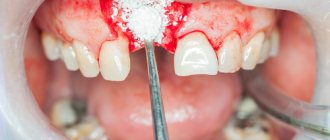
Stages of prosthetics
- Preparation
- assess the general condition of the oral cavity, take an x-ray, carry out hygienic cleaning (removal of plaque, tartar), treat caries, gums, change old fillings. - Preparation
- the front tooth is ground down for a crown, the stump is given the desired shape. According to indications, depulpation, treatment, and filling of dental canals are carried out. In case of severe destruction of the supragingival part, the root is strengthened with a stump insert. The prepared support is covered with a temporary plastic structure to protect tissues from destruction, chemical and temperature irritants. - Taking impressions
– the doctor takes impressions of the patient’s teeth using an impression tray with impression material or a digital scanner. The obtained data is transmitted to the dental laboratory. - Making a crown
- based on models, an orthopedic system is created in the laboratory using casting, pressing, and milling. At the manufacturing stage, the model is tried on the patient, and adjustments are made if necessary. - Installation
- the finished crown is fixed to the support with dental cement. The crown on the front tooth implant is installed using a cement or screw method.
Oral hygiene in children
There is a tradition of giving a silver spoon for the first tooth that erupts. We would add a toothbrush to this tradition. We recommend that you teach your child how to brush their teeth in a playful way from the moment the first tooth appears.
Doctors note that up to the age of 10, it is necessary to help children brush their teeth, since at a younger age children find it boring and do not understand why they should do it at all. There are also special liquids or tablets that allow you to check the remaining plaque on your teeth using contrast staining.
There are modern toothbrushes that can be synchronized with a tablet, then brushing your teeth turns into a game.
It is important to remember that the sooner you start brushing your child’s teeth properly, the healthier his teeth will be!
When is tooth lengthening necessary?
Lengthening the coronal area of a tooth is possible for various reasons: aesthetic, functional and restorative.
- The first group includes patients seeking to lengthen their teeth due to their cosmetic deficiencies (in particular, an inharmonious relationship between teeth and gums and the presence of a so-called gummy smile).
- Bad habits and endless stress have increased the percentage of people with increased tooth wear among residents of modern society. Such patients resort to surgical crown lengthening and orthopedic correction.
- Patients with a completely destroyed coronal part through surgical lengthening receive the effect of covering the hard tissues of the tooth. This allows you to fully distribute the load on the jaws when chewing and prevent fracture of the tooth root, as well as decementing of the orthopedic structure.
- Orthopedic or therapeutic restoration, for example, of a chipped fragment of the crown of a front tooth, is also essentially an extension of the clinical crown of a given tooth due to the free space between the antagonist teeth. This is the last group of indications for tooth lengthening, when, for example, a filling or ceramic veneer restores its height.
Modeling from plastic
Plastic is another material from which you can create a beautiful tooth model. This is a fairly dense, non-sticky material that does not require special preparation for work, but it is necessary to observe the conditions for its storage: temperature changes at which the material is stored can have a negative impact on its properties. Plastic is convenient because you can work with it for an unlimited amount of time, it does not harden, which makes it possible to work out the microrelief of the future model in more detail and clearly, and make the necessary adjustments during the work.
Hardening of the material occurs when it is placed in hot water or heated to 110-120 degrees in an oven for 5-10 minutes. The possibility of long-term work and fixation of the result by heating - this is the similarity between plastic and composite. Plastic cannot be reused. Well suited for creating phantom teeth models.
The first stage of working with this material will be to warm it up in your hands and shape it into a ball, then give the overall outline of the model, determine the main surfaces of the tooth model (Fig. 16) (M - medial contact surface, D - distal contact surface, V - vestibular surface, L - lingual surface), applying markings corresponding to the first-order fissure of the F-shaped form.
Rice. 16. Giving the overall outline of the model, applying markings corresponding to the fissure of the first order, F-shaped. M - medial contact surface. D - distal contact surface, V - vestibular surface, L - lingual surface
On the chewing surface with a tool, spatula or trowel, according to the applied markings, a fissure of the first order is deepened, five main tubercles are identified (Fig. 17) (1 - anterior lingual, 2 - posterior lingual, 3 - anterior buccal, 4 - posterior buccal, 5 - distal ).
Rice. 17. Tops of the main tubercles: 1 - anterior lingual tubercle, 2 - posterior lingual tubercle, 3 - anterior buccal tubercle, 4 - posterior buccal tubercle, 5 - distal tubercle
The equator of the 36 tooth model is also formed. The formation of second-order fissures occurs by modeling the longitudinal, medial, and distal ridges of the four main tubercles (Fig. 18-19).
Rice. 18. Modeling of the longitudinal (b), distal (a), medial (c) ridges, anterior lingual tubercle (1). 1 - anterior lingual tubercle, 2 - posterior lingual tubercle, 3 - anterior buccal tubercle, 4 - posterior buccal tubercle, 5 - distal tubercle
Rice. 19. 1 - anterior lingual tubercle: (a) longitudinal ridge, (b) distal ridge, (c) medial ridge. 2 - posterior lingual tubercle. 3 - anterior buccal tubercle: (a) longitudinal, (b) medial ridge, (c) distal ridge. 4 - posterior buccal tubercle: (a) longitudinal ridge, (b) distal ridge, (c) medial ridge. 5 - distal tubercle. 6 - additional tubercle. Rice. 19. 1 - anterior lingual tubercle: (a) longitudinal ridge, (b) distal ridge, (c) medial ridge. 2 - posterior lingual tubercle. 3 - anterior buccal tubercle: (a) longitudinal, (b) medial ridge, (c) distal ridge. 4 - posterior buccal tubercle: (a) longitudinal ridge, (b) distal ridge, (c) medial ridge. 5 - distal tubercle. 6 - additional tubercle
The distal tubercle has a less differentiated surface (Fig. 20).
Rice. 20. The final result of the simulation. 1 - anterior lingual tubercle. 2 - posterior lingual tubercle. 3 - anterior buccal tubercle. 4 - posterior buccal tubercle. 5 - distal tubercle
Models made of plastic can be stored for a long time; they will remind you of the results achieved in modeling.
Rice. 21. Lingual and medial contact surfaces of the mandibular molar model. M - medial contact surface. D—distal contact surface. V - vestibular surface. L - lingual surface
Rice. 22. Vestibular and chewing surfaces of the mandibular molar model
Contraindications to tooth lengthening
Contraindications include: difficult or impossible access; anatomical restrictions (for example, lengthening teeth from the oral side in the area of large molars is almost impossible); the ideal shape of the gingival contour is a contraindication to surgical lengthening. Orthodontic tooth lengthening is limited mainly by the need for space and the ability to attach an additional structure. The ratio of the crown and root of the tooth should not exceed 1:1. This is why teeth with short roots, for example after apex resection, are also not suitable for lengthening procedures.