From this article you will learn:
- how much does it cost to do a dental x-ray,
- what you need to know about radiation doses,
- Is it possible to take a dental photograph during pregnancy?
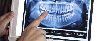
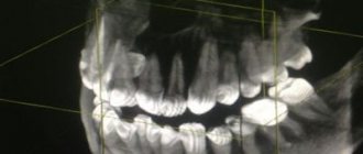
Dental X-ray is a traditional way to determine the quality of root canal filling, as well as to diagnose inflammation at the apex of the tooth root (with apical periodontitis). X-rays of human teeth in dentistry are most often carried out using targeted X-rays. A targeted photograph of a tooth received this name because it is small in size and shows the condition of only a few teeth and the bone tissue around them (Fig. 1-2).
Modern targeted X-rays produce a very low dose of X-ray radiation (compared to what it was 15-20 years ago). This is due both to the advent of ultra-sensitive photographic films, which require a significantly lower dose of radiation, and secondly, to the advent of special intraoral sensors that capture the image digitally and display it on a computer screen. Such digital images require several times less X-ray radiation - even compared to modern films.
Sighting and panoramic radiography in dentistry –
However, targeted images are not suitable for planning bite correction, assessing the condition of bone tissue before implantation, or planning the installation of future implants. They are not very convenient for planning treatment and prosthetics for a large number of teeth, and often do not allow detection of perforations and cracks in the tooth root. Focused photographs of the teeth may not be enough in cases where it is necessary to find problems with root canal filling in order to be able to properly treat the teeth.
Therefore, dentists very often refer patients to more complex options for x-ray examination of teeth and jaw bone tissue -
- dental computed tomography (CT),
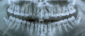
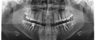
- orthopantomogram (Fig. 3),
- teleroentgenogram (for bite correction).
In this article, we will go into detail about the pros and cons of targeted dental x-rays, and what you need to pay attention to if you need to have a dental x-ray (for your own safety). For detailed reviews on the remaining specified methods of radiography in dentistry, read the links above.
Why do you need dental x-rays?
After the examination, the dentist will refer the patient for an X-ray examination:
- A targeted x-ray of a diseased tooth helps to clarify the degree of caries, distinguish pulpitis from periodontitis, find hidden carious cavities, and clarify the condition of root canals.
- Panoramic x-ray of teeth (orthopantomogram) - gives a detailed picture of the condition of all teeth, jaws, maxillary sinuses, temporomandibular joints. This study is the “gold standard” in dentistry, as images help diagnose hidden pathologies and inflammatory processes.
Indications and contraindications for the procedure
An X-ray is necessary in cases such as:
- severe toothache in the jaw when biting or chewing food;
- suspicion of inflammatory processes, cysts, granulomas, abscesses, deep caries;
- checking the quality of root canal treatment.
Diagnostics are also prescribed before orthodontic treatment, dental prosthetics, and x-rays of healthy teeth are taken before eliminating bite problems.
There are few contraindications to the procedure. These are diseases associated with immunodeficiency, pregnancy, pulmonary hemorrhages, and mental disorders.
How does the procedure and decoding work?
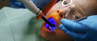
To take a 3D photograph of teeth, a standing or sitting patient needs to bite a special plate and fix his position in the device using a fixing stand. During the entire scanning time, you must remain absolutely still.
The tomograph sensor makes a series of revolutions around the patient’s head for 8-20 seconds, producing about 200 images in different projections. Processing digital data takes 5-15 minutes, after which the information is written to a disk or flash drive. No preparation is required, you just need to remove all metal jewelry from your neck, ears, and hair before the procedure.
Price for x-ray (image) of teeth
You can get an X-ray of your teeth in Moscow, find out prices, and make an appointment with a dentist by calling +7. Experienced specialists at Zubastik Dentistry will perform the necessary research and interpret the results in a high-quality manner.
In modern dentistry, increased attention is paid to dental x-rays. Radiography makes it possible to assess the general condition of the dentition, accurately determine the problem of a specific unit, and develop an optimal treatment program. You can take an X-ray of a tooth in Moscow at any Zubastik dental clinic. The procedure does not require special preparation.
Preparation and carrying out the procedure
Dental X-rays do not require special preparation. The specialist will guide you through each step of the x-ray process. He may leave the room briefly during filming. You will be required to stand or sit still for short periods of time. Spacers (film holders), if used, will move and adjust in the mouth to produce correct images.
Once the images are ready—immediately in the case of digital x-rays—your dentist will check them and determine whether or not there are any abnormalities.
Why do they send you for a dental x-ray?
The comfort and effectiveness of treatment of dental pathologies largely depend on an accurate diagnosis. An X-ray of a diseased or damaged tooth is prescribed by a doctor after a visual examination or during therapy. X-ray studies can be local or panoramic:
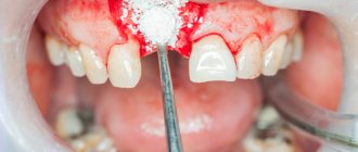
- A targeted spot X-ray of a problem tooth helps to notice a cyst or tumor at an early stage of development, to detect hidden caries under a filling, an abscess, or a broken root.
- A panoramic X-ray image of all teeth, unfolded in a plane, covers the maxillary sinuses, reflects a complete picture of the state of the dental system, and allows you to quickly identify the source of the inflammatory process.
Indications and contraindications
When examining a patient at an appointment, the dentist sees only obvious pathologies in the oral cavity. However, most problems are hidden in places inaccessible to the eye - spaces between teeth, canals, roots. In many cases, it is possible to correctly respond to a patient’s complaints only by having an X-ray of the teeth in hand. Indications for radiography are:
- sharp pain in the jaw when chewing;
- change in tooth angle;
- deep caries, including under fillings and crowns;
- inflammation and bleeding of the gums;
- mobility, crowding, fan-shaped discrepancy of teeth;
- checking the quality of tooth resection and canal treatment.
Prices for x-rays of problem teeth in Moscow are low. They allow you to take all the necessary images for the provision of medical services such as prosthetics, implant installation, and bite correction.
! X-rays are not recommended during pregnancy, low immunity, bleeding, or mental disorders.
Possible harm
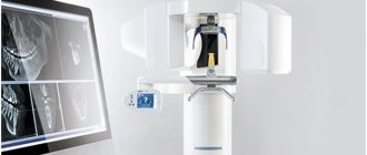
Cone beam dental computed tomography is the safest and fastest diagnostic method. Thanks to the use of a conical X-ray beam, the radiation dose received during the study is 10 times less than when using spiral CT. And the pulsating mode of the X-ray beam further reduces the radiation dose. The three-dimensional computed tomograph SOREDEX Scanora 3D is one of the safest devices in terms of X-ray radiation dose - only 0.035 m3v.
However, despite the safety of the study, CT also has contraindications. If we just talk about dental tomography, it is not performed during pregnancy (in the 1st trimester). 3D dental x-rays with contrast are prohibited for pregnant and lactating women, patients with endocrine disorders (diabetes mellitus, thyroid pathologies), renal failure and intolerance to iodine-containing drugs.
Types of dental x-rays
In the network of dental clinics "Zubastic" you can undergo the following types of x-ray examinations:
- traditional orthopantomography with recording of a detailed image of the jaws on a CD;
- intraoral bitewing radiography;
- dental computed tomography (CT) with obtaining a volumetric image of the maxillofacial area;
- teleradiography (TRG) of the skull in frontal and lateral projection.
How much dental x-rays, CT and TRG will cost can be found in the price list for services presented on the dentistry website.
Summary: important points
As a practicing dentist who knows the system from the inside, I want to draw your attention to the following points that are important for your safety. If the clinic has an X-ray machine, then a license must be obtained for it, the issuance of which presupposes the mandatory presence of a certified radiologist on the staff of the dental clinic. However, in reality, even in large clinics and public clinics, it is not always the case that x-rays will be taken by a trained specialist.
Even if he is, he may go on vacation or get sick, and a regular nurse (dental assistant) will take the pictures instead. This is a gross violation that leads to both the production of low-quality images and an increase in the radiation dose. In small clinics, the risks of receiving a poor-quality x-ray examination are much higher, and the first thing that makes you suspect a forgery is if the picture is taken not by a special employee, but by a nurse from the dentist you came to see.
The second very important point: if you see that the x-ray is not done in a special room, but the x-ray machine is located right next to the dentist’s chair, then you should change the clinic and the doctor. The fact is that all objects in this office (including the dental chair and doctor’s instruments) will have an increased background radiation, and this is no longer safe for health. We hope that our article on the topic: X-ray of the jaw and teeth was useful to you!
Sources:
1. Dental education of the author of the article, 2. Based on personal experience in therapeutic and surgical dentistry, 3. National Library of Medicine (USA), 4. “Digital and film radiography in outpatient dentistry” (Chibisova), 5. “X-ray diagnostics in dentistry" (Lutskaya I.K.).
Features of radiography
- All types of X-ray examinations in the clinic are carried out in a specially equipped room.
- To protect vital organs, the patient is wearing a special apron with lead inserts.
- The radiologist gives concise, clear instructions on how to behave during the procedure. Offers to remove metal jewelry.
- To obtain clear images, it is not recommended to move. At the request of the specialist, you need to hold your breath for a few seconds.
- In terms of time, the shooting process takes 1-2 minutes, transillumination – no more than 2-3 seconds. The sequence of the procedure depends on the type of x-ray.
When creating a panoramic image, the subject's head is fixed with a special device. The patient clamps the plate placed in a disposable cover with his teeth and remains motionless while the functional part of the X-ray machine rotates around his head.
Radiology and computed tomography
The role of correct diagnosis in dental treatment is difficult to overestimate. Both the success of treatment and the ability to save the patient from unnecessary inconveniences that accompany the dental treatment process fully depend on the accuracy and completeness of the diagnostic picture of the disease.
Diagnosis of the oral cavity
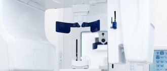
In our clinic, diagnostics are carried out using an ultra-modern panoramic X-ray machine Sirona, manufactured in Germany. When examining patients using a new orthopantomograph, the dose of ionizing radiation is 80% (!) less than when examining using traditional devices. It achieves excellent image quality through the use of panoramic dental X-ray equipment, which has a number of excellent features. This device has the ability to scan jaws layer-by-layer for the purpose of performing dental implantation operations.
Our doctors can instantly receive an accurate image of the patient’s teeth (panoramic image, etc.) on a computer monitor and can quickly process it - increase its size, enhance the contrast. The use of this device allows you to obtain high-quality and informative images of the upper and lower jaw in real time, and also allows you to perform 3D reconstruction of the image. This allows the doctor to detect the presence of a pathological focus at the earliest stages of the disease, monitor the quality of treatment, providing the patient with the most comfortable and effective treatment.
The equipment of the RuDenta clinic allows you to obtain complete diagnostic information of the highest quality, however, for successful treatment and further monitoring of patients, the quality of storage, recording and cataloging of the obtained diagnostic data is equally important. In the special computer patient registration system of the RuDenta clinic, a personal folder is created for each of them, where the entire history of treatment and diagnosis is stored, including all x-rays taken. Any of these images is available for viewing by specialists directly at their workplace.
We carefully collect anamnesis, which allows us to trace the history of the development of the disease, stages of development, and identify factors contributing to the progression of the process. Many systemic diseases are both triggering and supporting factors in the development and course of oral pathology. Timely identification of aggravating factors greatly contributes to the correct choice of treatment method.
X-ray of teeth in dentistry Zubastic: advantages
Expert X-ray equipment and highly qualified clinic specialists who are proficient in advanced techniques allow examinations to be carried out without risk to the patient’s health. We offer the best quality/price ratio for dental x-rays in Moscow, ensuring the safety of the procedure and quick results. Interpretation of CT images using artificial intelligence guarantees the complete elimination of diagnostic errors.
A good dental x-ray is the main source of reliable information about the condition of the dental system. It makes it possible to start therapy at an early stage of the disease, speed up treatment, and avoid complications and unforeseen consequences.
Features of the appointment of dental x-rays
If you are a new patient, you will likely undergo dental x-rays so that the new dentist can get a clear picture of your dental health. This is especially important if you do not have x-rays from your previous dentist. Children may need dental x-rays more often than adults because their dentists need to monitor the growth and development of their teeth. This is important because it can help the dentist determine whether baby teeth need to be removed to prevent complications, such as adult teeth growing behind baby teeth.