From this article you will learn:
- stages of caries,
- what are the signs of caries?
- how to identify caries yourself.
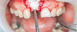
Caries is the process of destruction of hard tooth tissues with the active participation of cariogenic microorganisms of the oral cavity, such as streptococci, actinomycetes and lactobacilli. The main conditions for its development are irregular or insufficient oral hygiene, which leads to the accumulation of soft microbial plaque on the teeth, as well as food debris between the teeth. Depending on the depth of damage to the hard tissues of the tooth, caries is usually divided into several types, which, in fact, are successive stages of its development.
The initial stage is always spot-shaped caries (foci of demineralization in the form of white spots on the surface of tooth enamel), and if left untreated, it gradually goes through the stages of superficial, medium and deep caries. Symptoms of dental caries, where we include patient complaints and data from a visual examination of the carious cavity, will be slightly different at each stage, and below we will consider them in detail.
Use of a microscope in dentistry: indications, advantages, features of dental treatment
Dental treatment is a very delicate job, because the tooth itself is small, and the root canals inside it are even smaller. Therefore, the clearer the doctor sees the picture, the more effectively he will carry out all the manipulations. To perform their work efficiently, modern specialists use equipment, for example, they perform dental treatment under a microscope. This procedure is gaining more and more popularity today, and we offer to learn about all its nuances, advantages, disadvantages and features. Details in the article!
Prices
Of course, the cost of treating dental pathologies under a microscope exceeds the payment for the same disease, the treatment of which was carried out according to the standard protocol. Thus, the cost of treating some diseases is:
- pulpitis will cost 10 thousand rubles;
- periodontitis - 13-15 thousand rubles;
- granulomas and cysts – from 10 thousand rubles;
- caries - about 8 thousand rubles.
Typically, this price already includes payment for processing and filling of root canals. The approximate payment for retreatment is 4-6 thousand rubles, diagnostic measures - 1 thousand rubles. Prices at different clinics will vary slightly, as they largely depend on the following factors:
- clinic status;
- location;
- cost of drugs.
The high cost of the service is explained by the high cost of the device, as well as the need to improve the dentist’s qualifications. But when it comes to preserving the tooth and minimizing complications, such material expenses are justified.
The video provides additional information on the topic of the article.
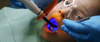
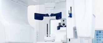
What is a dental microscope
A dental microscope is a high-precision optical device that can magnify an image of a tooth, as well as everything inside it, 20 times or more. This allows the endodontist or endodontist to carry out quite sophisticated work on cleaning canals, removing cysts or resection of the root apex. The dentist may also be able to see if there are abnormal or infected areas or may notice a foreign object inside the root canal.
Important! Many patients are afraid of using optical equipment in treatment because they think that the device will be placed in their mouth. However, this is not true. The device is located above the patient, at a distance of 20-30 cm from his face, which is convenient for both the patient and the doctor. The endodontist carries out therapy looking through the eyepieces, but the image from the device is also displayed on the monitor so that the assistant can observe the process and provide the doctor with the necessary tools.
Areas of application of a dental microscope
A microscope is a highly accurate and rather expensive device, so its use in conventional caries treatment is unlikely to be justified. But those who believe that this optics are used only during complex surgical interventions will also be wrong.
In dentistry, such sensitive equipment has its own area of application:
- identifying the structure of channels, their number and branches,
- filling of tortuous canals,
- diagnosis of caries in the initial stage,
- detection of cracks in the enamel or canal wall,
- detection and removal of foreign objects (pieces of instruments, remains of filling materials, etc.) from root canals,
- diagnosis and treatment of canal or root perforation,
- assessment of the tightness of fit of restoration materials or prostheses (performed both during fitting and during their installation),
- tissue treatment before prosthetics to prevent damage to soft tissues or loss of healthy tooth structure,
- quality control of restoration or orthopedic work,
- removal of cysts, granulomas,
- some types of surgical work, including maxillofacial surgery.
Where do the roots come from?
Caries (caries - “rotting”) is one of the most common human diseases, which is increasingly gaining momentum. About 42% of the world's population has dental caries [1]. However, this problem is far from exclusively human (Fig. 1).
Figure 1. Caries occurs quite widely in the animal kingdom, especially in ungulates. Above is a photo of a horse's teeth, below is a photo of a human.
Idade dos cavalos pelos dentes, “Complications in the treatment of dental caries in children”
It is now reliably known that the vital activity of oral bacteria leads to the occurrence of caries. Bacteria, like all living organisms, must eat, grow and reproduce, and also maintain their metabolism (metabolism) at the proper level. But not all bacteria in our mouths lead to tooth decay. Cariogenic bacteria include streptococci (Streptococcus mutans, Str. sanguis, Str. mitis, Str. salivarius) and some lactobacilli (Lactobacillus acidophilus) [2–4].
You can learn about who lives in the oral cavity and how individual representatives of its microflora affect our health from the article “Who lives in our mouth?” [5] special project “Biology, medicine and cosmetology of the oral cavity”. - Ed.
Like many other diseases, caries develops in several stages.
Anaerobic bacteria live in the form of a thin biofilm on the enamel of teeth - plaque. Fixing on the surface of the tooth, they produce extracellular heteropolysaccharides necessary to create comfortable living conditions for themselves, namely to reduce contact with oxygen. Often, the synthesis of such extracellular polysaccharides requires exclusively sucrose, as occurs in Streptococcus mutans (Fig. 2).
Figure 2. Streptococcus mutans is a species of gram-positive facultative anaerobic bacteria whose activity is directly associated with the development of caries. Streptococci, due to their structure (form chains) and the presence of a special receptor that allows them to attach to the smooth surface of the enamel, are most often found in dental plaque. Using sucrose, they synthesize sticky polysaccharides based on dextran, forming a biofilm on the tooth, thereby providing themselves with anaerobic living conditions.
"Wikipedia"
A person eats carbohydrates, and streptococci “eat” them along with him. Hence the direct relationship: the more sugar you ate, the more plaque formed. And the sweeter a person’s life, the better for streptococci: during the fermentation of carbohydrates (glycolysis), they receive energy for their vital functions, and from sucrose they build their protective coating - a “shell” from oxygen. The average time for visible and palpable plaque to form is 3–6 hours, and it takes about 24 hours for a dense bacterial biofilm to form.
For people, the activity of plaque bacteria has its disadvantages. They produce a large number of by-product organic acids, including pyruvic, formic, acetic and propionic acids [3]. And lactobacilli also produce lactic acid. The combination of all the released acids inevitably reduces the pH (increases the acidity) of that part of the oral cavity where the most active fermentation of carbohydrates occurs - in the folds of the enamel. At first, excessive acidity is neutralized by saliva, but the longer bacteria live on the tooth (you must understand that time flows differently for bacteria), the more heteropolysaccharides they secrete, and the more difficult it is for saliva to break through the growing shell of plaque. The critical pH value for tooth enamel is considered to be 5.5. It is at this value that caries begins to develop and demineralization of tooth enamel occurs (Fig. 3).
Figure 3. pH is very important for teeth. The normal pH of the oral cavity in the range of 6.8–7.4 is ensured by the buffering properties of saliva. A shift in the indicator to the acidic side to 5.5 is critical and indicates an active process of caries formation.
drawing by Anastasia Prokhorova
Indications for using the microscope
There are no specific diseases in dentistry that cannot be treated without high-precision optics, but the quality of medical care greatly benefits from its use. This is especially true for complex cases, for example, dental treatment with any anomaly in the development of the jaw or with an atypical structure of the root canals (when they are highly branched or tortuous). The use of electronic optics when re-sealing canals is justified, because this requires first unsealing1 and cleaning the canals without damaging their walls. The doctor must make sure that no particles of the previous filling, medication, pin, etc. remain in their branches.
The use of a microscope in the treatment of cysts, granulomas and other neoplasms at the apex of the roots allows for high-precision resection or cystectomy without damaging the surrounding tissues, and most importantly, preserving the tooth. This is a very important point when removing a cyst or root tip, because improper operation or injury to adjacent tissues can lead to re-infection and growth of the cyst.
And of course, such a device is used in all organ-saving and minimally invasive (low-traumatic) endodontic operations.
Progress of the procedure
Treatment using optical magnification is more comfortable for the patient. He reclines in a chair, which helps him relax better. In addition, the instruments that the doctor uses are much smaller than usual, and therefore do not cause discomfort or a gag reflex. This also allows the patient to swallow saliva normally. In addition, multiple magnification of the image allows the doctor to see the work area in great detail, and the patient does not need to open his mouth wide.
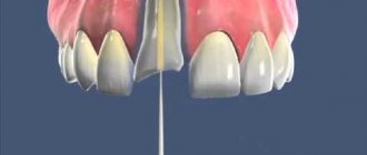
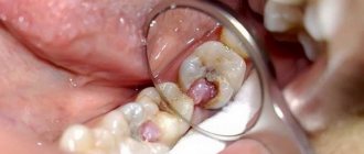
How to determine caries at home -
- The presence of pain syndrome, which we wrote about above.
- The cavity may have sharp edges and irregularities, which can be perfectly felt (in some cases) by the tongue.
- Bad breath may indicate the presence of caries. Food debris is retained in the carious cavity, which is difficult to clean; under the influence of the microflora of the oral cavity, they begin to rot, causing an unpleasant odor.
- Inspection of visually accessible surfaces of teeth (for example, in a mirror). At the same time, even if the surface layer of enamel is preserved, darkening may be visible underneath it. Such darkening may indicate caries.
Advantages of working with a microscope
Despite the fact that many dentists continue to treat teeth using conventional methods, many advanced clinics have already appreciated the quality of work using a microscope, because this method has a number of advantages:
- diagnostic accuracy: the use of high-precision optics allows you to diagnose caries at an early stage, when a drill is not required for its treatment. Multiple magnification allows you to examine hard-to-reach places (fissures, the junction of the crowns of adjacent teeth) and identify the beginning of pathological changes in the enamel,
- reduction of radiation exposure to the body: thanks to sensitive optics, the doctor can see with his own eyes the condition of the hard tissues of the tooth, its roots and canals, which means there is no need for radiography, which is especially valuable for pediatric dentistry,
- minimally invasive technologies: the endodontist can see the clinical picture in the smallest detail, which means that during his work he will not damage healthy tissue - gums, dentin, canal walls,
- high quality of work: when filling canals (especially if they have some anatomical features), it is very important to fill the entire cavity with filling material, leaving no voids or branches, so as not to create conditions for the development of microbes. In addition, the microscope allows you to verify the tightness of fillings or restoration overlays, to accurately fit the crown to the tooth,
- reduction of the rehabilitation period: often after canal filling, the patient experiences pain in the treated tooth for several days. This occurs due to the fact that nerve endings that have not been removed remain in the canal. The use of dental optics makes it possible to see and remove these particles, which relieves the patient of pain in the post-therapeutic period,
- monitoring the progress of work: since the high-resolution digital camera located inside the device displays the image on a large screen, the doctor can take a picture of any stage of work and any area of intervention. This allows him to adequately assess the quality of the work himself or invite colleagues to a consultation if a complex case requires consultation with another specialist - an orthopedist, surgeon, or orthodontist.
Reviews
A microscope is a universal dental equipment that gives a real chance to save a seemingly hopeless tooth and diagnose pathologies at the initial stage of their development.
It allows you to make the treatment process predictable, improve quality and accurately predict its outcome.
If you have similar experience and would like to express your opinion on the effectiveness and advisability of optical treatment, please share them in the comments to this article.
If you find an error, please select a piece of text and press Ctrl+Enter.
Tags toothache treatment dental treatment under a microscope
Did you like the article? stay tuned
Previous article
On the effectiveness of tooth restoration in dentistry using an anchor pin
Next article
Intraligamentary anesthesia is a step towards painless treatment
Complications after working under a microscope
As a rule, there are no complications after therapy where a detailed microscope was used. The risk is minimized, since the doctor has the opportunity to see the area of work in detail and control the process at all its stages. All complications arise when working without a microscope, because the doctor does not see the nuances, can injure neighboring tissues, accidentally pierce the walls of the canal, break the tip of the instrument in the root branch and not even notice it.
Attention! If after treatment with the use of optics a complication does occur, then this indicates a low qualification of the endodontist or a malfunction of the equipment.
Signs of caries and pulpitis (differences in symptoms) –
Typical signs of caries are the infrequent occurrence of short-term painful attacks under the influence of various irritants:
- thermal irritants (cold water or cold air),
- chemical irritants (for example, sour, salty or sweet foods).
At the same time, caries is characterized by the absence of spontaneous pain, i.e. pain during caries occurs only under the influence of irritants. And as soon as the irritant is eliminated, the pain should immediately disappear. The presence of spontaneous pain not associated with exposure to irritants indicates that the process from caries has already passed into pulpitis.
With deep caries (plus the above), pain may also occur when mechanical pressure is applied to the thinned bottom of the carious cavity. This can occur, for example, while eating - in the process of biting or chewing hard food. In this case, food enters the carious cavity and presses on its bottom, causing compression of the dental pulp (neurovascular bundle inside the tooth). In this case, removing food debris from the carious cavity usually relieves pain.
How to distinguish caries from pulpitis: differences in symptoms
- With caries, pain occurs only in the presence of irritants (thermal, chemical). When the irritant is removed, the pain immediately disappears.
- In acute pulpitis, pain is usually acute, spontaneous and not associated with any irritants.
- But with chronic pulpitis, pain can still be provoked by thermal irritants - cold or hot water. But the difference between caries and chronic pulpitis is that with caries the pain goes away immediately after the irritant disappears, and with chronic pulpitis the pain does not go away immediately, but only after 10-15 or more minutes.