» Anesthesia in dentistry » Anesthesia in the upper jaw For various operations on the upper jaw, dentists prefer to use infiltration anesthesia.
This method is most convenient because the thin layer of the compact lamina of the alveolar process of the upper jaw is porous. If you use modern painkillers that have a high diffusion ability, then with this method of their administration the desired anesthetic effect is achieved. Impregnation of tissues and alveolar processes with an anesthetic is carried out by injection, injecting the solution under the mucous membrane at an angle of 40-45o in the projection of the upper regions of the dental roots along the transitional fold of the vestibule of the oral cavity in the area that is supposed to be operated on. If this area is too large, the needle is moved along the transitional fold and the anesthetic solution is slowly injected. Because rapid administration of the substance can cause pain in the patient.
To anesthetize the palatal tissues, an injection of an anesthetic drug is made into the angle formed by the palatine and alveolar jaw processes, at a distance of 10-15 mm from the edge of the gum.
Regional (conduction) anesthesia is used quite rarely; it includes anesthesia on the tubercle of the upper jaw, in the area of the greater palatine, incisive and infraorbital foramen.
Intraoral anesthesia
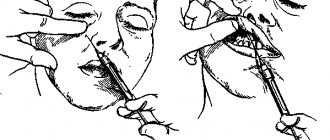
The patient's mouth is in a half-open state, the doctor inserts a mirror into the oral cavity, moving the cheek to the side. This ensures an excellent overview of the arch of the vestibule of the mouth, as well as tension in the mucous transitional fold near the molars. The needle is inserted into the mucosa above the projection of the apexes of the teeth at the level of the 2nd and 3rd molars. If there are no molars, then the needle should be behind the zygomaticalveolar ridge and move up, back and inward. The insertion angle should be 45°. It is necessary to control that the needle always moves along the bone with the beveled surface of its tip. During this movement, the anesthetic should be gradually injected. This method will help prevent injuries to the vessels of the pterygoid plexus. Having deepened the needle by 2-2.5 cm, the anesthetic solution is deposited. Thus, the molars located on the side of the vestibule of the mouth, the mucous membrane, periosteum and the outer posterior bone wall of the maxillary sinus are perfectly anesthetized.
Non-injection methods
For short, small-scale interventions, it is sufficient to superficially numb the soft tissues; this can be achieved using non-injection methods of local anesthesia in dentistry. These include:
- Application method . Applying a special gel or spray containing a certain anesthetic in a small concentration to the desired area of the oral mucosa. This method is often used in pediatric dentistry in the treatment of baby teeth, when treating gingival margins, opening small superficial abscesses, and also to prepare the patient for injection-type local anesthesia (to numb the injection site).
- Physico-chemical method . Injection of the required amount of anesthetic into soft tissues using electrophoresis. It is usually used for trigeminal neuralgia.
- Physical method (rarely used in recent years). Anesthesia of a specific area by applying a freezing reagent (chloroethyl), exposure to a laser beam or electromagnetic waves of a certain frequency.
All other techniques for performing local anesthesia in dentistry belong to the injection group, that is, they require an injection in a certain area of the oral cavity or even outside it.
Extraoral anesthesia
Having made a puncture with a needle in the area of the lower anterior corner of the cheek bone, the needle is then directed upward at an angle of 45° and again towards the tubercle of the upper jaw, bringing it to the bone. And then the anesthetic solution is deposited. The anesthetic effect occurs in approximately the same time as with the intraoral method of anesthesia.
However, during the process of anesthesia, a hematoma appears on the tubercle of the upper jaw, which is a consequence of injury to the veins of the pterygopalatine plexus by the needle.
Computed tomography studies of the path of solution propagation during tuberal anesthesia confirmed that such complications are most likely.
This tomogram, which was taken 5 days after intraoral anesthesia, shows that there are contours of a hematoma on the tubercle of the upper jaw in the pterygopalatine fossa. Approximately 40-60% is allocated to the fact that this cellular formation will fester and turn into phlegmon.
When using the tuberal method of anesthesia, injury to the veins of the pterygopalatine plexus is almost impossible to avoid, and there is also a fairly high risk of complications, in particular if the intraoral method is used. All this threatens the health and even the life of the patient. That is why it is recommended to use this type of anesthesia extremely rarely.
General principles of pain-relieving procedures
In order for local anesthesia in dentistry to bring the expected effect and not become a source of complications, it is important for the doctor to follow certain principles of its implementation:
- You should first assess the patient’s condition and find out if there are any allergic reactions to painkillers.
- The correct choice of anesthetic drug and place for its administration.
- The use of exclusively sterile preparations that are compatible with oral tissues.
- The temperature of the solution for administration should be close to normal human body temperature.
- The rate of drug administration should be minimal, and the patient should not experience any unpleasant sensations (burning, itching, pain) during the process.
- Only sharp needles should be used to avoid tissue injury.
- The area of the upcoming injection must be pre-treated with an antiseptic.
- The injection should not be unexpected: the patient must be prepared for local anesthesia.
Anesthesia of the infraorbital nerve
The purpose of this anesthesia is to block the branches of the inferior orbital nerve, which forms the lesser “crow's foot” in the area of its exit from the bony canal, as well as the middle and superior anterior alveolar branches.
The result of such anesthesia, performed at the lower orbital foramen, is pain relief:
- — Reztsov,
- - Fang,
- — Premolars,
- — The gums adjacent to the premolars in the area of the vestibule of the mouth,
- — Bone tissue of the alveolar process and nasal septum,
- — Shells of the mucous and bone structures of the walls of the maxillary sinus,
- - Skin of the infraorbital region of the lower eyelid and wing of the nose,
- — Mucous membrane and skin of the upper lip.
The projection of the infraorbital foramen, which is oriented towards during an anesthetic injection, is located 5 mm below the edge of the orbit, which corresponds to the axis drawn through the center of the eye pupil when the eye looks forward.
Dosage
In all cases of local anesthesia, it is necessary to calculate the dosage of the administered anesthetic in terms of the child’s body weight. For articaine preparations with a vasoconstrictor, the recommended dosage is 5 mg per 1 kg of weight. Before performing local anesthesia, the child’s weight is checked with the parents. In clinical practice, it is convenient to use a table with weight and the maximum permissible dose of the administered anesthetic (Table No. 1, 2).
Table No. 1
| WEIGHT | MG | ML | CARPOOLS |
| 10 | 44 | 1.5 | 0.8 |
| 15 | 66 | 2.2 | 1.2 |
| 20 | 88 | 2.8 | 1.4 |
| 25 | 110 | 3.6 | 1.7 |
| 30 | 132 | 4.4 | 2.4 |
| 35 | 154 | 5.1 | 2.9 |
| 40 | 176 | 5.9 | 3.2 |
| 45 | 198 | 6.6 | 3.6 |
| 50 | 220 | 7.3 | 4.0 |
| Mepivacaine 3% without vasoconstrictor. Maximum dose 4.4 mg/kg. 3% solution in 1 carpule 1.8 ml (54 mg). | |||
Table No. 2
| WEIGHT | MG | ML | CARPOOLS |
| 10 | 50 | 1.2 | 0.69 |
| 15 | 75 | 1.9 | 1.0 |
| 20 | 100 | 2.5 | 1.4 |
| 25 | 125 | 3.1 | 1.7 |
| 30 | 150 | 3.7 | 2.1 |
| 35 | 175 | 4.4 | 2.4 |
| 40 | 200 | 5.0 | 2.8 |
| 45 | 225 | 5.6 | 3.1 |
| 50 | 250 | 6.2 | 3.4 |
| Articaine 4% with a vasoconstrictor. Maximum dose 5 mg/kg. 3% solution in 1 carpule 1.8 ml (72 mg). | |||
Quite often, at outpatient dental appointments we encounter children suffering from obesity and metabolic syndrome, which is largely due to changes in the nutritional culture of the population. The dosage of the administered anesthetic in these cases has some peculiarities. In particular, if the doctor is going to administer anesthesia to an overweight child, the dosage of the administered anesthetic is calculated without taking into account adipose tissue.
Intraoral access
Using the thumb and forefinger of the left hand, push the upper lip up and out, and with the middle finger hold the projection of the infraorbital foramen. With this type of access, it is located at the intersection of two axes. One of them, horizontal, runs 5-7 mm below the lower orbital margin, and the second, vertical, runs on the corresponding side along the axis of the second upper premolar. The needle must be inserted, retreating 5 mm from the upper edge of the attachment of the transitional fold between the lateral and middle incisors. Then it is pushed upward, forward and outward in the direction of the lower orbital foramen until it stops at the bone. And only there the anesthetic solution is released.
Injection equipment
For local anesthesia in children, carpule syringes of various designs are used. Preference should be given to injectors intended for carrying out an aspiration test (Rabinovich S. A., Vasilyev Yu. L., Sokhov S. T., 2013; Tarasenko S. V., Kuzin A. V., Belyaeva E. A., Kurtyshov A. A., 2013). Local injection anesthesia in children carries the risk of intravascular injection of local anesthetic. This fact is explained by the high degree of vascularization of the tissues of the maxillofacial region of children. Thus, the frequency of intravascular administration of an anesthetic during mandibular anesthesia in adults is 10-15%, and in children - 20-25%. Syringes with a plunger in the form of an anchor and a corkscrew have the best technical characteristics. The choice of injection needle depends on the method of anesthesia. For conductive methods, needles with a diameter of at least 0.4 mm (27G) should be used. When conducting conduction anesthesia, 0.3 mm (30G) needles bend excessively in the tissues (deflection), which leads to the deposition of the anesthetic away from the intended end point of pain relief (Rabinovich S. A., Vasiliev Yu. L., 2011).
Needles 0.3 mm (30G) are advisable to use for infiltration anesthesia and periodontal methods of pain relief.
Do not forget that when performing local anesthesia, the needle may break off. This complication, as a rule, occurs when the child makes sudden movements: withdrawing the head, abruptly closing the mouth. In most cases, these severe complications occur when using 30G needles when performing mandibular anesthesia in children.
There is an opinion that the thinner the needle, the less painful the patient perceives the stage of puncturing the mucous membrane and advancing the needle into the tissues. This opinion can be classified as a misconception. There are studies confirming that the diameter of the needle does not affect the reduction in the degree of pain of the anesthesia (Malamed SF, 2002).
Anesthesia at the greater palatine foramen
This method blocks the innervation of the greater palatine nerve, thus achieving an anesthetic effect on the mucous membrane on the desired side of the hard palate, as well as on the alveolar process from the palate from the third molar to the middle of the short part of the canine. The anesthetized area can reach the lateral incisor, as well as the vestibular surface in the area of the third molar. In some patients the area extends to the second premolar.
The greater palatine foramen is located in the horizontal plate of the palatine bone and its pyramidal process at the base of the alveolar process, 5 mm anterior to the border of the soft and hard palate. Above the hole in the mucous membrane there is a small depression. The projection of the hole onto the mucous membrane of the hard palate is located at the intersection of two mutually perpendicular lines. The one that runs vertically goes through the middle of the line that connects the crest of the alveolar process and the center of the upper jaw, and the horizontal one runs through the middle of the coronal part of the third molar.
Anesthesia technique. The mouth should be opened wide, the syringe needle is directed from the opposite corner of the mouth and inserted 1 cm forward and inward in the direction from the projection of the opening of the palate into the mucous membrane. The needle is advanced until it comes into contact with the bone, and 0.5 ml of solution is injected. After a couple of minutes, the analgesic effect occurs. In the case when the solution is injected directly near the greater foramen of the palate, as well as into the opening of the pterygopalatine canal, the effect captures the posterior nerves of the palate emerging from its lesser foramen. As a result, the soft palate is numbed. Injecting the solution into this area may cause nausea and vomiting. Another side effect that may occur due to excessive injection of a solution under pressure is necrosis of the soft tissues of the hard palate. This can occur in patients with vascular atherosclerosis.
Anesthetics without a vasoconstrictor
Provide varying durations of pain relief for dental tissues. In particular, 2% lidocaine provides anesthesia of the dental pulp within 5 minutes, while the rate of onset of anesthesia is also 5 minutes, which is unsatisfactory for the doctor. Therefore, the use of 2% lidocaine without a vasoconstrictor is inappropriate for dental anesthesia.
3% mepivacaine, in comparison with other anesthetics, has a less pronounced vasodilator effect, which makes it possible to use it without adding a vasoconstrictor. The anesthetic provides pain relief for 10-20 minutes, while treatment must be carried out from the 5th to 20th minute during therapeutic interventions and from the 10th to 20th during tooth extraction surgery.
Articaine 4% is currently available in the Russian Federation. This anesthetic is short-acting: anesthesia of dental pulp for 6 minutes, soft tissue for 45 minutes. Its widespread use in pediatric dentistry is limited due to its short action, which is not suitable for most interventions.
Anesthesia at the incisive foramen
Such anesthesia is carried out by neutralizing the nasopalatine nerve in order to anesthetize the anterior region of the mucous membrane of the hard palate in the area of the anterior teeth.
The incisive foramen is located between the front incisors 7-8 mm from the edge of the gum at the intersection of the lines that connect the distal edges of the median palatal suture and the necks of the canines.
Anesthesia technique: the patient is in a chair, his head is thrown back, his mouth is open wide. The needle is inserted into the mucous membrane near the incisive opening to a depth of 3-4 mm. The anesthetic solution is released slowly. The process of inserting a needle into the papilla itself is very painful, so thin needles are used for such injections, and additional pain relief is performed. The anesthetic effect is achieved within a few minutes.
How to improve the quality of pain relief?
The patient, for his part, can also prepare for the upcoming intervention and thereby improve the quality of pain relief. To do this you need to follow these simple rules:
- Postpone a visit to the dentist if you have infectious diseases or (for women) during menstruation.
- Be sure to inform your doctor about allergic reactions to medications.
- The day before visiting the dentist, refrain from drinking alcohol and visiting the sauna.
- The evening before your visit, you may take a small dose of a sedative to relieve tension.