Target point of infraorbital anesthesia : the infraorbital foramen, which includes the peripheral branches of the infraorbital nerve. The location of the target point can be determined in 3 ways: 1. The infraorbital foramen is located 5-7 mm below the intersection of the lower edge of the orbit with the vertical line that passes through the pupil of the eye looking forward (see Fig. 70, A).
Rice. 70. Location of the infraorbital foramen (explanation in the text).
2. In the middle of the lower edge of the orbit, a bony protrusion or groove is palpated - the junction of the zygomatic process of the upper jaw with the zygomatic bone - 5-7 mm below this protrusion is the infraorbital foramen (see Fig. 70, B). 3. The infraorbital foramen is located 5-7 mm below the intersection of the lower edge of the orbit with a vertical line drawn through the middle of the second upper premolar (see Fig. 70, B). It must be remembered that the axis of the infraorbital canal is directed forward, in the middle, down and crosses the axis of the canal of the opposite side above the gingival papilla between the upper central incisors (Fig. 71, 72).
Rice. 71. Direction of the lower orbital canals and the place of their intersection between the upper central incisors
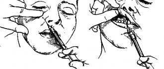
Rice. 72. Insertion of a needle into the canal during intracanal infraorbital anesthesia (V.I. Zausaev et al., 1981)
Modification of intraoral infraorbital anesthesia:
advancing the needle between the canine and the first premolar. In a similar way, you can advance the needle at the level of the canine, first and second premolars (Fig. 74).
Rice. 74. Modifications of intraoral infraorbital anesthesia. Injection site: 1 - between the central and lateral incisors; 2— above the fang; 3— above the first premolar
It was noted that the more distal the injection site, the weaker the analgesic effect due to the fact that the axis of the infraorbital canal does not coincide with the direction of the injection. Therefore, the best anesthesia will be in position 1 (injection point between the central and lateral incisors), the worst position is 3 (injection point above the first or second premolar). When passing the needle at the level of the canine, first and second premolars, it is impossible to enter the infraorbital canal; pain relief occurs due to the diffusion of the anesthetic in the area of the infraorbital foramen. It should be noted that with these modifications there are practically no complications due to injury to blood vessels and nerve endings when the needle is advanced. Therefore, in recent years, the following anesthesia has gained significant popularity.
Execution method
Infraorbital anesthesia provides the possibility of using one of two methods, the choice between which is determined by the specific anatomical structure of the maxillary region, as well as the goals that need to be achieved as a result of the procedure. The drug is administered both externally and internally, but in both cases the doctor requires concentration and attention.
The intraoral method involves determining the position of the desired hole, after which in the projection area the soft tissues are pressed against the bone base, and at the same time the lip is retracted upward. The needle insertion site should be located 5 mm above the fold that marks the transition between the premolar and the canine. After establishing contact, the doctor injects about 1 ml of the drug into the canal, after which he probes the position of the mouth with the end of the needle. The free position means hitting the desired area - the tip is immersed to a depth of up to 10 mm, where another part of the anesthetic is released. The result appears within 4-5 minutes from the moment the drug is administered.
In situations where it is not possible to place a needle in the area between the incisor and the canine, the injection is made at the level of the canine, the first or second small molar. However, in this case, entry into the canal is excluded, and the analgesic effect is the result of diffusion of the composition. It is also worth noting that the intraoral method of anesthesia is characterized by a number of disadvantages, including the technical complexity of implementation, as well as restrictions on the procedure for diagnosed frontal periostitis.
The extraoral method is implemented through the external tissue, in the area located next to the nose. The projection of the position of the infraorbital foramen is built mentally, after which the doctor presses on the tissue with the index finger and retreats about 5 mm downwards, towards the center. The needle is inserted in three directions - until it comes into contact with the periosteum tissue into which the drug is injected. Next, the procedure similar to the first method is repeated.
The use of active anesthetics makes it possible to administer the composition only in the area under the orbit. At the same time, the effectiveness of anesthesia at the site of innervation of the alveolar branches virtually remains unchanged.
Technique for intraoral infraorbital anesthesia between the canine and the first premolar:
1. Using the index finger of the left hand, fix the projection of the infraorbital foramen to the bone; with the thumb, pull the upper lip up to the index finger (Fig. 75, B). 1. The needle is inserted 5-7 mm above the transitional fold between the canine and the first premolar, moved back, up and in the middle so that it approaches the infraorbital foramen (Fig. 75, B).
Rice. 75. Stages of performing intraoral infraorbital anesthesia for an injection between the canine and the first premolar : A - the path of advancement of the needle during intraoral infraorbital anesthesia between the canine and the first premolar, the zone of dental anesthesia for infraorbital anesthesia is indicated (diagram); B: Stage I - soft tissues are fixed with the index finger and thumb; B: Stage II - the needle is inserted 5-7 mm above the transitional fold between the canine and the first premolar, advanced to the mouth of the infraorbital foramen
The following steps are similar to the above intraoral infraorbital anesthesia method. With this anesthesia, the solution is injected into the mouth of the infraorbital foramen, which prevents injury to the neurovascular bundle. It should be noted that when using a strong anesthetic, there is no need to administer an anesthetic solution intracanal.
Conduction anesthesia. Part 1
When performing palatal anesthesia, the solution deposition site is located in front of the greater palatine foramen at a distance of 5-10 mm.
To do this, with the patient’s mouth wide open, the needle is inserted 10 mm in front and inside of the projection of the greater palatine foramen onto the mucosal surface. The needle is then advanced upward, posteriorly, and externally until it contacts the bone, after which an aspiration test is performed. A small amount of solution should be administered: 0.3-0.5 ml. Anesthesia of soft tissues develops within 3-5 minutes. The following complications may occur when performing a greater palatine nerve block: - If the solution is injected close to the greater palatine foramen and/or an excessive amount of solution is injected, it may spread to the soft palate. This will turn off the muscles that carry out swallowing and anesthetize the tissues of the soft palate, which causes the sensation of a foreign body in the mouth. As a result, the patient experiences nausea and vomiting. — The introduction of a solution under significant pressure can cause a reflex spasm of blood vessels, severe mechanical compression and even rupture, which leads to necrosis of soft tissues. The risk of this complication is especially high in elderly and senile patients with atherosclerotic phenomena and a tendency to increased fragility of blood vessels. Incisal anesthesia
Performed during intervention on the frontal group of incisors and alveolar process. The needle is inserted into the incisive papilla, located at the intersection of the midline and the line connecting both fangs, and then its end is inserted into the incisive opening and advanced along the incisive canal to a depth of 0.8-1.0 cm. At this depth, 0.5 ml of anesthetic solution. The anesthesia zone covers the gums in the area of the upper incisors and the mucous membrane with the periosteum of the anterior part of the hard palate to the line connecting both canines. Infraorbital (infraorbital) anesthesia is carried out in two ways - intraoral and extraoral. With the intraoral method, the needle is inserted into the transitional fold above the lateral upper incisor of the corresponding side, and then moved upward and laterally to the palpable infraorbital foramen. This hole is located 0.5 cm below the middle of the lower orbital margin. The syringe is placed obliquely at the level of the upper central incisor of the opposite side. As the needle moves to a depth of 1.5-2.0 cm, 1.5-2 ml of anesthetic solution is injected at the topography of the infraorbital foramen. The area of anesthesia includes the anterior and middle superior alveolar nerves, which arise from the inferoorbital nerve. With the extraoral method of infraorbital anesthesia, the needle is injected above the infraorbital foramen to the bone, this hole is found with the tip of the needle, then the needle is inserted into it and advanced along the infraorbital canal to a depth of 0.8 to 1.0 cm, where it is released slowly 1.5- 2 ml of anesthetic. The direction of the syringe and needle is similar to that for the intraoral method. The anesthesia zone on the labio-buccal side covers the incisors, canine and first premolar, as well as the corresponding area of the mucous membrane of the gums of the upper jaw. In addition, the corresponding side of the upper lip, the wing of the nose and the front of the cheek are anesthetized. Conductive anesthesia in the lower jaw is called mandibular anesthesia, but this name does not correspond to the essence, since at the opening of the lower jaw its peripheral branches are turned off - the lower alveolar and lingual nerves.
Mandibular anesthesia
Can be achieved by various injection methods. The intraoral technique is most often used in practice using the finger and apodactyl (fingerless) approach. Anesthesia using a finger is carried out with the mouth wide open by injecting a needle to the bone along the upper edge of the terminal phalanx of the index finger of the left hand, located in the retromolar triangle of the corresponding side, while retracting the syringe to the premolars of the lower jaw of the opposite side; then the syringe is moved to the incisors, the needle is advanced 2 cm deep into the bone and 2-3 ml of anesthetic is injected. The apodactyl approach is guided by the pterygomandibular fold. With the patient's mouth wide open, the syringe is placed at the level of the small molars or the first large molar of the opposite side, and the needle is inserted into the outer slope of the said fold at the middle of the distance between the chewing surfaces of the upper and lower large molars (in the absence of teeth - at the middle of the distance between the ridges alveolar processes). The needle is advanced until it contacts the bone at a depth of 1.5-2 cm, after which 2-3 ml of anesthetic is injected. The anesthesia zone corresponds to the switching off of the lower alveolar and lingual nerves - the bone tissue of the alveolar process and the tooth of the lower jaw of the corresponding half (from the third molar to the second incisor), the mucous membrane of the floor of the mouth and the tongue on 2/3 of its surface. In this case, the buccal nerve is switched off by additional infiltration anesthesia along the transitional fold.
Torusal anesthesia according to M.M. Weisbrema simultaneously turns off the inferior alveolar, lingual and buccal nerves; practically convenient and efficient. The author called the fusion of flat bony ridges extending downward from the coronoid process and the condylar process of the mandibular ramus a protrusion, or torus. Three of these nerves pass through the loose tissue at the level of this elevation, which is why their anesthesia is called torusal. It is carried out as follows: a syringe with a 4 cm long needle is placed at the level of the second or third lower molar on the side opposite to anesthesia. The injection is made into the area of the groove located laterally along the pterygomandibular fold, 0.5 cm below the chewing surface of the upper molars. The needle is advanced all the way into the bone, where 1.7 ml of anesthetic is released; 1 ml of anesthetic is released when the needle is withdrawn to anesthetize the buccal nerve. The anesthesia zone is similar, it occurs faster (after 15 minutes), however, this approach is only feasible with the mouth wide open; when mouth opening is limited, this option of anesthesia of the inferior alveolar nerve is not feasible. According to research by I.I. Levene (1987), an anesthetic solution in an amount of 1.8 ml upon injection fills the pterygomaxillary space, regardless of the method of its administration, and therefore the author believes that the term “thorusal anesthesia” is not justified, and in fact there is no such method. In practice, dentists use this technique as anesthesia of the inferior alveolar nerve.
Suggested by P.M. Egorov’s method of “mandibular” anesthesia consists of a topographical and anatomical justification for the orientation of the needle insertion for more accurate delivery of the anesthetic to the inferior alveolar nerve. To do this, on the skin of the face in the area of the jaw branch on the side of anesthesia, the projection of the pterygomandibular space and the upper edge of the mandibular foramen is determined. For this purpose, with the mouth open, use a ruler to measure the distance between the lower edge of the zygomatic arch (in front of the articular tubercle) and the lower edge of the lower jaw, as well as between the anterior and posterior edges of the ramus. Two mutually perpendicular lines drawn through the center divide the ramus of the lower jaw into four quadrants. The projection of the pterygomandibular space above the mandibular foramen on the skin is determined using a finger, which is placed according to the resulting superolateral quadrant. The needle is inserted 1.5 cm below and outward from the hook of the pterygoid process of the sphenoid bone, that is, into the intermuscular triangle located below the middle edge of the external pterygoid, lateral to the internal pterygoid muscle and medial to the temporal muscle. Without touching the muscles, the needle is advanced through the intermuscular space to the area of the lower jaw branch, fixed by the tip of the middle finger of the left hand. Along the path of the needle, 2-3 ml of anesthetic solution is slowly injected at the inner surface of the lower jaw branch. Switching off the lower alveolar, lingual, and often buccal nerves occurs within 10-15 minutes. The area of anesthesia is typical for conduction anesthesia in the lower jaw. The rate of successful anesthesia is higher (up to 98%) than with the described methods of blocking the inferior alveolar nerve using the Gough-Gates technique. Of all the widely known methods of blocking the inferior alveolar nerve, the most effective is the one proposed in 1973 by the Australian dental practitioner Gow-Gates (1973). According to various researchers, effective pain relief when using this method is achieved in 90-97% of cases. Positive aspiration tests range from 1.6% to 1.9% of cases, which is almost 10 times less than with other methods of anesthesia. Local post-injection complications (hematomas, difficulty opening the mouth) occur so rarely that they are not even assessed by the authors in percentage terms. In addition, with one injection of 1.8-2.2 ml of local anesthetic solution using the Gou-Gates method, it is possible to achieve anesthesia of not only the inferior alveolus, but also the lingual, mylohyoid, auriculotemporal nerves, and in 65-75% of cases - and the buccal nerve. The given characteristics of the method indicate the indisputable feasibility of its widespread implementation in the practice of domestic dentistry.
Article continues here
Source rusmg.ru
Extraoral classical method of infraorbital anesthesia
Injection equipment: carpule syringe or disposable plastic syringe 2 ml, needle 25 mm long. Anesthesia technique 1. Using the index finger of the left hand, fix the projection of the infraorbital foramen to the bone (Fig. 76).
Rice. 76. Extraoral classical method of infraorbital anesthesia (V.I. Zausaev et al., 1981)
2. Stepping back from the projection of the hole downwards and in the middle by 5 mm, the needle is inserted into the bone, directing it up, back and in the middle. 3. Ahead of the needle, 0.5-1 ml of anesthetic is released. When the needle touches the bone, carefully look for the entrance to the canal. Sometimes the needle immediately falls into the canal. With experience it is often possible to immediately enter the channel. 4. Having entered the infraorbital canal, advance the needle 3 mm and, after an aspiration test, release 0.5 ml of anesthetic. Pain relief occurs instantly. 5. Anesthesia zone - see intraoral method (Fig. 77).
Rice. 77. Zone of anesthesia during infraorbital anesthesia
This anesthesia is used, as a rule, to inject an anesthetic into the infraorbital canal - for high-quality and long-term pain relief during traumatic interventions (surgeries). In case of significant inflammatory processes (periostitis, osteomyelitis) of the upper jaw, when the inflammatory infiltrate spreads to the entire infraorbital region, we used a modification of extraoral infraorbital anesthesia for pain relief.
Techniques
Mandibular conduction anesthesia can be performed intraorally and extraorally.
Intraoral administration techniques:
- palpation;
- apodactyl;
- appdactyl method according to A.E. Verlotsk.
Extraoral techniques:
- subzygomatic introduction in various modifications;
- insertion under the lower jaw;
- retromaxillary route of administration.
Next, we will consider the methods that are most often used in dental practice.