Treatment of non-carious dental lesions is the elimination of congenital and acquired pathologies in the development of enamel and dentin, which are non-infectious dental diseases. The destruction of enamel and dentin that progresses with age worsens the appearance of teeth and creates problems with chewing food, which is why it is so important to visit the dentist in a timely manner, i.e. at least 2 times a year to start treatment at an early stage. This group of diseases includes hypoplasia and hyperplasia of enamel, as well as erosion, wedge-shaped defect, pigmentation, dental trauma, hyperesthesia, necrosis of hard tissues.
- Hypoplasia
- Hyperplasia
- Hyperesthesia
- Wedge-shaped defect
- Hard tissue necrosis
- Pigmentation
- Erosion
- Fluorosis
- Injuries
- Advantages of treatment at GrandMed
- On a note
Non-carious dental lesions are a large group of diseases of a non-infectious nature that lead to the destruction of hard dental tissues. The destruction of enamel and dentin, which rapidly progresses with age, worsens the appearance of teeth and creates problems with chewing food. Non-carious lesions are diagnosed during an examination of the oral cavity, and treatment, the effectiveness of which directly depends on the timeliness of contacting a specialist, is aimed at restoring the mineral composition of tissues and eliminating the aesthetic defect of the dentition.
Dental lesions are divided into two groups:
- Congenital or occurring before teeth eruption (enamel hypoplasia and hyperplasia).
- Acquired or occurring after teething (erosion, wedge-shaped defect, pigmentation, dental trauma, hyperesthesia, necrosis of hard tissues).
About half of people in the older age group (45–65 years) suffer from acquired diseases.
Dental hypoplasia
The pathology is manifested by underdevelopment or absence (aplasia) of tooth enamel; It can be congenital, but also develops after the birth of a child due to insufficient intake of minerals into the body. Hypoplasia itself refers to non-carious lesions of teeth, but thanks to it, excellent conditions are created in the oral cavity for the development of microbes, including those that cause caries.
Causes of hypoplasia:
- metabolic disease;
- mechanical damage to the jaw bone;
- dystrophy;
- diseases of the gastrointestinal tract;
- developmental pathologies;
- During pregnancy, the woman had: severe toxicosis, rubella, ARVI, toxoplasmosis, conflict of Rh factors, as well as premature birth and birth trauma.
Symptoms of hypoplasia
Pathology can be systemic (affects all teeth) and local (affects several teeth). In the early stages, underdevelopment of the enamel (too thin a layer or complete absence), deformation of the teeth and the appearance of grooves, yellow and white spots on their surface are noted. Painful sensations are noted during meals, and later, in the absence of treatment, the tissues are destroyed (erased) and an abnormal bite is formed.
Treatment of hypoplasia
Since underdevelopment of hard tissues and tooth decay is associated with a lack of minerals, when treating hypoplasia, it is necessary to remineralize the enamel with special preparations. To improve the appearance, teeth that have retained their normal shape are whitened, and if this is not enough, the defects are filled or prosthetics are performed. Congenital dental hypoplasia in children, in most cases, can be prevented; for this, during pregnancy, a woman’s diet must be balanced and contain sufficient amounts of vitamins D, A, C, group B, as well as calcium and fluorine. In addition, the expectant mother, in order to avoid non-carious lesions of the child’s teeth, should pay due attention to oral hygiene.
Phenomena that occur after teething
- plaque of various origins, tooth pigmentation;
- increased abrasion of hard tissues;
- defects called wedge-shaped;
- erosion;
- traumatic lesions;
- hyperesthesia.
Changes in tooth color and the appearance of pigment spots on it may depend on several factors:
- taking special medications and food dyes;
- resorcinol-formalin method for treating pulpitis;
- application of silvering of root canals;
- poor quality filling;
- oxidation of instruments left during treatment;
- hemorrhages into the pulp (the enamel turns pink);
- jaundice (yellow color);
- pulp necrosis (dull enamel). Treatment depends on what caused the tooth discoloration.
Increased abrasion of hard tissues
Increased tooth abrasion is a loss of hard dental tissues, which can be caused by both internal (genetic predisposition, diseases of the endocrine system, etc.) and external factors (functional load on the teeth in the absence of some of them, malocclusion pathologies, unreasonable prosthetics). This pathology is accompanied by both functional changes and aesthetic defects.
This disease is quite common and affects about 12% of middle-aged people. Men are more susceptible to it than women.
The first sign of the disease is increased tooth sensitivity, which may decrease as the pathology progresses due to the formation of replacement dentin. Abrasion can occur right up to the neck of the tooth, and causes a decrease in the height of the lower part of the face and changes in the bite, which in turn provokes a change in the ratio of the components of the temporomandibular joint and disruption of its function.
Treatment in this case requires orthopedic completion in most cases. First, the diseases and causes that caused the pathology are eliminated. If other diseases, such as fluorosis, contribute to erasure, treatment is carried out for them as well. The sharp edges of the teeth are ground to avoid injury to the oral mucosa. The crown part of the tooth is restored using inlays or metal-ceramic crowns.
Wedge-shaped tooth defects
If the form of abrasion is localized, the doctor makes special caps with molded chewing surfaces soldered on them. When the height of the lower part of the face is reduced, the installation of prostheses, both removable and non-removable, is used. Wedge-shaped dental defects are often provoked by endocrine diseases, as well as certain pathologies of the central nervous system and gastrointestinal tract.
In this case, the defects are localized on the vestibular surfaces in the area of the crowns of the same teeth from different sides. At first it looks like the appearance of a gap or a kind of crack, but as the pathology develops, such gaps widen and take the shape of a wedge, hence the name of the pathology. Such a wedge has smooth edges, walls without roughness and a hard bottom. The formation of so-called secondary dentin avoids opening the tooth cavity. Further, as the pathology progresses, retraction of the gingival margin is formed, then the necks of the teeth are exposed and increased sensitivity of the tissues to the influence of the irritant occurs.
Treatment of a wedge-shaped defect can be carried out in different ways, and it most often consists of applying medications, filling formed cavities, making crowns from various materials, but it is easier to prevent the occurrence of pathology with the help of orthopedic treatment - timely correction of the bite by installing braces, crowns and grinding of teeth.
Erosion of hard dental tissues
Erosion of hard dental tissue is essentially a progressive loss of hard tissue, and the reasons for this are not fully understood. The disease begins with the formation of an oval-shaped or rounded enamel defect with a hard, shiny bottom, without roughness, formed on the most prominent area of the vestibular surface of the dental crown. Further, the erosion deepens and expands, this is accompanied by a change in the color of the enamel, often also by abrasion of hard tissues.
Treatment of erosion includes a list of measures to remove pigments, remineralizing therapy, filling with composite and glass ionomer materials, and deep fluoridation of teeth is recommended for prevention. Hyperesthesia is an increased sensitivity of dentin, which characterizes pain when the tooth comes into contact with irritants. The main treatment consists of closing enamel micropores and dentinal tubules with special preparations, and remineralization therapy of teeth, as well as recommendations for further dental care for prevention, the main one being the daily use of special toothpastes.
Dental hyperplasia
With hyperplasia, teeth acquire an unaesthetic appearance, becoming covered with growths due to excessive formation of hard tissue. The so-called “pearl drops” reach 5 mm in diameter and consist of tooth enamel, dentin, and connective tissue. Depending on the location, there are root, coronal and cervical (main area of localization) formations.
Causes of dental hyperplasia:
- metabolic disease;
- congenital developmental pathologies;
- mechanical injuries of teeth and jaw bones;
- diseases of the gastrointestinal tract;
- disorders of intrauterine development of the fetus.
Symptoms of dental hyperplasia
Excessive formation and deposition of hard tissues on the surface of teeth is a cosmetic defect, clearly visible when the frontal group of teeth is affected; pain or discomfort is not typical for the pathology. Sometimes “drops” consisting of enamel form inside the dentin of the crown or root parts of the tooth and do not show themselves in any way.
Treatment of dental hyperplasia
If “drops” worsen the appearance of the teeth, then they are polished, thus leveling the surface and giving it an aesthetic appearance. The pathology does not require any special treatment, but since cervical growths often cause inflammation of the gums, they are removed with a diamond bur and treated with drugs containing fluoride.
How gum and dental diseases manifest themselves
Regardless of the nature and complexity, dental diseases must be treated. People who visit the dentist at least twice a year protect themselves from the development of dental pathologies and dental diseases with regular visits to the doctor. Timely consultation with a doctor allows not only to carry out gentle treatment and prevent the development of the disease, but also to completely eliminate the consequences of the disease. Failure to see a doctor in a timely manner can lead to the development of complications, the appearance of concomitant diseases, damage to healthy teeth, tooth loss and disruption of chewing functions.
The following symptoms are reasons to consult a doctor:
- change in tooth color;
- The appearance of painful and uncomfortable sensations in the tooth or surrounding tissues;
- Sharp reaction of teeth to cold and hot foods;
- Mechanical damage to the tooth;
- Bad breath;
- Presence of tartar and abundant plaque.
Dental hyperesthesia
In most cases, hyperesthesia or increased sensitivity of teeth is not an independent disease, but a symptom of other non-carious lesions associated with changes in the structure and thinning of the enamel. The pathology is characterized by short-term acute or aching pain in one or more teeth that occurs when exposed to external irritants. Teeth with an intact pulp have increased sensitivity, both those that have lost part of their own tissues as a result of caries treatment, and those that maintain their integrity.
Causes of dental hyperesthesia:
- congenital structural features of enamel;
- thinning of enamel with age;
- exposure of the cervical part of the tooth due to receding soft tissue of the gums;
- violation of the integrity of tooth enamel by bacteria accumulating in the cervical area due to poor oral hygiene;
- mechanical damage to enamel, including hard toothbrushes and toothpastes containing coarse abrasives;
- enamel damage associated with exposure to chemically active substances that disrupt the acid-base balance (consumption of sweet and sour foods, as well as carbonated drinks);
- cracking of enamel when eating too hot or cold food;
- grinding and strong clenching of teeth (bruxism) lead to microtraumas of the enamel;
- unprofessional teeth whitening and abuse of such procedures;
- lack of calcium and phosphorus in the diet, or poor absorption of microelements due to diseases of the digestive system;
- general demineralization of the body, for example, in women during pregnancy or menopause.
Symptoms of dental hyperesthesia
Increased sensitivity is indicated by fairly severe pain that occurs as a response to any irritation. The tooth “aches” from hot and cold food, when consuming sour and sweet foods.
Treatment of dental abrasion and hyperesthesia
In case of increased tooth wear, orthodontic treatment (bite correction) is carried out, and if the patient has exposure of the cervical area due to pathological receding gums, then surgery is performed. In the case of a mild form, hypersensitivity is relieved by special toothpaste, application of fluoride-containing applications to areas with damaged enamel, as well as restoration of demineralized enamel with filling material. Prevention of hyperesthesia consists of maintaining oral hygiene (including regular professional cleanings) and a balanced diet, that is, sufficient intake of calcium, phosphorus and vitamins K, E, D. After sour and sweet foods, the mouth should be rinsed with water.
Dental diseases
Caries
Perhaps caries can be considered the most common dental disease, occurring in one degree or another in more than 75% of the population. Only a specialist can unambiguously determine the reasons for the development of caries, since this is associated with many individual characteristics of the patient: lifestyle, age, the presence of concomitant dental diseases and other pathologies, diet, habits, etc.
It was previously thought that tooth decay could be caused by poor oral and dental hygiene; this is still the main cause, but not the only one. Children are often told that eating a lot of sweets can cause tooth decay, but without a number of additional reasons this will not happen.
The most common causes of caries development are:
Insufficient oral hygiene. People who neglect oral hygiene procedures after meals face the problem of caries in 90% of cases. Insufficient or non-systematic brushing of teeth and failure to use dental floss contribute to the formation of persistent plaque on the surface of the teeth, which subsequently turns into tartar and contributes to the loss of microelements from tooth tissue.
Poor nutrition. Strict diets low in proteins and microelements, lack of foods containing calcium in the diet can cause changes in the qualitative composition of saliva, insufficient or excessive salivation, imbalance of oral microflora, thus provoking the destruction of hard dental tissues.
Enamel pathology. Inadequate development of dental tissues can cause insufficient supply of minerals from saliva to the enamel, which interferes with the normal development, formation and functioning of the tooth.
Signs of caries are a group of symptoms that can be identified even through self-observation:
- change in tooth enamel - darkening, acquiring a brownish or black tint;
- pain manifested by brushing teeth, using dental floss, chewing food;
- sensitivity of tooth enamel to cold, hot foods and drinks;
- chronic bad breath, regardless of hygiene rules;
Periodontitis
Periodontitis is an inflammatory disease of the tissues surrounding the tooth, characterized by the gradual destruction of the connection between bone tissue and the root, increased mobility of the tooth, up to its complete loss.
Periodontitis has an infectious nature of occurrence. The infection penetrates between the tooth and the gum, gradually disrupting the connection between the tooth root and the bone, increasing the mobility of the tooth in place; over time, the connection between the root and the bone weakens, which can threaten tooth loss. Periodontal disease can affect completely healthy teeth that are not affected by other dental problems.
Once an infection is identified, eliminating its causative agents is not particularly difficult, but in this case the consequences of periodontitis are important. Since after eliminating the infection, soft tissues are restored faster, rather than the ligaments that hold the tooth root in the bone, the resulting pocket is filled with granulation tissue, which contributes to the appearance of pathological mobility and the risk of tooth loss. Thus, the treatment of periodontitis consists of two main parts: destruction of infection, restoration of bone tissue and ligaments that hold the tooth in the bone.
After a course of treatment for periodontitis, the patient needs to pay special attention to hygiene procedures for a long time, monitor the condition of the teeth and regularly visit the dentist. Often, as a preventative measure, the doctor prescribes special care for teeth and gums using specialized teeth cleaning and oral hygiene products.
It should be remembered that even a completely stopped inflammatory process, under unfavorable conditions, can develop again.
Preventive measures are: maintaining hygiene of the oral cavity, teeth, gums; timely visits to the dentist; systematic self-monitoring of dental health.
Periodontal disease
Periodontal disease is an inflammatory disease of periodontal tissue, characterized by inflammation of the gums, destruction of the connection between the gum and the body of the tooth, leading to loss of teeth from initially healthy teeth due to severe mobility.
Periodontal disease has an infectious nature. The infection penetrates between the tooth and the gum, gradually disrupting the connection between the tooth root and the bone, increasing the mobility of the tooth in place; over time, the connection between the root and the bone is interrupted, the tooth dies and falls out. Periodontal disease can affect completely healthy teeth that are not affected by other dental pathologies.
Once an infection is identified, eliminating its causative agents is not particularly difficult, but in this case the consequences of periodontal disease are important. Since after the infection is destroyed, the soft tissue of the gums is restored faster, rather than the connection between the tooth root and the bone, the resulting cavity is filled with soft tissues, contributing to excessive mobility of the tooth and, again, its subsequent loss. Thus, the treatment of periodontal disease consists of two main parts: destruction of infection, restoration of bone tissue and connection of tooth roots with bone tissue.
After a course of treatment for periodontal disease, the patient needs to pay special attention to hygiene procedures for a long time, monitor the condition of the teeth and regularly visit the dentist. Often, as a preventative measure, the doctor prescribes special care for teeth and gums using specialized teeth cleaning and oral hygiene products.
It should be remembered that even completely cured periodontal disease can develop again under unfavorable conditions. Preventive measures are: maintaining hygiene of the oral cavity, teeth, gums; timely visits to the dentist; systematic self-monitoring of dental health.
Wedge-shaped (abfraction) defect
Non-carious lesion of teeth, in which a defect is formed on the neck of the tooth that looks like a wedge. In the early stages, a wedge-shaped defect is difficult to diagnose, since the tooth retains its structure and color, so dentists, as a rule, detect deep lesions accompanied by darkening of the tissue and pain felt by the patient while eating. Often, a wedge-shaped defect develops against the background of periodontal tissue diseases (periodontitis and gingivitis) and is usually diagnosed in older and middle-aged people, mainly on canines and premolars (small molars).
Causes of wedge-shaped defect:
- deterioration of tooth nutrition due to age-related problems with blood supply;
- metabolic disorders, including due to thyroid diseases;
- improper distribution of the load on the teeth, for example, chewing on one side;
- bruxism or teeth grinding;
- improper brushing technique;
- brushing your teeth immediately after eating acidic foods;
- increased stomach acidity;
- gum diseases, including gingivitis and periodontitis.
Symptoms of a wedge-shaped defect
The disease is non-carious in nature, that is, it is not associated with pathogenic microflora, and therefore develops rather slowly. The enamel in the gingival zone is mainly affected: in the early stages, the strongest tissue of the human body becomes thinner with the formation of a pit, but remains smooth and retains color. Gradually, the enamel is completely destroyed, dentin is exposed, and the teeth react sharply to sour and sweet, hot and cold foods, “aching” pain also appears when brushing your teeth.
Treatment of wedge-shaped defect
In case of significant damage, they resort to filling the defect cavity with a material that has a certain degree of elasticity. If the tooth is not strengthened in this way, its supragingival part may break off. Another effective way to protect a tooth with a wedge-shaped defect is to perform microprosthetics and install a veneer on it. In case of a small depth of the defect, remineralization (saturation with calcium and fluoride) of the enamel is carried out in a dental clinic. It is recommended to maintain the mineral composition of teeth yourself using toothpaste and mouth rinse selected by a specialist. In addition, it is important to master the correct technique for brushing your teeth and avoid using hard-bristled brushes, as well as toothpastes with abrasives, that injure enamel and gums.
Destroyed crown part of the tooth
The crown is the visible part of the tooth, which consists of dentin and enamel. It bears the entire main load when chewing food, as well as the effects of an acidic environment, bacteria and a number of other factors. Despite its strength, the coronal part (like any bone tissue) is susceptible to destruction even if the rules of dental care are followed. This may occur gradually over time or be caused by injury and mechanical damage.
In any case, it is necessary to pay attention even to small chips, cracks in the enamel, and especially to incipient caries. Dentin does not have the ability to self-heal, so even minor defects that initially do not cause any discomfort subsequently lead to visible destruction. Suffice it to say that if you ignore the problem, then after just six months the coronal part may be half destroyed.
For information about prices and treatment times, call:
+7
or fill out the feedback form:
Causes of damage to the crown of the tooth
- Caries. The most common cause, which invariably leads not only to the destruction of the supragingival part of the tooth, but also to complications such as pulpitis. Caries can also be internal, which makes its timely detection much more difficult.
- Mechanical injuries. The second most common factor causing both minor and deep damage. Even constant brushing of teeth with hard brushes or the predominance of solid foods in the diet contribute to the gradual wear of the enamel. Visible damage causes impacts and strong pressure on the teeth, which can cause chips.
- Temperature effect. Constantly alternating cold and hot promotes the formation of cracks in the enamel, which over time can deepen and affect the dentin.
- Depulpation and secondary caries. Treatment of large carious cavities (especially with secondary intervention) leads to thinning of the coronal part. Removing the pulp (dental nerve) also causes it to decay more quickly.
- Poor nutrition. Insufficient supply of minerals, trace elements and vitamins, as well as metabolic disorders also gradually destroy the crown part of the tooth.
- Remineralization of enamel. Weakening and thinning of enamel occurs not only due to a lack of calcium or fluoride, but also due to hormonal imbalances or various diseases. This is why tooth decay often occurs during pregnancy and with diseases of the endocrine system or digestive organs.
How to treat a destroyed coronal part?
Treatment and recovery methods are determined depending on the degree of damage and its area.
– Installation of a seal . If tooth decay occupies about 20-30% of the area, then filling is performed with composite materials. First, the cavity is expanded and cleared of carious damage (if any), after which the coronal part is built up with composite materials. In some cases, it may be necessary to remove the pulp, even if the area is small - the carious process penetrates into the deeper layers and the nerve becomes inflamed.
– Restoration with a crown . With more serious damage, installing a filling often becomes ineffective - for example, if only one wall remains. In this case, a metal-ceramic crown is installed, which completely restores the coronal part with its chewing and aesthetic function. In case of severe damage, there is a need to strengthen the root. For this purpose, a pin is first installed, and then a crown is attached to it.
– Implantation . If not only the crown part is destroyed, but also the root is damaged, then the tooth will have to be removed. The most effective, reliable (and in some cases the only) method of restoration will be dental implantation. An implant is implanted into the root, onto which, after complete engraftment, a crown is installed. The capabilities of modern orthopedics make it possible to restore a tooth so that it is no different in shape, color and individual characteristics from the patient’s dentition.
Necrosis of hard dental tissues
Necrosis or death of hard tissue is a lesion of enamel and dentin, manifested by the formation of multiple defects on the surface of the teeth. The disease is non-carious in nature, develops after teething and is equally common in men and women. Necrosis causes a change in cell structure, which manifests itself in the destruction of hard tissues and pulp vessels. For successful treatment, it is important for the dentist to establish the cause that caused the necrosis, so the patient must undergo a comprehensive examination, including an endocrinologist and gastroenterologist.
Causes of necrosis of enamel and dentin:
- hormonal imbalance;
- digestive disorders;
- work in hazardous industries and contact with toxic substances;
- exposure, including radiological treatment
Symptoms of necrosis of hard dental tissues
At the early stage of this disease, the enamel loses its shine and becomes covered with chalky spots, which gradually acquire a dark brown color. The matter is not limited to a change in color - the hard tissues under the spots soften, destruction of enamel and dentin occurs, which ends in the destruction of the supragingival part. The process of necrosis involves several teeth at once, which become sensitive to external irritants. The foci of the disease are located, as a rule, in the cervical part.
Treatment of hard tissue necrosis
The pathology is non-carious in nature and its symptoms resemble erosion and a wedge-shaped defect. In the early stages, calcium-containing preparations are applied to strengthen hard tissues and reduce sensitivity. In case of severe damage, orthopedic treatment cannot be avoided, during which the affected tissue is removed and the tooth is covered with a crown. At the same time, disease therapy and the elimination of negative factors that led to the development of necrosis must be carried out.
Phenomena occurring before teething
- hypoplasia, enamel hyperplasia
- endemic fluorosis;
- anomalies of tooth formation;
- color anomalies;
- genetic disorders.
Enamel hypoplasia is a disorder that is caused by changes in the cells from which enamel is formed. In these cells - ameloblasts, a change in mineral metabolism occurs and the trophism of hard tissues is disrupted. It develops in the fetal state or in childhood. It entails deformation of the pulp, dentin, and provokes malocclusion. Enamel hypoplasia affects up to 14% of all children.
Enamel hyperplasia involves excessive development of tooth tissue. Most often observed on the neck of the tooth, it may affect the contact surface of the teeth. Enamel hyperplasia does not cause functional impairment, but the orthopedist will have to take this feature into account when creating metal-ceramic and porcelain prostheses.
Dental fluorosis is considered a chronic disease that is caused by excess fluoride intake. As a rule, it occurs when drinking water containing a large amount of this element. Fluoride removes calcium from the body, as a result of which the mineralization of teeth is disrupted, they become fragile, and various associated anomalies appear.
Anomalies of hard dental tissues can be hereditary. This is due to diseases affecting the development of enamel and dentin. Often accompanied by changes in the color and shape of teeth.
Treatment of hypoplasia
Treatment for hypoplasia may vary depending on the degree of the disease and consist of bleaching and other measures, as well as remineralization therapy and subsequent prevention. Hyperplasia is the excessive formation of tooth tissue, in which so-called enamel drops of different sizes are formed, often located at the border of enamel and root cement in the neck area, less often in another place. Treatment is most often not required, but if the pathology has affected the front teeth, grinding and thorough polishing of the tooth surface can be used.
Endemic fluorosis
Endemic fluorosis is a lesion of hard tooth tissue due to the consumption of water containing more than 2 mg/l of fluoride compounds. In this case, treatment is prescribed depending on the period of residence of the patient in the area in which such water is used, as well as on the diet and social situation. It can consist of either remineralization of teeth in mild cases of disease, or restoration using composite materials or the use of orthopedic structures.
Anomalies of tooth formation
Anomalies in the formation and pathological processes during tooth eruption occur with developmental disorders in general, as well as diseases of the endocrine and nervous systems, and require complex treatment. Changes in tooth color depend on many factors - taking medications of a certain group, including by the mother during pregnancy, as well as other phenomena.
Pigmentation of teeth
A non-carious pathological condition in which teeth become brown, yellow, gray, pink, black, red or another unusual color or shade. In most cases, pigmentation develops against the background of demineralization of the enamel, accompanied by hypoplasia and hyperesthesia. Pigmentation occurs under the influence of general (diseases, medications) and local (smoking, food colorings) factors and occurs in 85% of people, more often in men than in women. Treatment of this non-carious dental disease involves regular professional cleaning and whitening, but often the only way to restore the aesthetics of the dentition is prosthetics.
Causes of tooth pigmentation:
- demineralization of enamel;
- chips and other injuries to hard dental tissues;
- poor oral hygiene;
- treatment using iodoform and resorcinol-formalin, as well as “silvering” of teeth;
- rupture of the neurovascular bundle (pulp);
- smoking;
- taking certain medications and foods with added food coloring;
- hemolytic disease;
- uroporphyria;
- Excessive production of pigment in the body.
Pigmentation, like many other non-carious lesions, develops not only after, but also before teething. Thus, pigmentation in a child is often a consequence of the mother taking tetracycline antibiotics during pregnancy, which, penetrating the placenta, are deposited in the bones and tooth buds. Since the composition of dentin and enamel remains virtually unchanged over time, one cannot expect that yellow-brown pigmentation will disappear with age.
Symptoms of tooth pigmentation
The natural color of enamel is milky white, all other options indicate pathological pigmentation. The color of teeth affected by non-carious diseases varies. Thus, with a hemolytic disease accompanied by bilirubin deposition, the teeth are painted in various shades of gray, blue, green and brown, while “tetracycline” pigmentation is yellow-gray. Red staining is characteristic of uroporphyria, and gray staining is characteristic of pulp necrosis. In cases of exogenous pigmentation (associated with external factors), enamel and dentin are colored dark brown by nicotine, and pink by resorcinol-formaldehyde paste (not currently used in dentistry).
Treatment of tooth pigmentation
If, based on the patient’s complaints, clinical examination data and the results of additional studies (radiography and thermography), pathological pigmentation of the teeth is diagnosed, then treatment begins immediately. The therapy consists of sanitation of the oral cavity, including thorough removal of dental plaque (professional cleaning with ultrasound and using the Air Flow air-abrasive system). In case of deep pigmentation, intracanal and external bleaching is carried out using special preparations. However, not all types of pigmentation are eliminated by bleaching. Thus, the procedure will not eliminate tetracycline discoloration and color changes that appear as a result of oxidation of metal pins installed in the channels and silver amalgams. In these cases, pigmented teeth are covered with crowns and veneers (if their condition allows this). When treating non-carious pigmentation in children, consultations with a pediatrician and a genetic specialist are often required.
Signs of cervical caries
Caries in the cervical zone, just like other varieties of this pathology, goes through 4 stages in its development:
- spot stage;
- superficial stage;
- average caries;
- deep carious lesion.
Stage 1 – spot stage
At the spot stage, the disease is asymptomatic, almost imperceptible to the sick person.
The main signs of the development of the pathological process at this stage are:
- loss of shine of tooth enamel in the affected area;
- the formation of a pigmented or white carious area with a smooth surface.
Stage 2 – superficial caries
With superficial caries, the clinical picture of the disease is complemented by the gradual destruction of enamel in the cervical area, while the carious spot loses its smoothness and becomes rough. The patient may complain of increased sensitivity of the tooth to temperature, mechanical and other influences.
Stages 3 and 4 – medium and deep caries
Further development of the disease is characterized by the appearance of carious cavities in place of the spots. With moderate caries, dentin and enamel are damaged, and with deep caries, all dental tissues are damaged.
Erosion of dental tissue
A non-carious lesion that is difficult to diagnose in the early stages and seriously worsens the appearance of the frontal teeth. It develops gradually; in the early stages, negative changes are almost invisible, so the pathology is often diagnosed when destructive changes become obvious: the tooth darkens and begins to hurt. If erosion is not treated, then there is a threat of penetration of pathogenic microorganisms, including those that cause caries, into the deep layers of dentin, which is fraught with the development of pulpitis, and subsequently periodontitis.
Causes of tooth erosion:
- mechanical damage (scratches, abrasions) resulting from the use of toothbrushes with hard bristles and pastes or powders containing coarse abrasives and operating on the principle of sandpaper;
- changes in the structure of the enamel and the appearance of microcracks on it under the influence of acid contained in food (marinades based on vinegar and lemon juice, fruits, carbonated drinks, freshly squeezed juices);
- malfunctions of the thyroid gland and digestive system, leading to metabolic disorders, including loss of calcium contained in enamel and dentin.
Symptoms of enamel erosion
The first sign of erosion is the appearance of spots on the enamel that lack their natural shine. The formations are non-carious in nature and are clearly visible, since due to their porous structure, pigments contained in food easily penetrate into them. As the pathology develops, the spots transform into round or oval-shaped depressions with a smooth bottom. Loss of tissue leads to increased sensitivity of teeth, which begin to respond with pain to thermal, chemical and mechanical stimuli. In addition, due to the loss of enamel and dentin, the teeth are deformed and look worn at the base.
Treatment of tooth enamel erosion
Erosion is a lesion diagnosed by a dentist during a visual examination of the oral cavity. In the early stages of the disease, therapy is carried out aimed at stopping the destruction of dental tissue. To do this, applications of preparations containing phosphorus and calcium are made, followed by treatment of the affected area with a fluorine-containing composition. In case of significant tissue destruction, the defects are filled, thereby restoring the integrity and aesthetics of the teeth. However, even in this case, remineralizing procedures cannot be avoided - they are done to stop the erosive process and prevent the filling from falling out. If the teeth are so damaged that it is impossible to restore them using artistic restoration techniques, prosthetics are performed with crowns or bridge structures. Erosion can be prevented to some extent by using a soft brush and low-abrasive cleaning pastes without bleaching agents for hygiene procedures, always rinsing your mouth after sour and sweet foods, and also limiting the duration of eating sour and sweet foods to 5 minutes.
Causes of cervical caries
To date, more than 400 theories have been developed regarding the causes of caries.
- Meanwhile, most of them are based on the assertion that the main factor contributing to the development of the disease is non-compliance with hygiene rules, which entails the formation of soft or hard plaque on the surface of the tooth enamel.
- Plaque plaques provide a favorable environment for the growth, reproduction and development of pathogenic bacteria. In the process of life, pathogenic microflora produces lactic acid, as a result of which demineralization of tooth enamel occurs.
- The increased risk of developing caries in the cervical area is due to the fact that at the base of the tooth the layer of enamel is thinner than on other parts of its surface.
- In addition, the cervical area is considered difficult to access for care.
- It should be noted that caries rarely occurs indirectly. The most important pathogenetic link in its development are gastrointestinal pathologies, decreased local and general immunity, errors in diet and other malfunctions in the body, and external factors.
Fluorosis
A non-carious lesion that develops as a result of large doses of fluoride compounds entering the body. As a rule, we are talking about a cumulative effect - a microelement enters the body over a long time before causing pathological changes in hard tissues, not only tooth enamel and dentin, but also bones. Even slightly exceeding the level of fluoride in drinking water can be fatal for people with calcium deficiency, especially for children during the formation of the skeleton and molars (baby teeth are rarely affected by fluorosis). There are several forms of the disease, differing in the shape of the spots and the degree of tooth decay. Thus, streaked, spotted and chalky-speckled forms are characterized by the appearance of white stripes, yellowish, brown and dark brown spots on the enamel, and erosive and destructive forms are accompanied by deep lesions of hard tissues, leading to tooth destruction.
Causes of fluorosis:
- consumption of water containing fluoride more than 1.5 mg (6.0 mg is a critical indicator leading to pathological changes) The optimal concentration of fluoride, which has a pronounced preventive effect on teeth: 0.6 - 1.5 mg/l.;
- work in production in conditions of excess fluoride in the air;
- lack of calcium with excess fluoride.
Symptoms of fluorosis
Teeth become brittle, their enamel wears off, and dark brown (brown) stains appear, which over time turn into erosions. The non-carious nature of the disease is confirmed by the fact that other hard tissues of the body also suffer from it, as evidenced by bone pain and decreased joint mobility. The development of fluorosis is often indicated by muscle weakness, as well as disturbances in the functioning of the liver and autonomic nervous system.
Treatment of dental fluorosis
First of all, you need to eliminate the source of excess fluoride intake - change the water and working conditions, and then begin to put your teeth in order. Treatment is determined by the form of fluorosis - sometimes, in order to return the teeth to an aesthetic appearance (in some cases after grinding off the enamel), it is enough to carry out bleaching and remineralization with preparations enriched with phosphorus and calcium, and if the process has gone deep, the teeth are restored with crowns or veneers. By the way, residents of Russia more often suffer from a lack of fluoride in water than from its excess. But it is better to find out exactly the microelement content in the water you drink, and not to use fluoride-containing toothpastes and mouth rinses if there is enough fluoride in it.
In tap water in St. Petersburg, the fluorine content is 0.15 - 0.20 mg/l of water, which is significantly lower than the optimal level.
To make an appointment with a doctor
- home
- Symptoms
- Bad breath
- Caries. etiology, stages of development, prevention
Treatment and prevention of caries
Caries is a pathological process in the oral cavity consisting of demineralization followed by destruction of hard tooth tissues and the formation of a defect under the influence of cariogenic microflora. In other words, caries is a process of tooth destruction associated with the erosion of the mineral substances that form the enamel under the influence of microorganisms and the formation of a void. The process of destruction of enamel occurs gradually: bacteria located in the oral cavity, “digesting” food rich in carbohydrates, release a huge amount of organic acids that contribute to the destruction of apatite crystals, a constituent element of enamel. Next, the microbes penetrate into the deeper layers of the tooth, provoking the development of destructive processes.
Causes leading to caries:
- poor nutrition with a predominance of carbohydrate foods
- the presence of microbes due to inadequate oral care
- insufficient content of vitamins, minerals, proteins, fluorine and other microelements in the body
- hereditary factor
- state of the dental system during the period of formation, development and eruption
Caries has several stages of development:
- Spot stage. This stage is characterized by the absence of any sensations on the part of the patient and is detected during a routine examination of the oral cavity.
- Superficial caries. This stage is characterized by the presence of roughness of the tooth surface and short-term pain when exposed to mechanical or temperature stimuli.
- Medium caries is characterized by the formation of a carious cavity.
- Deep caries is characterized by the occurrence of painful sensations when touching the formed carious cavity. Deep caries is a borderline condition of pulpitis.
Caries therapy, depending on the stage of development of this disease, has a number of nuances. Thus, the stain stage requires remineralization - restoration of a sufficient amount of minerals through the use of special solutions. The remaining 3 stages of the disease require treatment of the carious cavity followed by filling it.
Preventive measures to prevent the occurrence of caries include:
- balanced nutrition with its correction towards increasing the consumption of vitamin-mineral complexes and protein foods instead of carbohydrates;
- proper hygienic care of the oral cavity;
- regular (at least once every six months) visits to the dentist.
The relevance of seeking dental services
An oral examination is an important part of an initial visit to the dentist. It is during this procedure that all oral defects, the prevalence and intensity of dental diseases, the effectiveness of treatment of oral diseases (if any), the level of quality of hygiene and the need for therapy are identified, based on the individual characteristics of the body, methods and therapeutic treatment regimens. An initial appointment with a dentist allows you to create an individual program of treatment and preventive measures for each patient, taking into account the characteristics of oral diseases and the patient’s capabilities. Examination is an integral part of diagnosis and a preventive measure for the development of the pathological process. In this regard, it is necessary to make it a rule to visit the dentist once every six months in order to maintain oral health at the proper level.
Theories of caries occurrence
Dental caries is a pathological process associated with demineralization and softening of the hard tissues of the tooth, leading to the formation of a hole in the tooth. Many factors play a huge role in the occurrence of caries. In particular, the development of the disease is influenced by the quantity and quality of saliva, the quality factor of food and drinking water, hereditary factors and the conditions for the formation of enamel.
There are several theories about the occurrence of caries.
- Chemical-parasitic, discovered in 1884 by Miller, which was subsequently laid as the basis for the modern concept. The pathogenesis of the disease in this theory consists of 2 stages, consisting of the gradual destruction of inorganic substances of enamel and dentin under the influence of lactic acid in excess produced as a result of fermentation of carbohydrate food residues with the transition of destructive processes to the organic substances of dentin under the influence of proteolytic enzymes of microorganisms.
- Physico-chemical, discovered in 1928 by Entin, which considers the theory of the occurrence of caries on the basis of the physico-chemical properties of the tooth and saliva. This theory is based on the study of the direction of movement of osmotic currents of 2 media: blood inside the tooth and saliva outside it. This theory makes it possible to actively use in practice methods of exogenous prevention and treatment of caries in the spot stage.
- Biological, discovered in 1948 by Lukomsky, which is based on the importance of endogenous factors affecting tooth development, such as: tooth vitaminization, the ratio of salts Ca, P and F, the amount of ultraviolet radiation. The lack or absence of these factors leads to disruption of mineral and protein metabolism, and, consequently, tooth destruction. The theory did not find experimental confirmation and was declared untenable.
- Proteolysis-chelation, discovered in 1956 by Schatz and Martin. According to this theory, which has many weaknesses (lack of protein breakdown in the stain stage, lack of connection between the localization of caries and the occurrence of the process, and lack of explanation of the frequency of damage to certain parts of the tooth), the susceptibility of enamel to caries damage is explained by the instability of calcium-protein complexes.
- The trophoneurotic theory, discovered by Platonov, which is based on a violation of the nervous regulation of the tooth, has today confirmed its inconsistency by the fact of the normal functioning of a tooth with removed pulp for up to 17 years.
- The modern concept refers to the appearance of caries as a polyetiological disease, which, based on its occurrence, contains a number of reasons: the state of the microflora of the oral cavity, the nature and diet with the percentage of fluoride in food and water, the qualitative and quantitative composition of saliva, the general condition of the body and resistance to extreme influences.
Cariogenic situation in the oral cavity. Factors, clinic
The caries situation in the oral cavity makes the tooth more susceptible to the effects of acids and is characterized by a decrease in the resistance of dental tissues. The cariogenic situation in the oral cavity is caused by a number of local and general reasons.
Common factors include:
- Quality of drinking water and food
- The presence of somatic diseases and changes in the functional state of organs and systems during the formation and maturation of teeth
- Hereditary factor
Local factors include:
- Presence of dental plaque and plaque
- Violation of the qualitative composition of oral fluid
- Presence of food residues in the mouth
- Increased sensitivity of dental tissue associated with deviations in the biochemical composition
- The state of the dental system during the period of formation and eruption of teeth
The cariogenic situation is manifested in the following symptoms:
- Presence of abundant plaque and tartar
- Dental defects such as crowding or malocclusion
- Bleeding gums.
Prevention of a cariogenic situation is ensured by:
- presence of pellicle
- optimal chemical composition of dental tissues, saliva with its mineralizing activity
- quantitative composition of oral fluid
- permeability of tooth enamel
- uniform distribution of chewing load
- cleaning the surface of the teeth with careful oral hygiene, the presence of plaque, tartar
- full maturation of the tooth during its formation and formation.
Mechanism of caries development
Due to the frequent consumption of carbohydrate foods with insufficient oral hygiene, microorganisms have the ability to fix on the pellicle with the formation of dental plaque. Sticky food promotes hardening of this substance in pits, fillings, dentures, fissures, followed by fermentation and rotting. The soft porous structure of dental plaque quickly turns into dental plaque due to the accumulation of end products of microorganisms in it, under which organic acids accumulate in fairly large quantities, affecting the demineralization of the dental area. In addition, dental plaque contains streptococci, which, in turn, contribute to the rapid development of the pathological process.
The formation of dental plaque depends on:
- anatomical structure of the tooth, the structure of its surface
- diet and chewing intensity
- qualitative and quantitative composition of saliva and gingival fluid
- oral hygiene
- the presence of fillings, dentures, dental anomalies.
The formation of dental plaque depends on:
- food composition (eating soft foods and large amounts of sugar accelerates its development)
- salivation and mechanical impact (during sleep, a plaque forms faster than during eating)
Plaque microorganisms produce heteropolysaccharides that have a negative effect on tooth enamel:
Glycans help bacteria adhere to each other and the tooth surface, causing plaque to thicken
levans and dextrans are recognized as sources of a large number of organic acids that cause demineralization processes.
Multiple caries leads to excessive activity of streptococci and lactobacilli with high enzymatic activity of microorganisms, which is a cariogenic situation.
Clinical picture of caries
Classification of caries plays an important role in determining the clinical picture and choosing treatment tactics for the disease.
Caries classification:
- By location: in the spot stage, superficial, medium, deep
- According to anatomical characteristics: caries of enamel, dentin and cementum of the tooth root
- By localization: fissure, cervical, approximal
- Downstream: slow (fast) flowing, stabilized
- According to process activity: compensated, sub(de)compensated.
Initial caries, in other words, the stain stage is characterized by complaints of a sore throat. The tooth does not respond to any other irritants. Demineralization is manifested in the formation of a stain, which is detected during examination by a dentist. Loss of enamel shine, changes in the tooth surface, methylene blue coloring and quenching of luminescence when exposed to ultraviolet light are signs of caries at this stage.
Superficial caries is characterized by pain in response to various types of short-term stimuli. Examination of the tooth reveals a minor defect in the enamel, manifested by the roughness of its surface and the presence of a stain. Superficial caries can be accompanied by inflammation of the periodontal papilla, manifested in swelling, hyperemia and bleeding of the gums. To diagnose superficial caries, in addition to examination, complementary examination methods are recognized: repeated examinations after 3–6 months, translumination, electroodontodiagnostics and x-rays.
Average caries is characterized by pain in response to irritants of any origin, which disappears after the irritating factor is canceled. The integrity of the enamel-dentin junction is disrupted and a small carious cavity is formed, filled with softened dentin. The acute or chronic course of this pathological process is determined by the shape of the cavity, the entrance hole into it and the presence of dentin.
Deep caries is characterized by painful sensations to various irritants. Examination reveals a deep carious cavity with overhanging enamel edges and softened pigmented dentin inside. Diagnostically, deep caries is revealed by pain during probing and percussion (tapping) of the tooth.
Dental injuries
With any injury to the dental system, you should immediately contact a dentist (if you suspect a fracture of the jaw bone, contact a surgeon). The specialist will conduct an examination, study the X-ray data and give an opinion on the possibility of restoring the tooth or the need for its removal. There are different types of dental injuries, both adults and children suffer from them, and in most cases the problems could have been avoided if the patient had shown more attention and caution.
Causes of dental injuries:
- mechanical damage received during a fall, fight, accident;
- a foreign object that has entered the oral cavity with food;
- habit of biting nails and biting threads when sewing;
- using teeth for purposes other than their intended purpose, for example, to crack nut shells;
- installation of a metal pin of the wrong size or errors made during the manufacture or installation of an orthopedic inlay into the tooth root;
- untimely treatment of caries, periodontitis, hypoplasia, fluorosis and other dental diseases.
Symptoms of dental injuries
The most common tooth injury is a partial or complete fracture of the crown (supra-gingival part); bruises and dislocations of teeth are less common. The loss of the crown is visible to the naked eye; the root may also suffer from the injury, receiving a longitudinal or diagonal fracture, but only a doctor can determine this. If the matter is limited to a bruise and the tooth visually retains its integrity, its trouble, in particular the rupture of the pulp (neurovascular bundle), is indicated by aching pain that intensifies when the jaws are closed. If the injury causes hemorrhage, the supragingival part of the affected tooth sometimes takes on a red tint. Dislocation of the latter relative to the socket indicates dislocation. If a tooth falls out of its socket, the dislocation is called complete (typical for the frontal group, especially the central incisors).
Treatment of dental injuries
The most important question that arises during an injury is whether the tooth can be restored. In case of partial loss of the supragingival part, but maintaining the integrity of the root, artistic restoration with filling material is possible (if at least 50% of its own tissues are preserved). If the supragingival part is completely destroyed or little remains of it, then the tooth is restored using an inlay, onlay or crown. It is worse if the root could not withstand the load and cracked - in this case, the tooth can only be removed and an implant installed in its place, and this can be done (in the absence of inflammation) at the same time, combining the removal operation with implantation and prosthetics. The same treatment is relevant for complete dislocation, but if the tooth has simply moved, it is returned to the socket and wait for healing. During the treatment of injured teeth, it is recommended to exclude too hard foods from the diet.
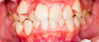
Non-carious lesions of teeth
In addition to caries, the most common dental disease, there is another diverse pathology of hard tissues - non-carious lesions of teeth.
Non-carious lesions of dental tissue occur without tissue softening and without the participation of microorganisms. These processes are based on a violation of the mineralization of hard dental tissues under the influence of external or internal factors. These pathologies occur in approximately 15% of patients, but no more than 5% seek dental care for them, since non-carious dental lesions, as a rule, do not cause pain or other subjective sensations, and often only worsen the appearance. However, if left untreated, a number of complications may occur, in particular early tooth loss.
Hypoplasia of teeth and enamel is most often the underdevelopment of enamel or tooth tissue in the tooth buds under the influence of disturbances in mineral and protein metabolism in the body of the fetus in the womb or in the body of a child. The enamel immediately after eruption has a chalky tint, and then becomes loose and is quickly lost. Hypoplasia is irreversible. Hypoplasia of permanent teeth occurs during the period of their mineralization under the influence of certain diseases (acute infectious diseases, measles, scarlet fever, rickets, diseases of the gastrointestinal tract, dystrophy, brain disorders) at the age of 6 months to 1.5 years. Hypoplasia can appear in the following forms: discoloration (white or yellowish spots on the teeth with a shiny surface and painless upon probing), wavy enamel, absence of enamel in a certain area. Spots with hypoplasia have pronounced symmetry and are characterized by stability (they do not change their shape and color). When the child’s body weakens, caries may occur at the site of the defects. Treatment of dental hypoplasia consists of filling the affected areas of the teeth with composite materials, using sodium fluoride paste, and coating the teeth with fluoride-containing varnish. It is also necessary to normalize general metabolism.
Enamel hyperplasia – “enamel drops” (pearls). These drops are usually located in the cervical area of the crowns of both permanent and baby teeth and have a diameter of 2-5 mm.
Dental fluorosis (endemic fluorosis) is a disease observed in individuals (mainly children) who live for a long time in areas with high fluoride content in water and soil (more than 1 mg/l). Fluorosis is a peculiar form of hypoplasia. Depending on the concentration of fluoride ions, stains that appear on the surface of teeth during fluorosis can have different colors - from white to brown and even black. The more fluoride in drinking water, the more often fluorosis occurs and the less often caries occurs. A predisposing factor to the development of fluorosis is a decrease in the body's reactivity (infectious diseases, endocrine disorders). Fluorosis spots are stationary, dense, with a shiny surface, painless and smooth upon probing. With fluorosis (as opposed to hypoplasia), caries almost never occurs, due to the fact that fluorapatite is deposited in the spots (hence their high microhardness and resistance to acids). Treatment of fluorosis. In the early stages (change in enamel color), bleaching is recommended, followed by remineralizing therapy; calcium and phosphorus supplements are prescribed. It is necessary to increase children's intake of proteins, milk, fruits, vegetables, and limit the consumption of fatty foods. In winter, fish oil, multivitamins, and ultraviolet irradiation are prescribed. It is important to maintain the correct daily routine and conduct general hardening of the body. For complex forms of fluorosis, methods of cosmetic dental restoration or covering teeth with artificial crowns are effective.
Marble disease is a congenital familial osteosclerosis (an abnormal increase in bone density). In this case, the bones of the entire skeleton are affected. After teething, teeth have a chalky tint, and then the enamel becomes loose and is quickly lost. The process can develop into a malignant form.
Wedge-shaped defects are a type of damage to dental tissues located near the walls of the teeth, on the cheek and labial surfaces. The defect has the shape of a wedge with the base towards the neck of the tooth and the apex towards the cutting edge or chewing surface. A wedge-shaped defect, as a rule, does not bother the patient much: pain occurs rarely (only briefly from thermal and chemical irritants), the tooth cavity is not affected and is not opened, the defects slowly deepen, and softening is not determined (this distinguishes the defect from caries). The causes of the wedge-shaped defect have not been fully established. There is a point of view that it occurs under the influence of mechanical factors (for example, a toothbrush). It is sometimes believed that since a wedge-shaped defect begins after exposure of the tooth wall, it is one of the manifestations of periodontal disease. There is evidence of the role of endocrine disorders, diseases of the central nervous system and gastrointestinal tract in the occurrence of a wedge-shaped defect. Treatment of a wedge-shaped defect is aimed at strengthening the hard tissues of teeth through the use of remineralizing therapy (applications of calcium, phosphorus, fluorine, use of fluoride varnish, fluorine gel, etc.). In advanced cases, filling with composite materials or orthopedic treatment (artificial crowns) is recommended.
Increased abrasion of dental tissues . The wear of tooth enamel is a completely natural process that occurs in all people by the age of 45-50. However, in a certain group of people, pathological abrasion occurs already at a young age. Not only the enamel is erased, but also almost completely the crowns of the teeth (most often the front teeth). The causes of the disease are: the habit of grinding teeth during sleep, disorders of the parathyroid glands, almost complete loss of chewing teeth (which increases the load on the front teeth). Treatment – orthopedic or orthodontic (in order to properly distribute the load on the teeth).
Erosion of hard dental tissues is the progressive loss of enamel and dentin on the vestibular surface of tooth crowns. Areas of erosion have an irregular rounded shape. The causes of tooth erosion are considered to be the mechanical effects of toothbrushes, endocrine disorders (in particular, increased function of the thyroid gland - thyrotoxicosis). It occurs mainly in middle-aged people and lasts 10-15 years. There are different degrees and stages of the process. Treatment is aimed at additional mineralization of hard dental tissues. Erosion is filled with composite materials and artificial crowns are made. Calcium and phosphorus preparations, multivitamins with microelements are prescribed internally.
Necrosis of hard dental tissues initially manifests itself in the loss of enamel shine, then chalky spots appear, gradually turning into dark brown. Softening appears in the center of the spots, the enamel becomes brittle and easily chips. Necrosis differs from erosion by softening in the center of the spot. Teeth are extremely sensitive to any irritants. The causes of necrosis are endocrine pathologies (hyperthyroidism, dysfunction of the gonads), pregnancy, diseases of the central nervous system, chronic intoxication of the body, hereditary factors. Treatment is aimed at eliminating tooth sensitivity, remineralization, and normalizing endocrine disorders.
Dental injuries. Acute dental injuries occur when a tooth is exposed to traumatic factors - impact, increased load during chewing, constant pressure from improperly made orthopedic structures. Injuries are more common on children's baby teeth (luxation, tooth fracture, crown fracture). Acute dental injuries include: tooth bruise, tooth dislocation (complete and incomplete), fracture of part or the entire crown, combined injury, tooth germ injury. If a tooth is bruised, it is necessary to rest it, eliminate contact with opposing teeth by grinding off the cutting edge, and exclude solid foods from the diet. It is necessary to monitor the condition of the pulp, and in case of irreversible changes, remove it and fill the canal. Tooth dislocation with healthy gum tissue rarely occurs. However, if there is periodontal pathology, in particular, bone tissue resorption, tooth dislocation can easily occur even when chewing hard food. In case of incomplete dislocation of a tooth, it is necessary to create rest for which the tooth is splinted. If the pulp ruptures, it is removed and the canal is filled. If part of the crown breaks off, it is restored using composite materials. Sometimes special pins are used.
Bibliography:
- Lutskaya I.K., Nichiporovich G.S. Frequency of enamel and dentin cracks in permanent teeth. Dental Journal 2006, number 2. Page. 87-91.
- Nichiporovich G.S. Modern ideas about the increased sensitivity of hard dental tissues. Dental Journal, 2006, number 2. Page. 92-95.
- Leus P.A. Non-carious diseases of hard dental tissues. Educational and methodological manual. Minsk, BSMU-2008, 56 pages.
- Borovsky E.V., Leus P.A. Erosion of hard dental tissues. Dentistry 1971. T. 50 No. 3.p. 1-5.
- Fedorov Yu.F. Clinic and treatment of hyperesthesia of hard dental tissues. - M 1970, 140 pp.
- Leus P.A., Kozel O.A. Disturbances in the development of tooth enamel. Educational and methodological manual. Minsk, BSMU-2004-29 pages.
Author: Shulyak Alesya Vadimovna
Advantages of the GrandMed clinic
- Dentistry has created an excellent diagnostic base that allows identifying non-carious dental pathologies in the early stages. Doctors have modern equipment at their disposal (radiovisiograph, orthopantomograph, computed tomograph), and all necessary clarifying tests can be done in the clinic’s biochemical laboratory.
- We employ professionals of the highest level, whose experience and qualifications allow us to treat complex non-carious pathologies, which often require the use of special techniques and non-standard approaches. Clients are provided with high-quality therapeutic and orthopedic care, including prosthetics on implants. So even in the most advanced cases, our patients can count on restoration of the beauty and functionality of their dentition.
- This disease is treated using local anesthesia. Patients of our clinic do not experience pain and feel comfortable during the procedure after injection of one of the modern anesthetic drugs that do not have side effects (Ultracaine, Ubestezin, Scandonest). You should not be afraid of the injection itself - it is performed quickly and accurately, and therefore is absolutely painless.
- Our clinic adheres to the highest sanitary standards, which is confirmed by the use of the latest sterilization equipment, which completely eliminates infection during treatment; Dentists have all the necessary consumables and disposable dental instruments at their disposal.
Note: computer necrosis of teeth
Experts started talking about this non-carious lesion of teeth relatively recently; it definitely did not exist in the 20th century, however, like people who spent most of their working and free time at the computer. The death of dental tissue threatens patients who are ready to sit at a computer for more than 8 hours a day, without days off and a full night’s rest. After 3–5 years of living in this mode, a person (and these are mainly young people under 35 years old) are guaranteed to have problems with the front teeth located in the path of the electrostatic radiation of the monitor.
Symptoms of computer necrosis of teeth
Experts, comparing computer and post-radiation lesions, find much in common between them: dying areas in both cases spread over the entire surface of the supragingival part of the teeth, which acquires a dark, almost black color. The focus of necrosis is filled with softened brown tooth tissues; the dentist easily removes them with special instruments, and the patient does not feel pain. As for the teeth of the chewing group, which are exposed to relatively less computer radiation, they also do not look the best: the enamel loses its shine and acquires a gray tint. Studies have shown that not only hard tissues undergo negative changes, but also the pulp. Thus, during electroodontometry, the neurovascular bundle does not respond to an electric current of 25-30 μA. In addition, changes in the functioning of the salivary glands are observed.
Treatment of computer necrosis of teeth
The “newness” of the disease complicates diagnosis, since the dentist may never have encountered it. An X-ray examination is required (sight image, orthopantomogram, computed tomogram), in the images the teeth look translucent and have unclear contours, which indicates a deficiency of mineral substances.
Treatment of computer necrosis is complicated by the fact that during the removal of damaged tissue with a bur, the tooth falls apart; in such conditions, it is impossible to preserve the vitality of the pulp, and often the tooth itself. If recovery is possible, it will take a lot of time, as it will be carried out in several stages, including local and general therapy, preceding filling or prosthetics. At the first stage of treatment, dead tissue is removed and healthy tissue is remineralized, and the patient is recommended to take mineral-vitamin complexes containing calcium, antioxidants, and vitamin A. After the condition improves, therapeutic linings are placed in the dental cavity and a temporary filling is performed. The result of this stage of treatment, which takes up to two months, is the restoration of dentin, after which the tooth is finally filled.
Prevention of computer necrosis of teeth
Computer necrosis of teeth can be prevented by a rational approach to working at the computer. Firstly, comply with sanitary standards, which require organizing a work area of at least 6 sq.m., located in a room with an area of 20 - 24 sq.m. The monitor should be located no closer than 60 - 70 cm from the face; if there are several computers installed in a room, the distance between them should be at least 2 m. Every two hours you should take a break of 15–20 minutes, during which the room is ventilated. Secondly, monitor your diet; if you don’t get enough antioxidants, vitamins and microelements with food, then take vitamin-mineral complexes. Thirdly, use new models of monitors that have minimal electrostatic effect on the body.