What is transmitted genetically
Most diseases of the dental system are not 100% genetically dependent. However, there are signs that are passed on from generation to generation and can ultimately affect the condition of the teeth. These include:
- composition, thickness and shade of enamel;
- shape of dental units;
- bite geometry;
- timing of the eruption of primary and permanent teeth;
- composition of saliva and oral microflora.
Along with these characteristics, the child also inherits a predisposition to certain pathologies. But there is no fatality in this. With a reasonable, competent attitude to oral care, you can avoid the development of one or another disease, the likelihood of which is genetically predetermined.
Publications in the media
Dental developmental disorders - structural anomalies and malformations of teeth - can appear in isolation or in combination with structural anomalies and malformations of organs and systems of the entire child's body.
Etiology. Dental tissue defects are caused by hereditarily fixed changes in the genetic code, but can be caused by environmental factors. Pathogenesis. Teeth during the period of their follicular development, i.e. Before eruption, several stages pass: laying, formation and calcification of crown enamel, formation and calcification of dentin. At each stage of tooth development, conditions can be created that lead to disruption of the formation of its shape, mineralization and color.
Classification • All disorders are divided into: •• anomalies of the structure of dental tissues, transmitted by inheritance •• anomalies in the number, size and shape of teeth, due to the hereditary transmission of the sample •• anomalies of the structure and malformations of dental tissues, which arose as patterns of the pathogenesis of systemic pathology in the child’s body (hereditary, congenital, acquired) •• structural anomalies and malformations of dental tissues caused by the influence of external factors. • Each group has individual clinical and radiological features, determined by the cause of occurrence and the mechanism of development.
Hereditary disorders of the development of hard dental tissues. Men and women suffer equally often. Both temporary and permanent teeth can be affected. • Enamel defects (most often dysplasia) are an isolated symptom (130900, Â). Combinations with cataracts and stenosis of the Sylvian aqueduct (600907, r), nail defects and hypohidrosis (ameloonychohypohidrotic syndrome, *104570, Â), also a sign of a number of hereditary diseases, for example: •• Renal tubular acidosis type II •• Albright’s disease •• Lacrimal duct defect (149700) •• Citrate transport protein defect (190315) •• Dysosteosclerosis (224300) •• Cranioectodermal dysplasia (218330) •• Tumor calcification (114120) •• Mucopolysaccharidosis, type IVB (253010) •• Neurosensory hearing loss, hypoplasia enamel, nail defect (234580) •• Goltz-Gorlin syndrome (305600) •• Menkes syndrome (261900) •• Naegeli syndrome (161000) •• Polyglandular autoimmune syndrome, type I (*240300) •• Stimmler syndrome (202900) • • Tuberous sclerosis (191092) •• Epidermolysis bullosa (226440). Oculodental-digital dysplasia (164200, genes ODDD, SDTY3, ODOD, 6q22 q24) •• Amelogenesis imperfecta (impairment of the process of enamel formation) is the most common pathology. Usually manifested by grooved (wrinkled) enamel or its sharp thinning. Erupting teeth are small, cylindrical, yellowish or brown in color. Amelogenesis imperfecta is inherited independently and is part of a number of hereditary syndromes ••• Amelogenesis imperfecta, type 1 (301200, genes AMELX, AMG, AIH1, AMGX, Xp22.3 p22.1, À) ••• type 2 (104500, gene AIH2 , 4q11 q21 À) ••• type 3 (301201, AIH3 gene, Xq22 q28 À) • with taurodontia (104510, À) ••• with nephrocalcinosis (204690, r) •• Enamel abnormalities are also observed in the following phenotypes: •• • platyspondylia with amelogenesis imperfecta (601216) ••• Kohlschutter syndrome (*226750, r): epilepsy, dementia and amelogenesis imperfecta ••• photoreceptor dystrophy and amelogenesis imperfecta (217080, r).
• Defects in dentin formation are numerous ; traditionally, the groups of dentinogenesis imperfecta and dentin dysplasia are distinguished. In addition, dentin defects occur in a variety of other osteogenesis disorder syndromes •• Dentinogenesis imperfecta. The structure of the enamel is not changed, but due to the imperfection of dentin, its connection with the enamel is fragile, which leads to enamel chipping ••• type I (*125490, 4q13–q21, DGI1 gene, Â). Synonyms: opalescent dentin, opalescent teeth without osteogenesis imperfecta ••• dentinogenesis imperfecta, type II, Capdepont teeth ••• type III (125500, Â). Clinically: Rapidly abraded dental crowns, smooth amber-colored dentin, open bite, very large pulp chambers and root canals of primary teeth, small or absent pulp chambers of permanent teeth •• Dentin dysplasia is a dental anomaly in which the teeth are clinically, morphologically and color-wise normal , but x-ray examination reveals short roots, obliteration of the pulp cavity and canals, increased mobility and premature tooth loss ••• type I (*125400, Â) •• type II (*125420, Â) - occlusion of the pulp cavity, the structure of the pulp in the form straws ••• Dentin dysplasia and cortical osteosclerosis (*125440) - compacted long bones, compaction of the alveoli of the teeth, narrow or sometimes closed medullary canals, thickening of the cortical layer.
• Mesoectodermal odontopathy (Stainton–Capdepont disease; simultaneous disruption of the formation of enamel, dentin and pulp). The teeth erupt at the usual time, have a normal size and shape, but their color is changed (from gray to blue-gray and yellowish-brown). The length and shape of the roots are changed. The tooth cavity and root canals are obliterated. Characterized by rapid wear of teeth (especially temporary ones). Their surface is shiny, flat and smooth (“polished”). At the same time, teeth practically do not react to mechanical, chemical and temperature stimuli.
• Oculodental-digital dysplasia (ODDD gene, 6q22–6q24, Â - *164200, r -*257850) - microphthalmos, coloboma or iris abnormalities in combination with congenital dental defects and finger abnormalities. Clinically: microphthalmos, microcornea, glaucoma, dental anomalies (enamel hypoplasia, small teeth), camptodactyly of the little fingers, syndactyly of the fourth and fifth fingers, absence of the middle phalanges of the toes, hypotrichosis, small nose, hypoplastic wings of the nose, hyperostosis of the skull bones, orbital (bone) hypotelorism, jaw overgrowth, spinal cord compression, spastic tetraparesis, progressive spastic paraplegia, the effect of paternal age on the frequency, pathology of the white matter of the brain on MRI, calcification of the basal ganglia. Synonyms: oculo-dental-digital syndrome, ODD (oculo-dento-digitalis) syndrome, Meyer-Schwickerath and Weyers syndrome. Note: a mutation at this same locus is the cause of syndactyly type III.
• ADULT syndrome (from Acro-Dermato-Ungual-Lacrimal-Tooth, 103285, Â). Clinically: hypodontia, early eruption of permanent teeth, ectrodactyly, obstruction of the lacrimal ducts, onychodysplasia, freckles, hypoplasia of the mammary glands. • Treatment depends on the degree of tooth tissue loss due to increased abrasion. In case of minor loss, it is recommended to regularly (2 times a year) conduct courses of remineralization therapy; in case of severe loss, orthopedic treatment is recommended. Enamel hyperplasia is the excessive formation of tooth enamel during its development. Typically, enamel hyperplasia is observed in the neck of a permanent tooth in the form of drop-shaped formations. After teething, these formations do not progress and are not affected by caries; do not cause any unpleasant sensations. Treatment is indicated only for noticeable cosmetic defects; Grinding is carried out followed by remineralization.
Enamel hypoplasia is a developmental defect that manifests itself in a violation of the structure and mineralization of tooth tissue during the period of their formation. Usually, only the formation of enamel is disrupted, and in severe cases, dentin as well. Most often, enamel hypoplasia is observed on permanent teeth, very rarely on temporary teeth, which is explained by their formation in the prenatal period. The localization of areas of hypoplasia depends on the age at which the child suffered from a general disease, and its severity depends on the severity of the disease.
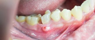
• Causes: severe infectious diseases, hypo- and avitaminosis, disorders of the gastrointestinal tract (dyspepsia), endocrine organs (especially the parathyroid glands), hemolytic jaundice, hereditary syphilis, etc. • Classification : •• systemic hypoplasia - a violation of the structure of the tissues of all or a group of teeth formed in the same period of time •• focal hypoplasia - a malformation of the tissues of one or more teeth that formed in the focus of inflammation of the tissues surrounding the follicle, in the area of tumor growth or exposed to trauma. • Manifestations. Hypoplasia of dental enamel manifests itself in the form of whitish spots or depigmentation of various sizes with a smooth shiny surface. They can be combined with point or cup-shaped depressions, grooves, and constrictions of the enamel of the crowns of the teeth. In these lesions, the enamel loses its inherent color, but retains its density. Sometimes a complete absence of enamel is observed in a limited area of the tooth, which looks like a cup-shaped depression. Rarely, enamel is absent on almost the entire surface of the tooth crown and then the tooth takes on a bizarre, ugly shape (Turner's, Hutchinson's, etc. teeth).
• Treatment and prevention . Treatment of dental enamel hypoplasia depends on the form and severity of the disease. For spotted hypoplasia, bleaching with medications or grinding is carried out. To prevent the occurrence of caries, remineralizing therapy is carried out. Small enamel defects are filled after preparation. Teeth significantly affected by hypoplasia are covered with artificial crowns. Prevention of dental enamel hypoplasia - harmonious physical development of the child, balanced nutrition, timely treatment of diseases of primary teeth. Fluorosis is an acquired malformation of dental tissues caused by excessive intake of fluoride during tooth formation (a kind of enamel hypoplasia). Most often it develops with an excessive fluoride content in drinking water (more than 1.5 mg/l), but it is also possible with an optimal fluoride content (0.8–1.2 mg/l) in children with a reduced immune status.
• Pathogenesis. Fluoride salts have a toxic effect on ameloblasts, which leads to impaired calcification of tooth tissue. Subsequently, the content of fluorine and calcium in the surface layers of the enamel increases, and, accordingly, the acid solubility of the enamel decreases. The severity of fluorosis depends on the dose of fluoride entering the body during amelogenesis. These teeth are not affected by caries.
• Manifestations: •• chalky spots and stripes on the vestibular surface of the teeth •• chalky and pigmented (to brown and black) spots •• erosive lesions of the enamel •• disturbances in the shape of the crowns of the teeth in combination with stains, erosions and fragility of the enamel (fractures of areas of enamel during mechanical influence).
• Treatment is prescribed taking into account the severity of the process. If the form is spotty, teeth are whitened. Enamel defects are filled or orthopedic treatment is performed (crowns, pinned teeth). • Prevention •• Public prevention: reducing the intake of fluoride from drinking water into the child’s body during the period of teeth formation (up to replacing the water source) •• Individual prevention: partial defluoridation of drinking water by boiling, settling •• Additionally, milk and dairy products are recommended throughout the year , careful hygienic care of the oral cavity.
ICD-10 • K00 Disorders of development and eruption of teeth •• K03.7 Discoloration of hard tissues of teeth after eruption
Structure of teeth
In the presence of a thin layer of enamel on the teeth and deep fissures (slit-like depressions on the chewing surface), the risk of early carious tooth destruction increases. But even with hereditary weakness of the enamel, it will be reliably protected:
- careful hygienic care;
- carrying out remineralization of enamel (saturation with calcium and fluoride salts);
- fissure sealing (sealing with a composite material immediately after teeth erupt).
At the Shifa clinic, children can always get help in strengthening hard dental tissues.
Diagnosis of fluorosis
A dentist can distinguish fluorosis at the initial stage from caries:
- Caries manifests itself asymmetrically, while fluorosis occurs on symmetrical teeth.
- Caries has typical places of manifestation: contact surfaces of teeth, cervical areas. Fluorosis, on the contrary, appears more often on the outer surface of the teeth.
- Caries, like fluorosis, can be multiple, but still, as a rule, with caries you can see only a few affected teeth, and with fluorosis there are more of them.
- The dentist will identify fluorosis by the characteristic shape of the lesions (striations, spots).
- Caries appears on the teeth gradually, but fluorosis is visible already at the moment of teething.
Meet our tooth fairies and check prices.
Composition of saliva
When parents complain about bad teeth, they often do not realize that the cause may be the composition of saliva, which determines heredity. It may vary in the ratio of mineral and organic components. And this has a direct impact on dental health.
With a reduced content of oligosaccharides in saliva, the population of microorganisms increases, which settle on the surface of the enamel, creating a predisposition to caries. To avoid this disease, you need to brush your teeth at least twice a day and use special mouthwashes.
An increase in the concentration of class A immunoglobulins (IgA) in saliva, which have an antibacterial effect, protects the enamel from cariogenic bacteria.
The specialists of the Shifa clinic will help you choose toothpaste and other hygiene products to create the right environment in the oral cavity. Dentists will also give recommendations on diets that reduce the negative effects of saliva.
Shape anomalies
There are pathologies in the development of dental units, due to which they acquire an unnatural shape. These disorders are named after the scientists who first described them. The following types of dental shape anomalies are found: spiny teeth, Pflueger teeth, Fournier teeth, Hutchinson teeth.
- Spiked or awl-shaped teeth. With this pathology, the teeth acquire a cone-shaped shape. Wide at the base, they gradually narrow and, towards the cutting edge, become sharpened into a spike shape. This problem is combined with microdentia. There are irregularities and stains on the surface of the teeth. The disease affects the front and lateral incisors. The disease occurs in childhood and is caused by genetic factors in combination with external factors. Among them are vitamin D deficiency and endocrine system problems.
- Hutchinson's teeth are characterized by a modified crown shape of the incisors. Externally, they have the shape of a barrel, since the neck is significantly thickened. The cutting edge of the teeth acquires an arched notch. The enamel layer also suffers, which is present only on the sides and absent in the center.
- Fournier's teeth are a form of systemic hypoplasia of dental units associated with metabolic disorders at the stage of intrauterine development of the fetus. The barrel-shaped shape of the teeth is preserved, but the notch of the incisal edge is absent. The color of the enamel is not disturbed, but the enamel layer is underdeveloped, which can be seen during microscopic examination.
- Pflueger's teeth are a disorder that affects permanent dental units. Their crowns take on a conical shape. Thickening develops in the cervical region, and the chewing surface is significantly underdeveloped. The chewing function of the teeth is completely preserved.
- Turner anomaly. With this pathology, there is no enamel on the teeth. They have an abnormally lumpy surface, and replacement tissue, dentin, forms in the exposed areas.
Systemic hypoplasia with a violation of the shape of the teeth has three degrees of development. The third degree is the most dangerous, in which the crown is severely deformed and the enamel layer decreases. In this case, the defective teeth are removed and replaced with dentures. It is also possible to restore affected teeth using reflective components.
Bite
Abnormal bite often causes:
- increased load on individual teeth, which contributes to their rapid destruction or damage to the ligamentous apparatus;
- crowding of teeth, leading to gum inflammation and dental caries.
The hereditary factor is detected in no more than 50% of cases of abnormal structure of the dental system. The remaining 50% is due to the following circumstances:
- artificial feeding;
- formation of bad habits at an early age (sucking pacifiers, fingers, lips, holding pencils or pens in the mouth);
- the habit of sleeping in one position, especially with your hand under your cheek;
- incorrect posture;
- lack of calcium and other trace elements in the body.
In preventing the development of pathological occlusion, the role of parents is great, who should pay attention to the early identification and elimination of provoking factors.
Dental fluorosis in children
Fluorosis is a chronic non-carious lesion of teeth, which is manifested by a violation of enamel mineralization. The causes of dental fluorosis in children are associated with excess fluoride in water or air if they live in an area where the fluoride content in these environments is, for some reason, elevated. Since fluoride is retained in a child’s body (in bones and organs) more than in an adult, it is children who suffer most from fluorosis, especially if they lived in such areas for up to three years. As a rule, fluorosis affects the buds of permanent teeth, less often milk teeth, since they are formed in utero, when the placental barrier protects the child from excess fluoride.
Fluorosis usually affects the central incisors, usually the upper jaw. First, white spots or stripes appear on their outer side (the streaked and spotted form are the initial stages of fluorosis), then gradually the enamel can be eroded (erased), and if the fluorine content in the water remains high, it can turn into a destructive form in which fluorosis affects not only enamel, but also dentin. The tooth becomes brittle, which leads to chips and breaks.
The importance of prevention
The child subconsciously adopts the behavior and habits of his parents. If older family members do not brush their teeth, eat improperly, or do not exercise, then dental diseases lie in wait for both adults and children. At the same time, the incorrect conclusion is often made that “bad teeth are inherited.”
Mineralization of dental tissue continues after teething. Any disease, weakened immune system, or unbalanced diet have a much greater impact on the hard tissues of teeth than hereditary predisposition.
Parents should monitor the health of their children by providing them with proper care and disease prevention. This will help avoid dental caries and the development of enamel hypoplasia, and reduce the risk of abnormal bite.
In case of poor heredity, timely prevention plays a key role. Regular dental care will reduce the risk of developing dental and periodontal disease in children. The specialists of the Shifa clinic are always ready to help. Our dentists regularly improve their professionalism, conduct diagnostics and treatment using modern equipment. This allows you to identify possible risks and prevent them in a timely manner. Contact professionals to ensure your child’s teeth are always strong and healthy!
Disorders of enamel maturation with hypoplasia and taurodontism
Some experts combine it with hypomaturation, others separate it separately. The formation of enamel tissue is disrupted during tissue differentiation. The pathology may be accompanied by grooved or thin hypoplasia.
The enamel turns into various colors - from white and yellow to unpleasant brown, becomes covered with opaque specks and acquires a pathological tendency to abrasion. The photographs show large cavities in the incisors and symptoms of taurodontism. In multi-rooted teeth, the pulp cavity is expanded and the crown is lengthened. The root is short, and the distance from the occlusal surface to the bifurcation zone is increased.
General information
Inferior amelogenesis is a severe damage to the enamel of the milk or permanent units, which consists of a violation of the formation or maturation of the protective layer. As a result, a number of morphological and clinical defects develop: hypoplasia, disorganization of prisms, hypomaturation, reduced hardness and thickness of enamel, discoloration of units, pathology of the shape and size of crowns and roots.
The listed disorders lead to the development of various defects in those affected by amelogenesis imperfecta:
- The appearance of pits, dots, grooves or absence of enamel in certain places of the tooth.
- Loss of shine, acquisition of yellow, chalky and other shades.
- Accelerated abrasion, bite pathologies.
- Untimely eruption.
- Violation of the structure of the root system, bifurcation area.
- Increased sensitivity to temperature, chemicals or mechanical irritants.
The functionality and aesthetics of teeth deteriorate.
Position anomalies
Incorrect position of teeth always develops as a consequence of another pathology and forms an incorrect bite. May affect one or both jaws. Incorrect position of teeth makes chewing food and oral hygiene difficult. This can lead to digestive problems and the development of cavities. There are several options for the pathological position of teeth within or outside the dentition:
- External or vestibular position means that the tooth is located outside the dentition, closer to the vestibule of the mouth. This position is typical for canines and incisors and develops as a serious cosmetic defect.
- Oral or internal position. The teeth are deviated inward closer to the tongue. The disorder is typical for canines, incisors and premolars. May cause tongue injuries and dysfunction of the temporomandibular joint.
- Mesial and distal position. Dental units are displaced forward or backward along the dentition, which leads to its shortening.
- Supra and infraocclusion are high or low positions of teeth relative to the occlusal curve. The cause of the disorder is underdevelopment of the alveolar process. It appears due to the presence of some obstacle that interferes with the normal formation of the tooth germ. Effective therapy is surgery.
- Tortoanomaly. The tooth is rotated around a vertical axis. The disorder is typical for incisors, canines, and premolars. The anomaly can cause injury to adjacent teeth. Dental units are rotated into the correct position using orthodontic structures.
- Transposition. The teeth change their location with each other. The disease affects canines that exchange places with premolars or lateral incisors. The disorder develops at the stage of tooth germ formation. The therapy is as follows: problematic teeth are removed followed by prosthetics.
- Crowding of teeth occurs due to lack of space when tooth germs are located too closely. Crowded teeth are closely adjacent to each other and rotate around their axis. Occurs in combination with macrodentia or with an undeveloped basal part. To eliminate the violation, some dental units are removed.
- Diastemas and tremas are wide spaces between teeth. The disorder develops as a consequence of an abnormal shape and size. The problem is treated with orthodontic methods or by installing veneers, which helps restore the aesthetics of the smile.
Misaligned teeth cannot always be cured using braces. Severe pathology, which is characterized by a skeletal disorder of the maxillofacial region, is treated with surgical reconstruction. The pathological fragment is transferred to the desired position and then secured. A fragment containing part of the dentition or the entire dentition can be used.
Type III dentinogenesis imperfecta
This form is rare and is considered an isolated hereditary form of wine opalescent dentin. With it, the crowns of the teeth take on the shape of a bell. Multiple openings are noticeable in primary teeth. The x-ray shows a contrasting contour of the tooth with internal enlightenment. This is explained by the fact that after the formation of the outer covering layer, dentin production ends.