According to antiplagiat.ru, the uniqueness of the text as of October 16, 2018 is 99.9%.
Key words, tags: X-rays, tooth extraction, orthopantomogram, periodontitis
A dental cyst belongs to the group of jaw cysts. Jaw cyst is a general concept that unites many pathologies. The formation of a cystic cavity in the jaw can be caused by an inflammatory process, or in rare cases, by a growing tumor. It can be in a “dormant” state and not manifest itself in any way, or, on the contrary, be in an active, acute phase, get sick, affect neighboring healthy tissues, and cause various diseases. In any case, the presence of cysts in the jaw and, in particular, the presence of a dental cyst, requires attention and serious consideration.
Tooth cyst: historical background
The word “cyst” is of Greek origin (“kystis”) and literally means “bubble.” A dental cyst is a cavity-like tumor-like formation. Previously, when X-rays were not taken, it was extremely difficult to see such formations and, as a rule, dentists encountered them during the extraction of teeth from patients. Thanks to the advent of X-ray diagnostics at the end of the 19th century (1895), it became possible to plan the treatment of dental cysts. And today, if a patient regularly visits the dentist’s office, we can successfully diagnose and eliminate dental cysts at those stages when they do not pose a danger.
What is the prognosis for the disease?
How successfully the situation in a patient with an odontogenic cyst will be resolved depends on at what stage the cyst was discovered, how severe the symptoms were and how it was treated.
As a rule, the use of surgical treatment gives a positive prognosis. Therapeutic treatment provides a positive prognosis only if it is started at the initial stage of the disease.
A negative prognosis may be associated with detection of the disease at a late stage: odontogenic cysts can provoke the development of serious pathologies that cause deformation of the maxillofacial tissues.
Tooth cyst: anatomy
A dental cyst is a benign cavity tumor-like formation that has a membrane and an internal epithelial lining, the cells of which produce fluid. Modern medicine still cannot accurately answer the question of how and where an incipient cyst takes epithelial tissue from. However, most scientists are inclined to believe that it comes from the remnants of the dental epithelium, the so-called islands of Malasse-Astakhov. And it develops as a result of chronic odontogenic inflammation, when microbes enter the jaw bone through the tooth canal. Further development of the process can go in two directions, the result of which are pathologies: an extensive destructive process that does not have a shell - a non-shell formation (such a process is not a cyst), or a dental cyst itself - a shell-like formation.
Types of dental cysts
It is customary to distinguish between 2 types of dental cysts - follicular and radicular. Follicular cysts are less common than radicular cysts. In the presence of certain factors, it grows from the dental follicle - the membrane around the growing tooth. When it is large in size and involves adjacent teeth, it is called a follicular dentigerous cyst.
Radicular (odontogenic inflammatory) cysts form on the roots of teeth as a result of pathological processes spreading beyond the root apex. By location, radicular cysts can be apical (root), lateral (lateral), apicolateral and interradicular.
The mechanism of formation and growth of a dental cyst
The cystic cavity, located in the jaw, has external resistance from the surrounding jaw bone. But the epithelial cells of the lining begin to produce fluid, which gradually fills the cavity, creating excess pressure.
This pressure acts on the surrounding bone tissue, causing its gradual peripheral resorption, giving the cyst the opportunity to grow even larger, increase fluid secretion, and therefore put even more pressure on the walls. This is why cysts can grow to very large sizes, sometimes asymptomatically, if not accompanied by periodic inflammation. This may be due to the patient’s good immune status, low pathogenicity of the microflora in the lesion and other factors.
Possible complications of a dental cyst
As the cyst grows, it can “push aside” nearby anatomical formations, such as the canal of the inferior alveolar nerve, and can disrupt the external contours of the jaw bones (plastic toy syndrome), thereby changing the contours of the face, that is, causing facial asymmetry. It may also involve other neighboring tissues in the pathological process. So, often, retrograde periodontitis develops in the teeth located next to the cyst. And if the cyst grows into the maxillary sinuses, chronic odontogenic sinusitis is formed, which can be asymptomatic for a long time, but, nevertheless, have a detrimental effect on health.
Diagnosis of a dental cyst
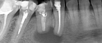
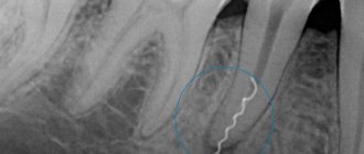
The choice of treatment method for a dental cyst is based on a multifaceted diagnosis, which may include:
— examination of the patient; - “targeted” x-ray of the tooth, which the patient indicates as the causative tooth; - orthopantomogram (panoramic image of the jaw bones), which allows you to see pathologies and the most likely places of their development in the upper and lower jaws, in the maxillary sinuses, in the canals of the nerve trunks; - in some cases - a computed tomogram, which allows you to see the facial skeleton and skull bones, joints, maxillary sinuses in three projections (frontal, sagittal and horizontal) and in a three-dimensional volumetric image.
Classification of pathology
Cysts localized in the tissues of the upper or lower row of teeth are very common. With their development, a cavity appears, the walls of which are covered with fibrous tissue, and the inner surface consists of an epithelial layer. The capsule holds clear or cloudy liquid.
Doctors distinguish the following types of pathology:
- Primordial. This is a cyst of the lower jaw. It appears in the area of the third molar. She has very thin fibrous walls. The inside of the capsule is lined with flat epithelium. According to its structure, the tumor may contain one or several small chambers.
- Follicular. Formed before tooth eruption. It can grow in the area of the alveolar margin. Includes cells that intensively produce viscous mucus. Because of this, the internal contents of the anomalous structure are quite viscous. A follicular formation is formed from the enamel organs of the unerupted unit. One or several teeth are found inside it. These may be formed crowns or tooth buds.
- Radicular. It occurs more often than others - in eighty percent of cases. Usually grows above the upper teeth. The diameter reaches from half to two centimeters. Often inflamed. Then the cells begin to hyperplasia, network-like processes are formed that extend into the thickness of the walls. The liquid contents of the radicular structures are rich in neutrophilic leukocytes. Often with this type of pathology, the patient develops sinusitis. This is due to the peculiarities of the localization of the inflammatory site.
- Retromolar. It is formed due to a long-term inflammatory process occurring in the thickness of the soft tissues. It is a consequence of complicated teething.
- Nasoalveolar. It occurs near the nasopalatine canal (the border between the upper row and the wing of the nose).
- Aneurysmal. Very rare in dental practice. Appears only on the lower jaw. This type of neoplasm is still poorly studied. It is known that there is blood or a red-pink liquid inside it. Many scientists agree that the symptoms of an aneurysmal cavity are a consequence of hormonal imbalance.
- Traumatic. Occurs due to recent facial trauma.
- Residual. The result of mistakes made by the doctor during tooth extraction, or the consequence of the patient ignoring the surgeon’s instructions.
The doctor decides how to treat a dental cyst, taking into account the cause of its development. It is very important to understand why the tumor occurred. If the root cause is not identified, the likelihood of relapse will remain high.
Removal of a dental cyst. Historically significant and modern methods of surgery for removing dental cysts
Initially, the most common method of treating cysts was their removal. For this purpose, 2 fundamentally different methods were used: cystotomy and cystectomy.
Cystotomy (or the PARCH-I method) has historical significance in the development and establishment of medicine. Today this method is practically not used, but previously it was indispensable for removing large cysts. To avoid complications and its severe consequences, a wide channel was created between the cyst cavity and the vestibule of the oral cavity by suturing the edges of the cyst shell with the oral mucosa.
Cystectomy (or PARCH-II method) involves complete excision/removal of the cyst. This surgical intervention is often simultaneously accompanied by the removal of the root apex, the source of the formation of the cyst containing infected apical deltas. Today this method is the most popular.
The main goal of cystectomy is complete sanitation of the cyst cavity, which is not possible without resection of the root apex. The fact is that the root pulp in its apical third has an apical delta with an unclear structure and very fine branching of the canals, even in single-rooted teeth. If such canals can be cleaned during tooth treatment, then during retreatment this is almost impossible, especially considering the narrowness of the lateral deltas. Removing the root apex, in addition, allows you to most effectively clean the cyst cavity behind the root. This is especially true for teeth 12 and 22, which have a more curved root system. The operation is carried out as follows: using a small incision on the gum, the surgeon very carefully reaches the surface of the bone located above the cystic cavity, removes the wall with a trephine or bur, making sure that the neighboring teeth are not damaged, and then removes the cyst shell and performs a resection of the root apex . Next, sanitation of the cystic cavity is carried out and its subsequent revision for residual cyst particles. After this, in some cases, retrograde filling is performed. The final step in the operation to remove cystic formations is filling the resulting cavity with osteoplastic material and suturing the wound.
In the case of the formation of a large cyst, amputation of the apex of the tooth root can be performed, or half of the tooth and damaged root can be removed (hemessection), and in the case of an interradicular cyst, this can be done by corono-radicular separation.
How to treat
In most cases, cysts cannot be treated without surgery. Only in unadvanced cases is it possible to get rid of the tumor by opening it and draining it.
The operations performed for the disease are called cystectomy and cystotomy. They both involve removing the formation. But in the first case, it is removed completely, and in the second, only the front wall is excised.
The main goal of a dental surgeon is to preserve the tooth growing in the area where the tumor is located. Unfortunately, this is not always possible. So, if the tooth root is immersed in the cyst cavity by more than 60% of its length, emergency removal is indicated. Such a unit can no longer be functional. Soon after excision of the tumor, it will still become loose and fall out.
Therefore, it is very important not to start the disease. Considering that it may not manifest itself for a long time, it is important to undergo a specialized examination at the dental office every year. This will increase the chances of successful early diagnosis.
Quality criteria for dental cyst removal
Despite the fact that cystectomy is a surgical intervention, the main indicator of the quality of treatment will be the sanitation of the root canals, including retrograde filling. Currently, in addition to eliminating the cyst shell itself, the unspoken rule has become the filling of its cavity with one or another osteoplastic materials, because this completely eliminates the possibility of blood clot suppuration, promotes rapid healing of the bone defect and improves the mechanical stability of the tooth.
Indications for tooth extraction with a cyst
As a rule, conservative treatment carried out on time or surgery to remove the cyst allows you to save the tooth. But this is not always possible. A tooth must be removed in the following cases:
— the presence of severe pain symptoms when drug treatment is ineffective; - purulent inflammatory process when it is impossible to drain it; — fracture of the coronal part of the tooth, without the possibility of restoration with anchor pins and/or stump inlays; — obstruction of root canals; — the presence of multiple damage to the roots of the tooth or large damage; the tooth is almost completely destroyed and orthopedic restoration is impossible; - no need for dental treatment due to the presence of a prosthetic plan agreed upon between the doctor and the patient.
Stages of non-surgical conservative treatment
If a decision is made on conservative treatment, we would like to warn you right away - this process is not quick, it will require time and money, but the tooth will be saved .
The details of therapeutic treatment are very individual, our specialists will optimize your treatment and select the best course of action.
General algorithm for a non-surgical, low-traumatic protocol:
- Examination A targeted image and computed tomogram to assess the condition and number of channels, the size of the inflammation. Microscopic diagnostics for visual examination of canals under magnification. Data is essential to create a perfect treatment plan.
- Consultation A joint consultation between an endodontist and a surgeon to determine the advisability of treatment and the possibility of re-treating infected tooth canals bordering the cyst. Selecting the optimal protocol, drawing up and approving estimates.
- Treatment is carried out in the microscopy department. The endodontist goes through all the canals to eliminate infection, unseals them, and treats them with antiseptics. Installs a calcium hydroxide-based drug into the canals and seals it with a temporary filling.
- Observation stage The drug remains in the canals for 2-3 weeks. At this time, the tooth is hermetically sealed with a temporary filling made of material for permanent restorations and is at rest. If improvement is observed in the control image, proceed to the next stage. Otherwise, the medicine is re-installed.
- Permanent filling After eliminating the infection, we remove the temporary filling, process the canals and seal them with sealant using 3D technology. We fix a permanent filling or a long-term temporary crown. You leave the clinic for six months for bone regeneration and tooth strength testing.
- Monitoring until the case is closed After 6 months, you are invited to a free examination and x-ray to monitor the restoration of bone at the root of the tooth. The next similar examination to confirm the absence of cyst recurrence is after 12 months. Preparation of guarantee documents, closing of the treatment case.
Treatment of a cyst is a long process. Be patient, this is the only way you can avoid surgery and keep your teeth intact. With the correct protocol, we are guaranteed to save the tooth and you will forget about the problem forever.
KarpovKonstantin Mikhailovich
Dentist-endodontist, 13 years of experience
Treats dental canals with a lifetime guarantee. Proficient in minimally invasive techniques for canal treatment and filling in 3D. Conducts computer and microscopic diagnostics.
More about the doctor
Price
The cost of treatment or removal of a dental cyst consists, like many other surgical interventions, of a number of factors. The complex of diagnostic procedures performed, types and methods of treatment, conservative preparation of canals, preoperative preparation, the operation itself, the osteoplastic materials used, postoperative observation - all this affects the final cost of treatment in these clinical situations.
A dental cyst is a serious pathology. In all cases, even in cases of complete absence of any symptoms, it poses a health hazard. With regular visits to the dentist, you can be sure: a dental cyst, if there is one, will be detected in time, and subsequent treatment will allow it to be eliminated without leaving a trace, preserving the health of the tooth.
According to antiplagiat.ru, the uniqueness of the text as of October 16, 2018 is 99.9%.
Key words, tags: X-rays, tooth extraction, orthopantomogram, periodontitis
What can you do to avoid getting sick?
You can reduce the likelihood of developing the disease by following simple rules:
- Brush your teeth twice a day. Use a high-quality brush and paste during hygiene procedures. Don't forget about the role of dental rinses and dental floss.
- Once a year, remove tartar in the dental office using an ultrasonic scaler. Professional hygiene is the best prevention of most dental ailments.
- Eat a balanced and healthy diet. Eat less sweets. Eat plenty of fresh fruits and vegetables.
- Check your dental health once a year. You need to visit a doctor, even if nothing bothers you.
- Always follow your dentist's instructions.
- Avoid traumatic injuries to the face. When engaging in potentially dangerous sports, always wear special protective equipment.
A jaw cyst is a dangerous tumor. Initially it is benign, but under certain conditions it can become malignant. Fortunately, this happens extremely rarely.
If the tumor grows very quickly, even a jaw fracture is possible. It shouldn't come to this. Treatment must be timely and competent.