Root cysts or gingival granulomas are a common problem today. Neglect of regular examinations, hygiene rules, the development of caries or periodontitis contribute to the development of cystic formations. The disease affects all segments of the population, regardless of gender and age. In fact, it does not matter on which jaw a radicular cyst occurs, because the process of appearance, treatment, and consequences develop according to the same scenario. To eliminate pathology, there are a number of new techniques that can completely eliminate the disease with a guarantee of no relapses.
Content:
- Classification of pathology
- Causes of the disease
- Symptoms of the disease
- Diagnostic measures
- How to treat
- What will happen if left untreated?
- What can you do to avoid getting sick?
A jaw cyst is a hollow benign formation localized in the jaw bone.
There is liquid content inside it. Very often, the structure develops unnoticed by a person for a long time and is accidentally discovered during an X-ray diagnosis for another reason. If a cyst in the jaw becomes inflamed, the patient immediately feels it. Suppuration can provoke the occurrence of periostitis, sinusitis, osteomyelitis, and fistula.
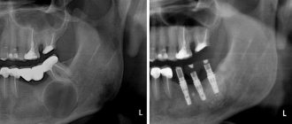
Unfortunately, most often the formation has to be removed surgically. The doctor performs a resection of the apex of the tooth root and at the same time fills the cavity cleared of exudate with a special biological composite composition.
What treatment methods for jaw cysts exist?
The choice of treatment method for an odontogenic cyst directly depends on the symptoms it causes, as well as the results obtained during instrumental and laboratory diagnostics.
If surgical treatment is chosen (cystotomy or cystectomy), the maxillofacial surgeon performs complete removal of the cyst. In some cases, it is necessary to remove the cyst along with the affected parts of the tooth root. Treatment is carried out in a hospital setting.
If the choice falls on therapeutic treatment, the doctor will carry out procedures aimed at reducing inflammation. This is a long process, taking at least six months.
The first step is to drain the contents of the cyst using a special drainage tube, which is inserted into a small incision in the tumor. As the contents drain out and the tumor shrinks, the size of the tube is adjusted downward.
After removing the contents of the cyst, the dentist cleans the root canals and administers medications that destroy the tumor tissue. At the end of all procedures, the doctor uses a special solution aimed at accelerating healing.
Treatment is monitored radiographically.
Both after surgical and therapeutic treatment, the patient requires preventive measures that will help avoid the re-formation of an odontogenic cyst.
Classification of pathology
Cysts localized in the tissues of the upper or lower row of teeth are very common. With their development, a cavity appears, the walls of which are covered with fibrous tissue, and the inner surface consists of an epithelial layer. The capsule holds clear or cloudy liquid.
Doctors distinguish the following types of pathology:
- Primordial. This is a cyst of the lower jaw. It appears in the area of the third molar. She has very thin fibrous walls. The inside of the capsule is lined with flat epithelium. According to its structure, the tumor may contain one or several small chambers.
- Follicular. Formed before tooth eruption. It can grow in the area of the alveolar margin. Includes cells that intensively produce viscous mucus. Because of this, the internal contents of the anomalous structure are quite viscous. A follicular formation is formed from the enamel organs of the unerupted unit. One or several teeth are found inside it. These may be formed crowns or tooth buds.
- Radicular. It occurs more often than others - in eighty percent of cases. Usually grows above the upper teeth. The diameter reaches from half to two centimeters. Often inflamed. Then the cells begin to hyperplasia, network-like processes are formed that extend into the thickness of the walls. The liquid contents of the radicular structures are rich in neutrophilic leukocytes. Often with this type of pathology, the patient develops sinusitis. This is due to the peculiarities of the localization of the inflammatory site.
- Retromolar. It is formed due to a long-term inflammatory process occurring in the thickness of the soft tissues. It is a consequence of complicated teething.
- Nasoalveolar. It occurs near the nasopalatine canal (the border between the upper row and the wing of the nose).
- Aneurysmal. Very rare in dental practice. Appears only on the lower jaw. This type of neoplasm is still poorly studied. It is known that there is blood or a red-pink liquid inside it. Many scientists agree that the symptoms of an aneurysmal cavity are a consequence of hormonal imbalance.
- Traumatic. Occurs due to recent facial trauma.
- Residual. The result of mistakes made by the doctor during tooth extraction, or the consequence of the patient ignoring the surgeon’s instructions.
The doctor decides how to treat a dental cyst, taking into account the cause of its development. It is very important to understand why the tumor occurred. If the root cause is not identified, the likelihood of relapse will remain high.
What is a cyst? Jaw cyst
A cyst is a cavity that is lined with epithelium and filled with fluid or soft material.
The formation of teeth (odontogenesis) is a complex process in which connective tissues and epithelium (enamel organ, dental follicle and dental papilla) participate.
The enamel organ refers to an epithelial structure derived from the oral ectoderm. The dental follicle and dental papilla are ectomesenchymal structures, because they are partly derived from neural crest cells.
For each tooth, odontogenesis begins with the apical (affecting the tip of the tooth root) proliferation of the epithelium of the oral mucosa, known as the dental lamina. The dental lamina gives rise to the enamel organ, a cap-shaped structure that subsequently takes on the shape of a bell. After the formation of the enamel organ, the dental lamina cord usually fragments and degenerates. However, small islands of dental lamina may remain after tooth formation. They are believed to be responsible for the development of some odontogenic cysts and tumors.
The enamel organ has four types of epithelium. The inner lining of the enamel organ is called the inner enamel epithelium and becomes the ameloblastic layer that forms tooth enamel. The second layer of cells adjacent to the inner enamel epithelium is the intermediate layer. Adjacent to this layer is the stellate reticulum, followed by the outer enamel epithelium. The enamel organ is surrounded by loose connective tissue known as the dental papilla. Contact with the epithelium of the enamel organ causes the dental papilla to produce odontoblasts, which form dentin. As odontoblasts lay down dentin, they induce ameloblasts to form enamel.
After initial crown formation, a thin layer of enamel organ epithelium, known as Hertwig's root sheath, grows at the apex of the tooth root. This epithelial expansion later becomes fragmented but leaves behind small nests of epithelial cells known as Malassez remnants in the space of the periodontal ligament. They are the source of epithelium for most periapical (radicular) cysts, but do not cause any odontogenic neoplasms, with the exception of squamous odontogenic tumor.
Causes of the disease
Already based on the classification, it becomes clear that tumors appear in the tissues of the oral cavity for a variety of reasons. Among the main provoking factors:
- Damage to hard dental tissues. Untreated caries, periodontitis, pulpitis are all diseases in which pathogenic agents penetrate the periodontium through unsealed root canals.
- Inflammation of the area adjacent to the area where the cyst is located. Quite often it turns out that a benign tumor formed after sinusitis or inflammation of the gums. This happens because the infection penetrates through the bloodstream into the bone tissue from the maxillary and nasal sinuses.
- Injuries, bruises, blows. The first time after them, a person may not notice anything. But later a nagging pain appears, indicating a problem.
Very rarely do doctors have to treat congenital cysts. It has not been established why they occur.
Causes of radicular cysts
The development of a radicular cyst in most cases is caused by an infectious-inflammatory process. The body forms a cystic formation to isolate the source of infection. The inflammatory process begins due to:
- chronic untreated caries;
- pulpitis;
- granulomatous periodontitis (over time, granulomas transform into cysts);
- tooth or jaw injuries;
- previous infectious disease (angina, otitis, sinusitis);
- reduced immunity;
- malocclusion;
- complications during the eruption of wisdom teeth.
Symptoms of the disease
In the first days of development of the pathology, there are no subjective symptoms. But the larger the cyst becomes, the stronger and more vividly it declares itself. Then a kind of gumboil may form in the gum area. At first, the protrusion does not hurt during palpation and hardly bothers the patient.
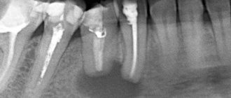
On an orthopantomogram, the doctor observes a spherical cavity with clearly defined contours. On an x-ray, you can see the root of the affected tooth with inflamed periodontium.
In advanced cases, when a large amount of purulent mass accumulates inside the cavity, the patient experiences the same symptoms as with osteomyelitis. Then the tumor volume increases. The gums swell and become very painful. Periodically, a small hole appears in its wall through which pus is released.
What is the difference between a cyst and a granuloma?
A dental cyst is often confused with a granuloma, another dangerous disease in which a round-shaped formation forms in the root area. It is important to distinguish between them, since the diagnosis directly affects the treatment plan and its prognosis. For granuloma, in most cases therapeutic treatment is sufficient. But a cyst requires more radical measures.
Here are the main parameters by which a dental cyst and a dental granuloma differ:
| Difference options | Cyst | Granuloma |
| Size | 0.9 - 3 cm | 0.5 - 0.8 cm |
| Structure | Cavity with fluid or pus | Solid formation covered with connective tissue |
| Clinical picture | The tooth becomes mobile | No mobility observed |
| X-ray result | The image shows a round capsule with clear boundaries | There are no strict outlines of education |
| Condition of periodontal tissues | The mucous membrane is usually not inflamed. In this case, bone loss occurs. | There is severe swelling and redness of the mucous membrane. But bone tissue does not decrease so much that it can be detected clinically. |
How to treat
In most cases, cysts cannot be treated without surgery. Only in unadvanced cases is it possible to get rid of the tumor by opening it and draining it.
The operations performed for the disease are called cystectomy and cystotomy. They both involve removing the formation. But in the first case, it is removed completely, and in the second, only the front wall is excised.
The main goal of a dental surgeon is to preserve the tooth growing in the area where the tumor is located. Unfortunately, this is not always possible. So, if the tooth root is immersed in the cyst cavity by more than 60% of its length, emergency removal is indicated. Such a unit can no longer be functional. Soon after excision of the tumor, it will still become loose and fall out.
Therefore, it is very important not to start the disease. Considering that it may not manifest itself for a long time, it is important to undergo a specialized examination at the dental office every year. This will increase the chances of successful early diagnosis.
How are odontogenic cysts diagnosed?
The leading method for identifying odontogenic cysts is radiography, which is capable of visualizing jaw cysts at an early stage of their development. On an x-ray image, the cyst is distinguished by the presence of clear boundaries, and the formation itself gives a characteristic shadow of a round or oval shape, immersed in the sinus of the tooth root.
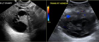
Ultrasound examination also helps to recognize odontogenic cysts.
As already mentioned, pronounced symptoms are characteristic of the late stage of development of a pathological formation, therefore it is difficult to diagnose a cyst at the initial stage, relying only on symptoms.
The final diagnosis is made on the basis of histological examination. It is important to differentiate an odontogenic cyst from other pathologies (adenomatoid odontogenic tumor, ameloblastic fibroodontoma and calcifying epithelial odontogenic tumor).
The CT scan method is widely used in the diagnosis of jaw cysts to confirm the presence of calcifications along the wall of the cyst, as well as tiny spots that are usually not found on x-rays. In addition, computed tomography is necessary during the surgical planning stage.
What will happen if left untreated?
Despite the fact that most patients do not have acute symptoms with the diagnosis described, the growth of the cavity can lead to very unpleasant and even dangerous consequences for health. This means:
- sepsis;
- phlegmon;
- premature loosening and loss of one or more teeth;
- inflammatory damage to the tissues of the periosteum;
- the formation of a malignant tumor due to cell degeneration;
- osteomyelitis;
- frequent occurrence of fistulas;
- high risk of fracture.
All these consequences are not harmless, which is why it is so important to detect pathology at the very beginning of its development.
Treatment
Treatment of radicular cysts is exclusively surgical. Surgical intervention tactics are chosen depending on the stage and characteristics of the disease.
The first technique is cystotomy. Such treatment is advisable in cases where the pathological focus is of impressive size, or there is destruction of the maxillary sinus. In the projection of the cyst, an incision is made in the oral mucosa and periosteum, and a hole is made in the bone. This allows access to the cavity. The entire contents of the cyst are removed, antiseptic treatment is performed, and a tampon soaked in iodoform is placed. The success of the operation depends on the quality of the revision of the cystic cavity. If the contents and necrotic tissue have been completely removed, no relapse is observed. The disadvantages of this treatment include a long rehabilitation period after surgery. The tampon installed during the operation must be changed to a new one after a week, and you will have to regularly visit the dentist for dressing changes.
The second tactic that is successfully used in the treatment of radicular cysts is cystectomy. If the cystotomy does not affect the membrane, with this technique the formation is radically removed along with the capsule. This tactic is used to treat small cysts with minor destruction of bone tissue. In the postoperative period, it is necessary to rinse the mouth with antiseptic solutions and preventive visits to the dentist to monitor the condition.
In some cases, surgical treatment of a radicular cyst combines both techniques. A competent dental surgeon, after examining a patient, will always choose the optimal treatment tactics for a particular patient.
During the operation, it is important to follow the rules of asepsis and antisepsis. This reduces the risk of postoperative complications. The purpose of surgery is to remove the source of infection from the body. The patient’s further condition depends on how thoroughly the pathological contents of the cyst are removed. The dentist takes these nuances into account and treats the radicular cyst, which does not give the disease any chance of relapse.
What can you do to avoid getting sick?
You can reduce the likelihood of developing the disease by following simple rules:
- Brush your teeth twice a day. Use a high-quality brush and paste during hygiene procedures. Don't forget about the role of dental rinses and dental floss.
- Once a year, remove tartar in the dental office using an ultrasonic scaler. Professional hygiene is the best prevention of most dental ailments.
- Eat a balanced and healthy diet. Eat less sweets. Eat plenty of fresh fruits and vegetables.
- Check your dental health once a year. You need to visit a doctor, even if nothing bothers you.
- Always follow your dentist's instructions.
- Avoid traumatic injuries to the face. When engaging in potentially dangerous sports, always wear special protective equipment.
A jaw cyst is a dangerous tumor. Initially it is benign, but under certain conditions it can become malignant. Fortunately, this happens extremely rarely.
If the tumor grows very quickly, even a jaw fracture is possible. It shouldn't come to this. Treatment must be timely and competent.
Rehabilitation period and prevention of dental cysts
Due to the fact that the cyst is an inflammatory infectious process, drug therapy necessarily includes taking antibiotics, which are selected by the attending physician. Vitamin complexes and immunomodulators act as compensating medications. Treatment of dental cysts with medications is carried out simultaneously with removal of the cyst.
The main ways to avoid the disease are to regularly visit the dentist and carefully monitor oral hygiene. For preventive purposes, you can periodically rinse with infusions and decoctions - for example, aloe, calendula, sage. Combined with strict adherence to the dentist’s recommendations and timely relief from chronic nasopharyngeal diseases, this will ensure dental health for years to come.
You also need to clearly understand that a cyst on the root of a tooth is one of the serious and complex diseases, the treatment of which requires time and highly qualified surgeons. Only this guarantees the absence of complications: re-infection, the occurrence of an abscess, pulpitis or fistula, damage to adjacent tissues and teeth.