A dental cyst is a benign neoplasm that is located inside the bone tissue of the jaw, in the upper part of the tooth root. A cyst is formed by isolating pathological cells from healthy ones by a dense membrane. It protects the roots of adjacent teeth and the body from further spread of inflammation.
The diameter of the cyst is from 0.5 to 3 cm. A dental cyst is dangerous because it develops into a malignant tumor. It also provokes various consequences in the form of:
- damage to neighboring teeth;
- tooth loss;
- destruction of the bone tissue of the jaw.
Content:
- Classification of pathology
- Causes of the disease
- Symptoms of the disease
- Diagnostic measures
- How to treat
- What will happen if left untreated?
- What can you do to avoid getting sick?
A jaw cyst is a hollow benign formation localized in the jaw bone.
There is liquid content inside it. Very often, the structure develops unnoticed by a person for a long time and is accidentally discovered during an X-ray diagnosis for another reason. If a cyst in the jaw becomes inflamed, the patient immediately feels it. Suppuration can provoke the occurrence of periostitis, sinusitis, osteomyelitis, and fistula.
Unfortunately, most often the formation has to be removed surgically. The doctor performs a resection of the apex of the tooth root and at the same time fills the cavity cleared of exudate with a special biological composite composition.
Etiology and provoking factors
The appearance of a cystic component is the body’s response to the inflammatory process in the tooth. Cysts form in patients with a long course of carious destruction, when the lesion reaches the root part of the tooth. But there are other diseases or conditions that can serve as a trigger for the formation of cavity formations:
chronic periodontitis; severe pulpitis; trauma to the tooth and gums: previous illness of an infectious nature; chronic stomatitis; inflammation of the maxillary sinuses; abnormal bite; catarrhal otitis; diseases with a pronounced decrease in immunity; complication during the eruption of wisdom teeth.
A radicular cyst of the maxillary bone is most often formed under the influence of a long-term untreated carious tooth. Sometimes the cause of the appearance of a radicular cyst in the upper jaw is unprofessional treatment of caries, violation of canal filling technology, or lack of competence of a specialist in the treatment of chronic forms of periodontitis. Lack of treatment for a radicular cyst can lead to serious complications not only from the maxillofacial anatomical zone, but also from all organs and systems of the body as a whole.
Classification of pathology
Cysts localized in the tissues of the upper or lower row of teeth are very common. With their development, a cavity appears, the walls of which are covered with fibrous tissue, and the inner surface consists of an epithelial layer. The capsule holds clear or cloudy liquid.
Doctors distinguish the following types of pathology:
- Primordial. This is a cyst of the lower jaw. It appears in the area of the third molar. She has very thin fibrous walls. The inside of the capsule is lined with flat epithelium. According to its structure, the tumor may contain one or several small chambers.
- Follicular. Formed before tooth eruption. It can grow in the area of the alveolar margin. Includes cells that intensively produce viscous mucus. Because of this, the internal contents of the anomalous structure are quite viscous. A follicular formation is formed from the enamel organs of the unerupted unit. One or several teeth are found inside it. These may be formed crowns or tooth buds.
- Radicular. It occurs more often than others - in eighty percent of cases. Usually grows above the upper teeth. The diameter reaches from half to two centimeters. Often inflamed. Then the cells begin to hyperplasia, network-like processes are formed that extend into the thickness of the walls. The liquid contents of the radicular structures are rich in neutrophilic leukocytes. Often with this type of pathology, the patient develops sinusitis. This is due to the peculiarities of the localization of the inflammatory site.
- Retromolar. It is formed due to a long-term inflammatory process occurring in the thickness of the soft tissues. It is a consequence of complicated teething.
- Nasoalveolar. It occurs near the nasopalatine canal (the border between the upper row and the wing of the nose).
- Aneurysmal. Very rare in dental practice. Appears only on the lower jaw. This type of neoplasm is still poorly studied. It is known that there is blood or a red-pink liquid inside it. Many scientists agree that the symptoms of an aneurysmal cavity are a consequence of hormonal imbalance.
- Traumatic. Occurs due to recent facial trauma.
- Residual. The result of mistakes made by the doctor during tooth extraction, or the consequence of the patient ignoring the surgeon’s instructions.
The doctor decides how to treat a dental cyst, taking into account the cause of its development. It is very important to understand why the tumor occurred. If the root cause is not identified, the likelihood of relapse will remain high.
Causes of the disease
Already based on the classification, it becomes clear that tumors appear in the tissues of the oral cavity for a variety of reasons. Among the main provoking factors:
- Damage to hard dental tissues. Untreated caries, periodontitis, pulpitis are all diseases in which pathogenic agents penetrate the periodontium through unsealed root canals.
- Inflammation of the area adjacent to the area where the cyst is located. Quite often it turns out that a benign tumor formed after sinusitis or inflammation of the gums. This happens because the infection penetrates through the bloodstream into the bone tissue from the maxillary and nasal sinuses.
- Injuries, bruises, blows. The first time after them, a person may not notice anything. But later a nagging pain appears, indicating a problem.
Very rarely do doctors have to treat congenital cysts. It has not been established why they occur.
Clinical picture
The development of the cystic component can develop for about several years and does not manifest itself symptomatically. If unexpressed but unpleasant signs of a cyst appear, treatment should be carried out immediately, since treatment of the tumor after its significant growth will be quite difficult. As the cyst grows, it puts strong pressure on the surrounding connective tissue, so its growth is rapid. In the photographs, the cyst looks like a small spherical formation. The main manifestations are:
the appearance of severe throbbing pain; swelling and redness of the gums in the affected area; feeling of tooth mobility; feeling of fullness in the apical space; bad breath: prolonged persistence of low-grade body temperature.
An early cyst can be diagnosed by X-ray examination, and later stages of development are detected by visual examination of the patient’s oral cavity. Usually the suspicion of the presence of a cystic cavity is justified.
Symptoms of the disease
In the first days of development of the pathology, there are no subjective symptoms. But the larger the cyst becomes, the stronger and more vividly it declares itself. Then a kind of gumboil may form in the gum area. At first, the protrusion does not hurt during palpation and hardly bothers the patient.
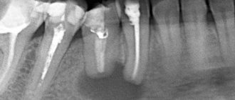
On an orthopantomogram, the doctor observes a spherical cavity with clearly defined contours. On an x-ray, you can see the root of the affected tooth with inflamed periodontium.
In advanced cases, when a large amount of purulent mass accumulates inside the cavity, the patient experiences the same symptoms as with osteomyelitis. Then the tumor volume increases. The gums swell and become very painful. Periodically, a small hole appears in its wall through which pus is released.
Diagnostics
- Taking an anamnesis
- the dentist interviews the patient and conducts an examination. - X-ray diagnostics
- to clarify the diagnosis, determine the size of the formation, the stage of development, an orthopantomogram and 3D tomography are prescribed.
The granuloma in the picture looks like a dark spot that does not have clear contours. A cystic capsule with clear boundaries is located at the apex of the root (surrounds it). The program uses 3D images to determine the condition of the dental system, helping the doctor plan treatment. Before the operation, the patient undergoes a standard set of blood tests (coagulability, group, Rh factor, glucose, infections).
How to treat
In most cases, cysts cannot be treated without surgery. Only in unadvanced cases is it possible to get rid of the tumor by opening it and draining it.
The operations performed for the disease are called cystectomy and cystotomy. They both involve removing the formation. But in the first case, it is removed completely, and in the second, only the front wall is excised.
The main goal of a dental surgeon is to preserve the tooth growing in the area where the tumor is located. Unfortunately, this is not always possible. So, if the tooth root is immersed in the cyst cavity by more than 60% of its length, emergency removal is indicated. Such a unit can no longer be functional. Soon after excision of the tumor, it will still become loose and fall out.
Therefore, it is very important not to start the disease. Considering that it may not manifest itself for a long time, it is important to undergo a specialized examination at the dental office every year. This will increase the chances of successful early diagnosis.
Treatment methods for dental cysts
Treatment of dental cysts is carried out using two methods:
- therapeutic;
- surgical.
If the cyst is small and is not accompanied by serious symptoms such as purulent abscesses and swelling, conservative treatment is possible. In this case, the cyst in diameter should not exceed 8 mm.
Treatment is carried out through the canals of the tooth. After opening them, an antiseptic treatment is performed and a specialist injects a copper-calcium suspension through the canal. Next, the tooth is filled with temporary materials. After a time determined by the dentist, a repeat x-ray is taken to see progress in treatment or lack thereof.
A copper-calcium suspension allows you to restore destroyed bone-jaw tissue.
Additionally, at the discretion of the specialist and in case of severe inflammation, antibiotic treatment may be prescribed. For this disease, this is an auxiliary measure, and in no case is the main treatment.
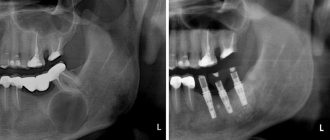
If there is no progress or if the cyst is large, treatment is carried out through surgery. The operation is performed by a maxillofacial surgeon under local anesthesia. During surgery, the top of the tooth root is cut off, after which the cyst is completely removed.
In some cases, only the anterior wall of the cyst shell may be removed. The recovery period after such an operation is longer.
Among the modern methods of treating cysts are operations using lasers. In this case there is practically no pain. In addition to removing the tumor, during the manipulations the tooth canals and the affected area are disinfected. Wounds with this method heal quickly, complications are extremely rare.
When treating a cyst, specialists try to preserve the tooth. After the suture has healed, the temporary cement filling is removed from the canals, and they are filled with permanent materials. If this is not possible, the tooth itself is removed after surgery.
What will happen if left untreated?
Despite the fact that most patients do not have acute symptoms with the diagnosis described, the growth of the cavity can lead to very unpleasant and even dangerous consequences for health. This means:
- sepsis;
- phlegmon;
- premature loosening and loss of one or more teeth;
- inflammatory damage to the tissues of the periosteum;
- the formation of a malignant tumor due to cell degeneration;
- osteomyelitis;
- frequent occurrence of fistulas;
- high risk of fracture.
All these consequences are not harmless, which is why it is so important to detect pathology at the very beginning of its development.
What can you do to avoid getting sick?
You can reduce the likelihood of developing the disease by following simple rules:
- Brush your teeth twice a day. Use a high-quality brush and paste during hygiene procedures. Don't forget about the role of dental rinses and dental floss.
- Once a year, remove tartar in the dental office using an ultrasonic scaler. Professional hygiene is the best prevention of most dental ailments.
- Eat a balanced and healthy diet. Eat less sweets. Eat plenty of fresh fruits and vegetables.
- Check your dental health once a year. You need to visit a doctor, even if nothing bothers you.
- Always follow your dentist's instructions.
- Avoid traumatic injuries to the face. When engaging in potentially dangerous sports, always wear special protective equipment.
A jaw cyst is a dangerous tumor. Initially it is benign, but under certain conditions it can become malignant. Fortunately, this happens extremely rarely.
If the tumor grows very quickly, even a jaw fracture is possible. It shouldn't come to this. Treatment must be timely and competent.
Prevention
- undergo regular dental examinations;
- undergo diagnostics if a cyst is suspected;
- treat teeth and gums in a timely manner, preventing inflammation from spreading to the dental canals;
- monitor oral hygiene;
- keep chronic diseases (especially nasopharynx) under control;
- do not self-medicate.
Following simple recommendations will minimize the risk of disease, help prevent complications, and provide high-quality therapy without resorting to surgery.