From this article you will learn:
- what is a dental cyst - symptoms, photos,
- why does it form?
- cyst on the root of a tooth - treatment.
The article was written by a dentist with more than 19 years of experience.
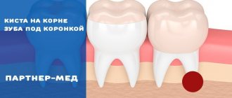
A dental cyst is a round-shaped inflammatory formation, which is a cavity in the bone tissue, which is lined from the inside with a fibrous capsule and filled with purulent contents (Fig. 1). In dentistry, the terms “root cyst of the tooth” or “radicular cyst” (from the word radix - root) are most often used to refer to this pathology. This is due to the fact that the most common place for its formation is the area of the apex of the tooth root.
The formation of a cyst on the root of a tooth is associated with an infection in the root canals. Most often it appears - 1) either in the absence of timely treatment of pulpitis or periodontitis of the tooth, 2) or is a consequence of poor-quality root canal filling. The formation of a cyst occurs gradually and over a long period of time. First, at the apex of the root, under the influence of infection in the root canals, a small apical granuloma is formed, which subsequently increases in size and transforms into a cyst filled with pus.
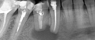
What does a dental cyst look like: photo
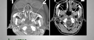
Pay attention to how a cyst on the root of a tooth looks on an x-ray - it looks like intense darkening at the apex of the tooth (for your convenience, we have limited this area with arrows). Such intense darkening at the apex of the tooth root indicates the presence in the bone tissue of a site of bone destruction in the form of a cavity. In Fig. 3 you can see what a cyst looks like at the apex of the root of an extracted tooth (like a sac filled with pus).
This article is written for patients and therefore will contain unnecessary medical details. But to complete the picture, it is worth adding that a dental cyst can be localized not only at the apex of the tooth root, but also on the lateral surface of the root, as well as between the roots of multi-rooted teeth - in the root bifurcation zone. But such localizations of root cysts are quite rare, and therefore we will examine in more detail the symptoms and treatment of a dental cyst localized at the apex of the root.
Type of cyst on the root of an extracted tooth (video 1) –
Laser treatment of cysts is the best therapeutic method of treatment
Treatment of dental cysts is possible! The specialists of the Bionic Dentis clinic prove this every day.
Dental clinic "Bionic Dentis" is the No. 1 clinic in Russia and the CIS countries for the treatment of dental cysts with a laser . We have the most experience in treating this disease and annually admit more than 1000 patients in this area of activity. The clinic's doctors are recognized in the professional community as experts in laser cyst treatment, conducting educational courses and training specialists in this technique.
Specializing in laser treatment, our dental center presents in the Russian Federation an exclusive technique based on the use of the latest generation dental laser SiroLaser Sirona SIEMENS (Germany).
Russia leads in the number of patients with dental cysts, because the level of root canal treatment in dental clinics in our country is prohibitively low. A public dentist, even acting according to the instructions of the Ministry of Health, works according to an outdated technique, which does not give a positive result in the long term and leads to the development of a dental cyst.
User Questions
QUESTION My dentist found a cyst in my front tooth under the crown. She says that while she is small, she can be treated with antibiotics. But he said it was better to remove the crown. Why can't you leave her? Alexei
ANSWER Hello, Alexey.
It is likely that your doctor wants to reduce the chewing load on the tooth. To prevent you from accidentally biting something hard or pushing the infection deeper, it is better to remove the tooth from the bite. Another option is to remove the crown. Don't worry, after treatment it will be put back in place. During the treatment process, the tooth will be covered with a temporary filling. 1Epifanov E.A. Medical rehabilitation for diseases and injuries of the maxillofacial area, 2022.
Methods for treating dental cysts
All methods of treating dental cysts can be divided into surgical and therapeutic.
Surgical methods for treating cysts include:
- Cystotomy.
- Cystectomy.
- Resection of the apex of the tooth root.
All these methods are associated with surgical intervention (operation) and require long periods of rehabilitation and healing, and are always accompanied by pain in the postoperative period and massive swelling.
Laser treatment of dental cysts was developed in Germany more than 10 years ago. Over the years, this technique has received worldwide recognition from dentists and patients. The high effectiveness of this treatment method has been proven by more than 900 clinical studies.
But the main problem with surgical treatment methods is that their use has a very high percentage of relapses. Approximately 45% of cases of recurrence of dental cysts.
What is the difference between a cyst and a granuloma?
There are other diseases in the oral cavity that are symptomatically similar to a cyst. Specifically, a granuloma is an outwardly swollen round projection in the root zone of a tooth. The doctor must correctly diagnose the disease, since the sequence of exposure, removal techniques, and pharmacology have significant differences. Granuloma is eliminated with the help of medications, as well as accompanying mouth rinsing with phytocomponents. The cyst has deeper roots and does not go away without surgery.
Unique. 100% channel processing. Laser technique
Let's look at what symptoms indicate that you have a dental cyst:
- Abscess on the gum.
- Flux on the gum.
- It feels like the tooth has grown and is taller than the rest.
- Pain from chewing hard food.
- Fistula on the gum.
These signs are a suppurating tooth cyst. If the described symptoms appear, you should urgently consult a dentist; the fact is that pus can quickly spread through the channels in the bone and reach such vital organs as the eye and ear, which leads to dire consequences.
However, a dental cyst would not be such an insidious disease if it immediately gave clear symptoms. The fact is that in 90 percent of cases, the cyst is completely asymptomatic. The patient does not even suspect that a dangerous disease is maturing in his jaw bone. Often, the patient undergoes a routine examination - a panoramic photograph, which reveals a dental cyst. Let us note right away: even if the cyst does not make itself felt and does not hurt, it must be treated urgently. If left untreated, it leads to serious complications:
- Suppuration of the cyst.
- Compression of nerves, which leads to paresthesia (numbness) of part of the face.
- Destruction of the jaw, up to a fracture of the lower jaw. If the cyst reaches a significant size, the bone becomes brittle and brittle. Even chewing hard food can cause a broken jaw.
- Cysts of the upper jaw in 99% of cases lead to sinusitis. This is a serious and unpleasant disease of the paranasal sinuses that is accompanied by unbearable headaches and nasal congestion.
X-ray diagnostics –
As we said above, the final diagnosis can only be made using an x-ray, on which a cavity formation in bone tissue will always look like a rounded darkening.
In the x-ray images below we have presented you with 3 options for localizing root cysts. The most common location is root cysts at the apex of the tooth root, but cysts on the lateral surface of the root or located between the roots of multi-rooted teeth are much less common. Radiographs of root cysts of different locations –
More details about the technique:
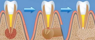
The dentist drills a small hole in the tooth on which the cyst is located. On the front teeth, this hole is located behind the tooth, on the lateral teeth, on top.
The doctor’s goal is to get to the root canals of the tooth, which are located in its center (the pulp of the tooth).
Once access to the nerve is achieved, the dentist removes the contents of the root canal, whether it is an inflamed tooth nerve or an infected tooth canal filling material after poor treatment.
Root canal treatment is not performed blindly. In our clinic, endodontists use a special device: a computerized root canal cleaning system from VDW GOLG (Germany).
Special titanium instruments - MTWO files - with progressive movements under the control of a microprocessor, millimeter by millimeter, clear the root canal, making it smoother, cleaner and straighter. The use of machine computer technology for canal processing in our clinic has a number of advantages:
- reducing the risk of tool breakage and jamming in the tooth canal.
- improving the quality of tooth canal treatment
- increasing the channel processing speed by more than 10 times compared to the manual method.
- eliminates the risk of uncontrolled penetration beyond the apex of the root canal and injury to periapical tissues.
Constant monitoring of canal clearing is carried out by a VDW apex locator (Germany) and the main stages of treatment are controlled using a radiovisiograph XIOS Sirona (Germany), which is installed in each office and allows you to do research without interrupting treatment.
A feature of the diagnostic equipment we use is minimal X-ray exposure to the patient and the resulting high-definition image.
Sometimes it takes 1 - 1.5 hours to clean and unfill the root canal.
After mechanical cleaning and expansion of the canal, they begin its ultrasonic treatment. Scientific research in recent years has proven that microscopic branches extend from a large root canal, which are the main reservoir for bacteria that cause dental cysts. It is not possible to clear these microtubules with files, but our clinic is equipped with a SiroSonic Sirona (Germany) ultrasonic root canal treatment system. This development by the German company SIEMENS ensures the penetration of a disinfectant solution into microscopic tubules and pores, killing microbes there and chemically sterilizing the root canal.
After sterilizing the root canal with chemicals and ultrasound, it is the turn of the SiroLaser laser dental unit. The dentist penetrates the cyst through the root canal and treats it with a laser. Thanks to the unique flexible light guides of our laser, the dentist can penetrate a dental cyst even through a curved canal. This innovation has made the SiroLaser the best in its class.
After laser treatment, the dentist performs laser sterilization of the root canals of the tooth and the cavity in the bone. This allows for 100 percent disinfection of infected tooth tissues, which eliminates the recurrence of a cyst in a given tooth.
After laser sterilization, the drug is installed. This long-acting substance stimulates bone tissue regeneration and tissue disinfection for a period of 4-6 months.
A lighted, especially strong temporary filling or temporary crown is installed on the tooth.
Treatment of dental cysts in 1 visit - we guarantee our patients the use of the world's best achievements in dental treatment.
Unlike the ineffective classical method of treating dental cysts by placing calcium in the root canals, when the patient needs to visit the dental clinic every week for six months, laser treatment of cysts at the Bionic Dentis clinic is carried out in one visit!
After 4-6 months, a radiovisiographic study is performed to confirm tissue regeneration and the healing process in the bone.
The second stage of dental treatment is no less important than laser treatment of a dental cyst.
Causes of the disease
Why did a cyst form under the crown? The main reason is a long-term inflammatory process at the roots (periodontitis). If pulpitis, and sometimes even caries and gingivitis, are not treated, then the microbes penetrate deeper into healthy tissue and “get” to the edge of the root - the apex or apex. In the case of pulpitis, the infection from the inside travels along the dental nerve and exits through the apex, infecting the surrounding tissues. In the case of gingivitis, the infection spreads from the outside through the edge of the gum and deep into the ligamentous apparatus - this is the periodontium, which, as it were, “ties” the roots to the bone socket of the jaw. The causes of the inflammatory process may lie in the following:
- incomplete preparation before prosthetics: the tooth could initially already have periodontitis, a cyst, but the dentist did not detect this - if he did not take an x-ray or the picture turned out to be unclear,
- poorly processed and sealed root canals: there are voids and microbes left in them that begin to penetrate into the periodontium,
- if during the passage of the canals the doctor screwed the instrument too deeply: the tip of the file went into the periodontium and brought with it microbes,
- a fragment of the instrument remained in the root canal,
- too much filling compound: the excess is “pressed” into the periodontium,
- inflammation under a crown with a pin or with a stump inlay: a tooth cyst can appear both due to microbes on the pin/inlay, and from displacement, breakage, or too much length of the internal structure under the crown. And also due to the low quality of the filling material under and around the pin,
- cyst at the site of an extracted tooth under a bridge (bridge): may develop due to infection during or after extraction, or if a bone fragment remains in the socket,
- poorly fitting prosthesis
- trauma: strong blow, bruise on the jaw area,
- poor oral hygiene,
- sinusitis, sinusitis: here the infection penetrates through the bloodstream to the roots and provokes inflammation,
- diseases on neighboring teeth,
- weak immunity, chronic diseases of the body.
3D filling of the root canal of a tooth after laser treatment of a cyst.
To complete the treatment of the tooth and prepare it for restoration, it is necessary to fill the root canals with a permanent material - alpha gutta-percha.
This is a biocompatible medicinal product that does not cause an allergic reaction, does not cause rejection and is ideal for obturation of the root canal.
The dentist re-treats the root canal with an antiseptic using an ultrasound machine. By doing this, he achieves the opening of the entrance to the micro tubules for the filling material.
An alpha gutta-percha pin is lubricated with a sealant and inserted into the root canal. After that, the gutta-percha is heated and pressed into the canal using the VDW BeeFile (Germany) three-dimensional root canal obturation system. Under the pressure of a tool - a plugger, molten liquid gutta-percha penetrates into micro tubules and instantly solidifies there, forming a three-dimensional network - a 3D system of a filled tooth canal.
This method of filling tooth canals is new for the Russian Federation, but this method is the gold standard for obturation of tooth canals in Europe and the USA. Our specialists use this method in 100% of treatment cases, which allows us to avoid recurrence of dental cysts .
As a result of this filling technique, even the most complex root canal systems will be 100% sealed.
There is a simplified analogue of filling canals with gutta-percha, using a standard gutta-percha pin. But it is not effective and cannot compare in terms of results with the original method.
After filling the root canals, a filling, inlay or crown is placed on the tooth.
Types of dental cysts
A molar cyst is distinguished by several characteristics:
- depending on the reasons for the appearance;
- according to location.
Distributing according to the reasons that provoke the appearance of a dental cyst, the following varieties are distinguished:
- A keratocyst appears on the lower jaw. Older people are susceptible to the disease. It can turn into a malignant form - it grows deeply into the body and destroys the bone.
- Odontogenic or radicular dental cyst is the most common type. After periapical inflammation and necrosis of the pulp, a granuloma of the dental root is formed in the upper part. The size of the purulent focus ranges from 3 mm to 3 cm. The process does not affect bone tissue and does not provoke tooth displacement.
- A cyst that forms at the site of teething is a retention cyst. It appears during the period of replacement of milk teeth with molars. A purulent fistula with bloody contents looks like a dark blue abscess. Education is formed if the process of eruption is slow. The purulent contents of the abscess should be opened and cleaned out.
- A calcifying odontogenic tumor occurs in the area of the support of the lower jaw. Experts do not have a consensus on the reasons for its formation - they have not yet been fully studied.
- A residual formation with pus occurs after tooth extraction as a result of careless work by the doctor. If a piece of root or a fragment of a tooth remains in the wound, a granuloma forms.
- A follicular cyst forms in place of soft tissues that resist the eruption of a new tooth. A cyst grows around a tooth hidden by the gum, enlarges and can spread to other teeth. The result of the development of a cyst can be tilting, tooth displacement or root resorption.
- A lateral periodontal cyst looks like a small formation, most often found on the lateral part of the root.
Depending on the location of the purulent tumor, there are:
- A cyst is formed on a tooth root. Three location options: basal, inter-root or peri-root zones. When a purulent capsule forms, the disease manifests itself well.
- The purulent focus is located in the maxillary sinus. Making a diagnosis is extremely complicated - you cannot do without an image of a certain area. In the advanced phase, the cyst provokes the appearance of sinusitis.
- At the initial stage, a cyst on the gum is asymptomatic. At the moment of filling with purulent contents, the bubble begins to grow rapidly. In treatment, medications are used, and they are also fought by puncturing the capsule, treating with saline solution and cleaning until complete healing.
- The formation of a cyst under the tooth crown occurs after unsuccessful disinfection during its installation after treatment. As a result of a loose fit, food debris gets under the crown, rots and creates a favorable environment for cyst growth. To treat it, you need to remove the crown.
- A cyst on the front teeth has practically no room for development and localization. It comes out at the initial stage of development.
- A wisdom tooth cyst forms at the site of a long-erupting “figure eight.” As a treatment for this category of teeth, a proven effective method is recommended - removal of the cyst and wisdom tooth together.
Laser treatment of dental cysts. Beware of fakes.
Recently, several clinics have appeared on the Moscow dental market that promise laser treatment for 3,000 - 5,000 rubles. These clinics, taking advantage of the patients’ ignorance, do not work according to the methodology and on equipment that is not intended for this method of treatment.
Be careful when choosing a clinic for this procedure.
In order for the patient to undergo the original treatment method, the following conditions must be met:
- The clinic must be equipped with the following equipment: computer system for root canal treatment VDW GOLD (Germany), dental laser SiroLaser Sirona, ultrasound dental system SiroSonic Sirona, device for 3D obturation of canals VDW BeeFile (Germany).
- Clinic doctors must be trained in laser technologies from SIRONA SIEMENS.
- Doctors must have a valid state certificate in the specialty: therapeutic dentistry.
Do not hesitate and ask the clinic administrator for all the necessary information regarding your treatment. This will give you the opportunity to choose only a certified clinic and receive a high-quality original method of treating dental cysts with a laser.
Every day we see patients with inflammatory diseases of the jaw bone tissue. We treat patients from Moscow, St. Petersburg and cities of Russia and CIS countries. Due to the fact that treatment takes place in 1 visit, patients from other cities can come to Moscow for treatment for just 1 day.
We have performed this cyst laser treatment on dozens of patients from Europe, since the technique we use is identical to that used in the EU , and the cost is much lower.
By choosing our clinic for cyst treatment, you receive highly technological treatment with a guaranteed result.
How do you know whether to treat or remove?
To make a decision on the method of treating the cyst and, in general, on the advisability of carrying out tooth-preserving measures, we first send the patient for a consultation, which is carried out by two specialists at once - an endodontist and a surgeon . After collecting all the necessary information and analyzing the images, a detailed clinical picture is drawn up and, together with the patient, the advisability of endodontic or surgical treatment is discussed.
When deciding on the “fate” of a tooth, a lot of factors are taken into account:
Education size
According to our treatment algorithm, it is first necessary to establish the exact dimensions of the area of inflammation at the root apex in order to make the correct diagnosis, calculate the estimate, number and sequence of procedures.
- The size of the formation is less than 5 mm - amenable to non-surgical therapeutic treatment.
- More than 5 mm in diameter - surgical removal of the cyst and correction of the root apex will most likely be required.
Root canal condition
Single-rooted teeth, easily passable canals, and undeveloped situations are treated cheaper and faster. Four-rooted teeth previously treated with the resorcinol-formalin method, perforations and cracks in the teeth that pose a threat to your health and budget are discussed in detail and individually before starting treatment.
Degree of coronal destruction
If a tooth is severely damaged, especially if the damage is below the gum level, it is worth thinking about the advisability of tooth-preserving measures. Even after ideal canal treatment, it will not be possible to recreate the shape of the tooth either with composite restoration or orthopedic structures. This means that all endodontic treatment will be reduced to zero, since there will be no reliable protection of the canals from above.
Localization of the cyst
The position of the cyst relative to arteries, veins, nerve fibers, as well as neighboring teeth is taken into account. It is not uncommon for detailed examination and treatment planning to reveal cysts growing into adjacent living teeth. In such cases, it is necessary to recheck the scope of work many times to prevent recurrence of cysts from neighboring areas. This is especially true for cases of follicular cysts (appearing from the growth zones of developing and growing “wisdom teeth”).
We
fight for every tooth - if there is even the slightest chance of saving it.
The specialists of our Center will be happy to save a tooth in any hopeless situation. We provide a lifetime guarantee for the treatment of cysts . However, sometimes the rescue budget is comparable to the implantation budget, which is achieved with much less effort and also with a lifetime guarantee. Let us make this important decision for you.
Levin Dmitry Valerievich Chief physician and founder of the Doctor Levin center
VIDEO: Method of treating a dental cyst with a SiroLaser SIEMENS laser (Germany) at the Bionic Dentis clinic
This video shows in detail all the stages of a dentist’s work when treating a cyst with a laser. The main points to pay attention to are: computerized root canal cleaning system, ultrasonic root canal cleaning, sterilization of the cyst and root canal with laser radiation.
Watch a video about laser cyst treatment in our clinic:
Consequences and complications of the disease
Why is a tooth cyst under a crown dangerous? If the pathology is left unattended, then eventually even a slowly developing neoplasm will provoke any of the following complications:
- spread of inflammation to the roots of neighboring teeth: they may change color, begin to hurt, become loose or fall out,
- tooth loss,
- spread of pus to the jaw and soft tissues: this can result in osteomyelitis, abscess, phlegmon, sinusitis, etc.,
- spread of infection throughout the body through the bloodstream: can cause damage to the brain, lungs, liver, kidneys and other organs,
- “degeneration” into a malignant neoplasm,
- sepsis.
Treatment of consequences and complications is much more difficult, longer and more expensive than treatment of a cyst. In many cases, the patient will additionally require rehabilitation1, and, for example, jaw bone augmentation after osteomyelitis. By the way, with this disease you can lose all your teeth in a row.
Complex on 4 OSSTEM implants with delayed loading - from RUB 170,000.
Complex implantation Osstem (South Korea) with delayed loading after 4-6 months.
Guarantee for the doctor’s work - unlimited Call now or order a call
Opening hours: 24 hours a day - seven days a week
Cost of treatment of a dental cyst with a SiroLaser SIRONA laser (Germany)
The cost of treating a dental cyst depends on the number of root canals in the tooth.
You can independently determine the number of canals in a tooth using this information:
1st canal in the teeth: Incisors, canines, premolars of the lower jaw, second premolar of the upper jaw.
2 canals in the teeth: First premolar of the upper jaw.
3 canals in teeth: Molars of the upper and lower jaws.
4 canals in teeth: First molars of the upper and lower jaw in 30% of cases.
Warning
: Invalid argument supplied for foreach() in
/var/www/u0695953/data/www/bionicdentis.ru/wp-content/themes/bionic/single-service.php
on line
59
| № | Procedure/manipulation | Price, rub.) |
Laser treatment of dental cysts was introduced in our clinic in 2007. During this time, doctors have accumulated vast experience in using this technique. We can safely say that the greatest experience in the Russian Federation and CIS countries is with the dentists and endodontists of our clinic.
The clinic has accumulated a huge collection of photographs and computed tomography scans of patients before and after treatment.
On this page you can familiarize yourself with the clinical cases of our real patients who underwent successful treatment.
Our statistics: 96% of cysts are successfully cured using the therapeutic method of laser treatment.
What are the stages of the operation?
The procedure consists of the following steps:
- The desired area is numbed using conduction or infiltration anesthesia.
- The gum is peeled off from the wall using a rasp.
- The tooth is loosened and removed using forceps or an elevator.
- The dental unit is removed from the socket. In difficult cases, the tooth is first sawed into pieces using a drill, and then each of them is removed separately.
- The extracted tooth and socket are examined by a doctor.
- The cyst cavity is cleaned, and the wound is treated with antiseptic drugs.
- The wound is stitched up.
- Using an X-ray examination, the doctor makes sure that fragments, particles of the tooth and the removed tumor do not remain in the jaw.
- Antibiotics, anti-inflammatory drugs and anesthetics are prescribed.
Two to three days after the operation, you need to re-visit your dentist to monitor the condition of the tooth socket.
Laser treatment of dental cysts at the premium clinic “Bionic Dentis”
We have created all the conditions for your comfortable treatment in our clinic.
Convenient location in the center of Moscow.
Personal approach to each patient.
Classic clinic interior.
The best European equipment.
Modern techniques and expert doctors.
Ozerov Petr Vladimirovich
Chief physician. Dentist, implantologist, orthopedist, surgeon. Laser dentistry specialist
More details
Werner Elena Vladimirovna
Dentist periodontist
More details
Eremina Anna Arturovna
Dentist therapist
More details
When is laser cyst removal not recommended?
Today, in all areas of medicine, laser treatment is considered the safest method. However, despite all its advantages, the action of laser beams can be dangerous for the body if the patient has contraindications.
The use of another method of removing a dental cyst is recommended for diseases or pathological conditions:
- diseases of the cardiovascular system;
- poor blood clotting rate;
- varicose veins of any form;
- immunodeficiency diseases (including diabetes mellitus);
- cancerous formations;
- postoperative period (in the treatment of any disease);
- individual negative reaction to the action of the laser.
During the procedure, only local anesthesia is used, so such operations are also contraindicated for patients with mental disorders. The doctor can consider each case separately, but to avoid complications, the dentist must be warned about all existing pathologies.
Clinical case of cyst treatment with laser. year 2014.
Patient Maria N, 36 years old, went to the dental clinic at her place of residence with complaints of gumboil, pain in the gums, a feeling of fullness, increased body temperature and general malaise. A panoramic photograph was examined at the patient’s clinic. Based on the results of the study, a diagnosis was made - a cyst of teeth 12 and 13. Dentists recommend that the patient remove both teeth and undergo a cystectomy.
The patient contacted several private dental centers, where she was also offered to remove teeth 13 and 12.
The patient did not agree with the proposed treatment and contacted the Bionic Dentis clinic for a consultation about the possibility of saving her teeth and treating the cyst on teeth 12 and 13 with a laser.
The clinic's dentist confirmed the possibility of treating the cyst with a laser.
The patient decided to undergo treatment at the Bionic Dentis clinic.
12 and 13 were treated using the SiroLaser SIRONA laser (Germany) according to the standard protocol.
Within 8 months, complete regeneration of bone tissue occurred and the cyst successfully disappeared.
3D root canal filling was performed. A restoration has been installed on the tooth - light filling FILTEC ULTIMATE
Types of neoplasms under the roots
In the dental classification, there are 3 types of cysts under the crown; let’s look at them in more detail:
- root or radicular: located in the apical part of the root. Appears due to infection during preparation before prosthetics or due to injury,
- primary or keratocyst: most often located near the lower molars - large chewing teeth. It does not appear in any way for a very long time; it is discovered by chance on an x-ray - in the corner of the jaw, on its body or branch. The bone tissue is greatly sparse,
- follicular or tooth-containing: develops from the tooth germ of a canine or wisdom tooth on the upper or lower jaw. Can cause ameloblastoma, a benign tumor
- residual: we can say that this is a secondary cyst, since it develops on the site of a previously poorly removed one,
- paradental or retromolar: most often located between the dystopic wisdom tooth and the roots of the adjacent one, on which the “figure eight” presses.
DENTAL PROSTHESIS ON 4 IMPLANTS - from RUR 170,000.
The price includes all procedures for installing Osstem implants (South Korea), including anesthesia and diagnostics.
Hurry up to sign up for a free consultation and fix your prices.
Call now or request a call
Opening hours: 24 hours a day - seven days a week
Results of cyst treatment with laser “BEFORE” and “AFTER”
We have collected a huge collection of dental x-rays and computed tomography scans showing the condition of the tooth with a cyst before treatment and after laser treatment.
Our experience in treating dental cysts with laser is the largest in Moscow.
You can be convinced that this technique can cure even teeth with large cysts.
All pictures are clickable.
Over 10 years of work, we have accumulated vast experience in treating dental cysts with laser.
During this time, the clinic's doctors became experts in the treatment of this disease.
We are proud that our dental colleagues from many Moscow and regional clinics trust us by referring their patients to us with complex cases, which we successfully handle!
By choosing the Bionic Dentis clinic to treat a dental cyst with a laser, you will save your tooth from removal. Cysts treated by us do not recur, and teeth last for many decades after treatment.
The relevance of traditional methods of treatment
You need to understand that the cyst is located deep under the crown - and none of the home or folk methods will help cope with it, but will only smooth out the manifestations a little. Those. the patient will feel a little better, but the pathology will not go away - it will continue to develop until complications begin. Therefore, the best option is to seek professional dental help. And the use, for example, of saline solution, tinctures of medicinal herbs, various natural oils, and alcohol tinctures is possible only with the permission of the dentist as an addition to the main treatment.
“The crown on the lower five began to wobble a little after 6 years of wearing it. I went to the dentist and thought about removing the tooth and placing an implant right away. But the doctor upset me that it was too early to think about it, because there was a large cyst under the root. I had to remove the tooth along with it anyway. Now I'm waiting for everything to heal. They said that it would take a few months for the bone to recover. In the meantime, I sometimes go for checkups so that there are no problems.”
Glorya_77, review from gidpozubam.ru