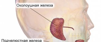
Salivary gland cysts
Symptoms of a salivary gland cyst
Depending on which gland it forms in, the symptoms of a salivary gland cyst can be distinguishable.
Small gland cyst
If a small gland has undergone a neoplasm, then the cyst is noticeable on the surface of the mucosa from the lower lip, and less often - on the mucosa of other parts. The small gland cyst does not exceed 1 cm in diameter and grows slowly. In appearance, it is a round elastic movable sphere protruding above the mucous membrane. It is practically imperceptible to the patient. If accidentally bitten or damaged by hard food, the cyst opens and secretes a viscous fluid, after which it closes again and mucus accumulates inside it again.
Sublingual gland cyst
Its location is under the base of the tongue. Such a cyst has a spherical or oval shape and a bluish light tint. When the cyst is located near the mylohyoid muscle, it takes on an hourglass shape.
A cyst of the sublingual gland can cause inconvenience because, as it increases in size, it provokes displacement of the frenulum of the tongue. Its incorrect position causes difficulties while eating and talking. Periodically, the cyst may spontaneously empty and be filled again with transparent or translucent secretion due to the fact that the gland continues its work.
Submandibular gland cyst
This cyst manifests itself as a fluctuating, round-shaped formation with a soft, smooth surface, located in the lower jaw, closer to the jaw joint. It can also spread to the sublingual area of the oral cavity and manifest itself as swelling of its bottom. In advanced cases, a submandibular gland cyst causes facial deformation. Like other types of similar neoplasms, this cyst can empty and fill again with liquid contents.
Parotid cyst
The parotid type of cyst, like others, has the shape of a ball with an elastic structure. A cyst forms on the mucous membrane inside the mouth near the auricle. Typically, a parotid cyst affects only one side of the mouth, which can cause facial deformity due to swelling of one cheek. The skin over the site of the cyst does not change color or structure. On palpation, the gland affected by the cyst does not show signs of fluctuation and does not cause pain.
In the absence of treatment and subsequent infection, the cyst can be complicated by abscess processes, accompanied by hyperemia of the skin and pain in the ear area. In this case, opening the mouth becomes very difficult, fluctuation and a slight increase in temperature in the tissues of the cheek are observed.
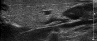
Causes of retention cysts
A retention cyst can form in both adults and children.
The cause of cyst formation is often a blockage of the excretory duct of a small salivary gland, which occurs due to an accidentally bitten lip. And since each gland has an excretory duct, due to a bite or injury it heals with the formation of a scar. If this scar gets on the excretory duct of the salivary gland, it becomes blocked. The secretion of the gland has nowhere to go, it accumulates in the gland and it begins to grow in size. Therefore, this cyst (retention) is nothing more than an enlarged salivary gland.
Removal of the cyst with resection of the hyoid bone
A midline neck cyst is a congenital pathology that occurs as a result of a violation of the intrauterine development of the baby. It is formed from the thyroid-lingual duct, which should disappear (overgrow) even before the baby is born, in the second month of the mother’s pregnancy, but in some cases this does not happen, and then the duct forms a closed cavity, which fills with fluid over time. Such a cyst often remains invisible, and can be detected not only in childhood or adolescence, but also in adulthood - either when it begins to grow and cause discomfort, or accidentally, during any medical examination of the neck for another reason. The growth of a cyst in adolescence or adulthood can be triggered by injury, infectious disease, or pathologies of the lymphatic or circulatory systems. Initially, cervical cysts are benign, but over time they can transform into malignant formations. In addition, suppuration can develop inside the cyst, and if the pus breaks out, a fistula is formed, but if the pus breaks inside the neck, then, in addition to the fistula, serious intoxication occurs in the body. If the cyst is not removed, then suppuration can be repeated many times, weakening the human body and increasingly threatening not only his health, but also his life. A cyst without inflammation can also cause discomfort - interfere with swallowing, change your voice, cause a sore throat and other unpleasant phenomena. There is no conservative treatment for cervical cysts, so the only option for eliminating them is surgery. If there is a need to carry out such an operation, it is extremely important to find a competent and experienced professional who will remove the cyst on the neck as carefully and safely as possible and completely, since if any fragments of the cyst remain unremoved, there will be a possibility of relapse.
As a rule, such an operation is planned, and before it, the doctor, as before any other operation, prescribes a detailed examination of the patient. As part of such an examination, a specialist may prescribe an ultrasound or tomography - computer or magnetic resonance imaging (CT or MRI, respectively). Urine and blood tests, puncture of the contents of the cyst, as well as other studies may be prescribed to exclude an oncological process and to obtain the maximum amount of information about both the formation and the general condition of the patient. If the patient comes with a suppurating cyst, before performing the operation, the doctor removes the pus from the cyst using a puncture method, prescribes treatment to eliminate inflammation, and only then proceeds to prepare for removal of the cyst. The operation is performed using general anesthesia in a sterile operating room. After the anesthesia is administered and begins to take effect, the doctor treats the surgical area with an aseptic agent and makes a skin incision in the projection of the cyst and up to the hyoid bone. Next, the specialist removes the tissue covering the cyst and reaches the formation itself. Since the thyroglossal duct passes through the middle part of the hyoid bone and fuses with it, when removing the cyst, it is necessary to remove a fragment of this bone to avoid the development of relapses. The doctor cuts the bone on either side of the cord of cyst formed from the duct, so as to avoid damage to the nerves leading to the tongue and located under the bone. Next, the doctor separates the bone fragment to be removed, as well as the remains of the thyroglossal duct from the remaining tissues, muscles and organs and removes the separated fragments along with the cyst as a single whole. The doctor brings the ends of the remaining fragments of the hyoid bone as close to each other as possible during suturing of the muscles. After this, the doctor stops the bleeding and sutures the wound, layer by layer connecting the tissues that were affected during the operation. A sterile bandage is applied to the wound.
After the operation, the patient remains under the supervision of specialists until his condition stabilizes, after which he can go home and only visit the doctor for observation and removal of stitches. During the rehabilitation period and until full recovery, the patient must carefully follow all medical instructions and restrictions, and then he will not be afraid of complications and will soon forget about the illness and completely return to a full, healthy life.
The multidisciplinary clinic “Medistar” brings together a galaxy of highly qualified specialists, true professionals in their field, conducting both diagnostic and advisory practice, and successfully treating ailments of any complexity. Our own laboratory, modern equipment for diagnostics and therapeutic manipulations allow us to go through all stages of the fight against the disease - from diagnosis to complete recovery, without leaving the medical center in search of the desired method of diagnosis or therapy.
Treatment methods for retention cysts
If this type of cyst appears, under no circumstances should you bite it, as its shell will collapse and removal of this cyst will be impossible.
The main treatment method for retention cysts of the sublingual salivary gland is surgical - cystotomy and cystectomy (rarely). The procedure takes a few minutes (10-15), and is performed under anesthesia and is absolutely painless.
During surgery on a retention cyst, the part located under the mylohyoid muscle is removed and the isthmus is ligated. After this, a cystectomy of the part of the cyst located in the oral cavity is performed.
Hypertrophy of the masticatory muscles
Along with a tumor of the salivary glands, there is hypertrophy of the masticatory muscles, which also leads to disproportions of facial features, but is sometimes caused by dental and neurological problems. To understand the true causes of the deficiency, it is necessary to conduct a detailed examination and determine whether the glands are directly enlarged or just the muscles.
When it comes to muscle hypertrophy, botulinum toxin injections come to the rescue, blocking excessive muscle activity and over time they decrease in size.
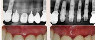
Cyst in the oral cavity: symptoms
At the initial stage, a cyst in the mouth develops asymptomatically. The danger of the disease lies in the fact that the pathological process can be detected at a late stage, accompanied by various consequences. A neoplasm in the oral cavity can be determined by an x-ray examination, which is carried out at the Yusupov Hospital using European equipment.
A cyst of the oral mucosa has characteristic signs:
- pain in the palate, gums or cheeks, which intensifies when chewing or biting food;
- feeling of pressure in the gums;
- the appearance of a small tubercle in the affected area, which, as the disease progresses, enlarges and fills with pus;
- the appearance of flux;
- increased body temperature, malaise and swollen lymph nodes.
Accidental trauma and damage to the cyst can lead to infection of the oral cavity. If these signs appear, you should consult a dentist who will help solve this problem. The Yusupov Hospital has the necessary equipment for diagnosing and treating cysts in the mouth. Specialists at the surgery clinic of the Yusupov Hospital provide emergency care to patients during exacerbation of the disease.
Causes of formation of salivary gland cysts
Salivary gland cysts appear as a result of blockage of the salivary ducts. Pathology of patency can be caused by various factors:
- various types of injuries;
- Poor, untimely or completely absent oral hygiene;
- unhealthy diet;
- bad habits;
- various types of infectious diseases of the oral cavity and teeth;
- difficulty or disturbance, followed by cessation of secretion outflow;
- the appearance of a plug as a result of thickening of the secretion, disrupting the patency of the excretory canal;
- the presence of various tumors that put pressure on the duct;
- the presence of scars narrowing the canal.
Removal of cervical lymph nodes (cervical dissection)
When removing a malignant tumor of the salivary gland, the cervical lymph nodes are usually removed, a so-called cervical dissection is performed. The lymph node is usually small, surrounded by a bean-shaped capsule, and lymph fluid flows through it. There are a large number of lymph nodes in the neck.
The first metastases of salivary gland cancer are usually found in the cervical lymph nodes and therefore it is believed that their removal is necessary in the case of cancer. Typically, surgery to remove the salivary gland, which also involves a cervical dissection, takes 1 to 2 hours longer.
Facial nerve
The facial nerve—the motor nerve—provides facial expressions for the corresponding half of the face.
Damage to the facial nerve during resection of the parotid salivary gland ©headandneckcancerguide.org
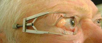
When the facial nerve is damaged in the area of the parotid gland, denervation of all the facial muscles of the corresponding side of the face occurs - the muscles weaken, the face turns into a motionless mask: the eyebrow, eyelids, corner of the mouth sag, ectropion develops - inversion of the lower eyelid, it becomes impossible to close the eye and smile.
Result of facial nerve damage
The facial nerve can be damaged not only by tearing it, but also when working with a surgical instrument - as soon as the nerve is pressed harder, it ceases to perform its function. Therefore, in my practice, I use a microsurgical instrument to isolate the facial nerve.
The surgeon’s task is not only to remove the area affected by the tumor, but also to improve the quality of life, so it is extremely important to preserve the integrity of the facial nerve.
For malignant tumors of the parotid gland, it is sometimes necessary to remove a segment of the facial nerve according to indications. Then, to preserve facial expressions, it is necessary to replace the removed segment with an insert of another nerve: the greater auricular or sural nerve, which is taken from the lower leg.
If the facial nerve was accidentally or intentionally severed and then restored, then motor function will be preserved, but not always to its full extent.
Important: Make sure your surgeon is experienced and knows how to repair the damaged nerve, including transferring a segment of the sural nerve from the leg.
Cyst in a child’s mouth on the gum
The development of cysts in the mouth in childhood occurs frequently, which is explained by improper dental care, injuries received from a fall, as well as untimely treatment of caries. A cyst in a child’s mouth on the gum may also appear during teething.
Treatment of cysts in childhood should be carried out by a dentist, who will determine the type of formation and the reasons for its development. The capabilities of modern medicine make it possible to painlessly treat cysts in children at the initial stage of development.
However, some parents, after a cyst in the child’s mouth was discovered, look at photos, study thematic forums and use traditional methods of treatment. Self-medication for this disease can lead not only to tooth loss, but also to further spread of the source of infection and the formation of fistulas.
The Yusupov Hospital treats patients over 18 years of age. If a cyst is found in a child’s mouth on the gum, experts recommend seeking help from a pediatric dentist.