Gum drainage in dentistry
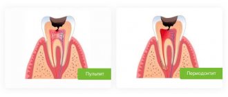
Inflammation of the gums can develop very slowly, or it can go through all stages - from mild swelling to severe suppuration - literally in hours.
This is why any disease in which an infection enters the soft tissue is dangerous.
And it is for this reason that, having noticed that the gums are swollen (the inflammatory process has begun), you should rush to the clinic.
Only here can a doctor guarantee safe treatment (and given that dangerous inflammation occurs near the facial muscles, nerves, bone tissue, and most importantly, the brain, safety is a priority).
Why cut the gum?
Pain in the gums continues for a long time - this is a natural accompaniment of tooth growth. Contact your doctor if you notice any of the following symptoms:
- Severe pain when chewing food or occurring spontaneously, without reason.
- If you have bad breath, this is a sign of pus under the hood.
- If the gums not only hurt, but also became red and swollen.
- The temperature has risen, severe weakness and headaches are felt.
Dentists will offer to cut the gum, be strong - this is not such a terrible procedure as it seems. After the incision, the rot will come out, the gums will heal, the pain will subside and the tooth will no longer cause concern. In different patients, the gums healed at different times: it usually takes from two days to two weeks.
What to do if the drainage falls out of the gums
If the drainage randomly falls out of the gums, you should contact a specialist to determine the cause. This is often caused by improper installation of the system, weak fixation, intensive brushing of teeth and frequent rinsing.
In this case, you should pay attention to whether the swelling has subsided or not, whether there is suppuration.
The decision to return the system is made by the dentist; under no circumstances should you install drainage yourself.
Incorrect actions by the patient can lead to the development of complications and relapse of the infectious-inflammatory process. In this case, a rapid overgrowth of soft tissue occurs, blocking the exit to the outside for pus and excess fluid. As a result, the gums hurt, a lump forms, where purulent secretions accumulate abundantly.
If a child pulls out a drain, you should immediately contact a specialist for an examination of the oral cavity and further observation and treatment.
Types of drainage
Drainage has its own types:
- Active - installed for the outflow of liquids through a special tube or hole from the bottom up;
- Passive - on the contrary, is installed according to the top-down principle.
The use of any of the presented types of drainage depends on the patient’s well-being, the degree of complexity of the operation performed, and the presence or absence of signs of inflammation. It is worth noting that failure to use wound drainage greatly contributes to the deterioration of suture healing time and can lead to the entry of microorganisms and infections into the wound, which are especially dangerous for patients in the postoperative period.
You can ask all questions on the topic by phone: +7-(928)-900-32-69
After the procedure
After the procedure, you will be placed in a recovery room. You will need to stay in bed until the sedative medicine wears off.
In the recovery room, the nurse will continue to monitor your condition, heart rate, breathing and blood pressure. They will watch your catheter for possible bleeding.
About your catheter
A black mark will be placed on your catheter at the top of the disc (see Figure 4). The nurse will show it to you. This mark should always be at the same distance from the top of the disc. If the distance changes, the catheter has moved. You should call the interventional radiology department to have a staff member check the position of the catheter.
Figure 4. Black mark at the top of the disc.
The outer end of the catheter is connected to a 3-way stopcock (see Figure 5). It is called a 3-way stopcock because it has 3 connection points and a stopcock that can be turned to control the flow of bile. The drainage bag will be attached to the connection point opposite the catheter. The third connection point has a safety valve cap through which you can inject liquids. This valve is called a needleless connector.
Figure 5. 3-way faucet
A drainage bag will be attached to the catheter. You will see bile (a yellowish-green liquid) draining into the bag. The fluid may be blood red on the first day or the first two days. The color will eventually turn golden yellow or greenish depending on where the catheter is placed in your body.
The CathGrip® is a device that will prevent the catheter from leaving your body if you accidentally pull on the catheter.
to come back to the beginning
After surgery for hydronephrosis
After surgery for hydronephrosis, you will need to stay in the hospital for a couple of days. What should you expect after surgery?
Intravenous catheter
For the surgical procedure and for the first time after it, you will have a venous catheter for administering fluids and various medications. In some cases, a central venous catheter may be installed.
Pain syndrome after surgery to eliminate hydronephrosis
The severity of pain depends on the type of surgical intervention. Open surgery with a large lateral incision is associated with severe pain. Laparoscopic surgery for hydronephrosis can significantly reduce the intensity of pain after surgery. Prolonged epidural anesthesia has a good effect. If this is not possible, intravenous administration of painkillers can be established. The choice of analgesics, narcotic or non-narcotic, depends on the severity of pain.
Drawing. Epidural catheter.
Nausea and vomiting
On the first day after surgery for hydronephrosis, you may experience nausea and vomiting, this may be due to the anesthesia or pain. Nausea and vomiting are not a sign of an abnormal course of the postoperative period. Do not be upset; as a rule, after one or two days these phenomena go away on their own. Antiemetic medications may be prescribed to significantly ease your condition.
Urinary catheter
After surgery, you will also find that you have a urinary catheter. It ensures the outflow of urine from the bladder and plays an important role in the smooth course of the postoperative period. Typically, the catheter is removed 2-3 days after surgery for hydronephrosis.
Drawing. Urinary catheter.
Drainage after surgery for hydronephrosis
Drainage is a tube, one end of which is located in the area of the operation, and the other opens outward. Drainage is of great importance in the first days after surgery for hydronephrosis. If bleeding or leakage of urine occurs, pathological discharge, blood or urine, is separated through the drainage to the outside, which makes it possible to promptly detect a complication and take measures to eliminate it. Normally, a light discharge with a small admixture of blood flows through the drainage. After two to three days, when the period of possible complications is over, the drainage is removed.
Drawing. Wounds and drainage after surgery for hydronephrosis.
Mobilization and breathing exercises
Early mobilization - restoration of physical activity - is an important aspect of the prevention of respiratory and thrombotic complications after surgery for hydronephrosis. Laparoscopic intervention allows for earlier mobilization; the patient is allowed to sit and stand up in the evening after surgery. Open surgery for hydronephrosis makes early mobilization difficult due to the rather traumatic lateral incision affecting several layers of muscle. Breathing exercises can reduce the risk of respiratory complications.
Prevention of thrombotic complications
Thrombosis (blockage) of the deep veins of the lower extremities and blockage of pulmonary vessels is a serious postoperative complication, the risk of which can be reduced by a number of preventive measures. First of all, this is the above-mentioned early mobilization. Wearing special compression stockings and using medications that prevent blood clotting inside the vessels are of great importance.
Nutrition after surgery
Surgery does not directly affect the gastrointestinal tract. But in the first hours or days after the operation, the work of the gastrointestinal tract may be slightly suppressed due to the extensiveness of the operation, the use of narcotic painkillers, etc. In the absence of nausea and vomiting, drinking is allowed in the evening after the operation, and the next day a gradual expansion can begin diet. Additionally, fluid and nutrients may be given through a venous catheter.
Constipation after surgery
Bowel depression after surgery may be associated with constipation and bloating. If constipation occurs, laxatives will help.
Ureteral stent
A ureteral stent is a hollow tube located in the ureter, with one end opening into the renal collecting system and the other into the bladder. The ureteral stent plays a primary role in the healing of the sutures between the pelvis and the ureter after surgery for hydronephrosis. The stent ensures unimpeded flow of urine from the kidney to the bladder, thereby minimizing the negative impact on the anastomosis.
The presence of a ureteral stent is also associated with negative events. Firstly, its presence in the body can cause severe discomfort, even pain. The second unfavorable phenomenon is a high risk of developing urinary infections, accompanied by fever, lower back pain, urinary disorders, and the appearance of leukocytes and red blood cells in the urine. Less commonly, phenomena such as migration of the stent, when it occupies an incorrect position in the urinary system, encrustation and blockage of the stent with urine crystals can develop. These complications can be avoided by timely treatment of the infection with antibiotics and uroseptic drugs, as well as by consuming at least 3 liters of water per day to better “wash” the stent.
The stent is removed approximately 6 weeks after surgery for hydronephrosis. It is removed by cystoscopy, a procedure using an optical instrument inserted into the bladder. Extraction occurs on an outpatient basis, under general or local anesthesia.
After discharge from hospital
You will be able to go home 3-5 days after surgery for hydronephrosis.
Pain syndrome
If pain persists after discharge from the hospital, you can take non-narcotic pain medications.
Taking a shower and bath
In the first days after surgery for hydronephrosis, it is not recommended to shower; hygiene procedures should be limited to wiping with a damp sponge. You can shower when you return home, but water baths should be avoided for the first two weeks after leaving hospital.
Sutures after surgery for hydronephrosis
If the operation for hydronephrosis was performed laparoscopically, then the wounds are sutured with self-absorbing suture material; removal of the sutures is not required. After complete healing, you will be left with barely noticeable skin defects. In open surgical interventions, the wound is closed with special staples or sutures, which are removed after 10-14 days. Unfortunately, the cosmetic effect of open surgery is not as good.
Drawing. Scar after open surgery for hydronephrosis.
Drawing. Cosmetic effect of laparoscopic surgery for hydronephrosis.
Return to work after surgery
Return to work after laparoscopic pyeloplasty for hydronephrosis is possible three to four weeks after discharge from the hospital.
The recovery period after open surgery lasts longer. Restoring physical activity, playing sports - all this must be done gradually, starting with minimal loads and gradually increasing them. Back to articles
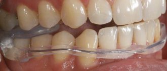
In what cases is drainage installed on the gum?
There are many pathologies in dentistry for which drainage is installed on the gums. These include:
- flux - is a manifestation of periodontitis and is accompanied by severe swelling of tissues
- neoplasms in the roots of teeth - drainage is installed on the gums if abscesses and cysts are diagnosed
- alveolitis - inflammation of the tooth socket
- in order to accelerate the healing of the wound surface after complex tooth extraction
- if necessary, regularly inject medications into the lesion
Gum drainage, photos of which you can see on thematic websites, plays a significant role in dentistry. It ensures the removal of pathological fluids and access of drugs even into the deep periodontal tissues.
There are few contraindications to drainage. The procedure cannot be performed if you are allergic to the anesthetics used during the procedure or have a blood clotting disorder.
How long should drainage remain in the gum?
Patients are most concerned about the question: how long should drainage remain in the gum? This period is determined individually depending on how quickly the swelling of the periodontal tissue subsides. It is the disappearance of this symptom that indicates complete cleaning of the wound and the beginning of its healing.
Only a doctor can determine exactly when to remove the drainage from the gums, taking into account the nature of the pathology. As a rule, the wearing period is 4-5 days. This is enough to clean the wound from pathological fluids. Sometimes the drainage falls out earlier, but complications are not observed - this means that the wound cleared earlier.
If the drainage has been in place for 5 days, and after its removal the wound swells again and causes pain, it is important to immediately consult a doctor. Additional diagnostics and identification of the cause of this condition are required.
Suction drainage from the pleural cavity
Suction tube
For suction drainage of the pleural cavity, various rubber and synthetic tubes are used.
For the most commonly used drainage, a rubber tube about 40 cm long with several side holes at the end is used. This tube is placed along the lung (from the base to the apex) and passed over the diaphragm from the pleural cavity to the outside. The drainage is attached to the skin with a knotted U-shaped suture. When the suction drain is removed, the threads are tied again, thereby sealing the hole in the chest. A three-barrel suction catheter (Viereck) is advantageous, allowing free passage of the tube inserted inside.
Insertion of suction drainage
In the chest between the two pleural layers, the intrapleural pressure is lower than atmospheric pressure. If air or liquid gets between the pleural layers, then the normal physiological state can only be restored by long-term suction drainage. A closed drainage system is used to suction pleural fluid for recurrent pneumothorax and to treat empyema. This drainage is now usually inserted into the intercostal space through a trocar. The thickness of the drainage tube is determined in accordance with the consistency of the substance being sucked out (air, as well as watery fluid or serous, fibrinous, bloody, purulent fluid).
On the drainage, mark with paint or thread the place to which it will be inserted. The size of the trocar must correspond to the size of the drainage. It is advisable to have at least three trocars of different sizes with suitable tubes of 5, 8 and 12 mm in diameter. Before inserting the trocar, you must make sure that the selected drainage tube passes through it easily.
The site of the skin incision is filtered with novocaine to the pleura. A test puncture in the designated area makes sure that the desired air or liquid is really there. The assistant gives the patient the necessary position: the patient must sit and lean on the highly raised operating table so that the puncture area protrudes as much as possible, and the selected intercostal space is, if possible, expanded. A scalpel is used to cut the skin over an area slightly larger than the size of the trocar. Then the trocar is inserted with a strong movement along the upper edge of the rib into the pleural cavity. After removal of the trocar, unimpeded release of fluid or free entry and exit of air indicates its correct insertion. Drainage is performed and the trocar tube is removed. If you are not convinced that the drainage is in the right place, you should, in order to prevent the trocar from puncturing the lung, heart or large vessel, perform the puncture again, taking all measures to localize it under X-ray control.
Before closing each thoracotomy hole, a drainage is introduced into the pleural cavity, which is brought out above the diaphragm through a separate hole in the intercostal space. Through a hole measuring about 1-2 cm, a forceps is inserted into the pleural cavity under the control of the eyes and under the protection of the left hand to ensure the correct position of the drainage from the inside. The drainage is pulled through the chest wall with a forceps from the inside to the outside. Pay attention to the fact that the drainage section, free from holes, is at least 5 cm in the chest cavity. If the fixation of the drainage to the skin is broken, then it slips out, and the first side hole appears outside the pleural cavity above the skin. In this case, the closed system turns into an open one, suction becomes ineffective, and pneumothorax often occurs.
Suction systems
There are so-called individual (“bed side”) and centralized suction systems. The suction action due to the hydrostatic effect can be obtained by a tube lowered under water, a water or gas pumping device (in this case the action is based on the valve effect) or an electric pump. Both individual and central systems must ensure individual regulation. If the release of air from the lung is insignificant, then due to its simplicity, the Biilau drainage system is still successfully used today, which can be sufficient to straighten the lung. A glass tube immersed under water (disinfectant solution) is equipped with a valve made from a finger cut off from a rubber glove, which protects against reverse suction. The Biilau system uses the physical law of communicating vessels to move bottles under the bed to create a suction effect.
The Fricar air pump best meets modern requirements. This device can work for many days without interruption and without heating up. The strength of the suction effect can be precisely adjusted.
Central suction devices are triggered by an oxygen canister system or a powerful suction pump. The system of outgoing tubes, if necessary, supplies hospital departments located on different floors. Depending on the need, the required number of hospital beds can be connected. An oxygen-powered system has the advantage that the suction and supply of oxygen to individual hospital beds is provided by the same tubing system. The suction effect is provided by a valve tube mounted along the oxygen flow. In this case, however, the effect produced by the central suction pump is not achieved.
Individual adjustment can be carried out using a dosimeter tap connected to a well-functioning pressure gauge, or through the so-called. three bottle system. The latter can be easily prepared by yourself. This system also has the advantage that it can easily and reliably create a very low suction effect (from 10 to 20 cm of water column). It is rarely possible to achieve such low pressure values using factory pressure gauges.
Indications for suction drainage
Spontaneous and traumatic pneumothorax, hemothorax
Spontaneous pneumothorax occurs at a young age, more often as a result of rupture of single pulmonary alveoli in the apex of the lung, in older people - as a consequence of rupture of alveolar vesicles with diffuse emphysema. Due to the fact that the number of patients with emphysema is constantly increasing, the number of cases of spontaneous pneumothorax is becoming more frequent. The same applies to traffic accidents that result in closed injuries in the chest cavity, which often occur with pneumothorax or hemothorax.
Correctly performed pleural puncture for spontaneous pneumothorax is practically safe, and its benefits can hardly be disputed. If the flow of air from the damaged lung is completely stopped and the perforation site is closed, then it may be possible to completely remove the air that created the pneumothorax with a simple closed puncture. If pneumothorax recurs after puncture (even repeated), then drainage with long-term suction should be used. Recurrence of pneumothorax, even after prolonged drainage with suction, can only be reliably eliminated by surgery.
Traumatic pneumothorax most often results from rib fractures. When a rib fragment injures the lung, most often a significant amount of air comes out of it, and a tension pneumothorax occurs. At the same time, subcutaneous or even mediastinal emphysema may occur. Spontaneous pneumothorax can also occur when the pulmonary alveoli rupture or due to blunt force on an emphysematous lung. Therefore, in patients with pulmonary emphysema, chest injuries are often associated with the occurrence of pneumothorax, often severe tension pneumothorax. The principles of treatment for spontaneous and traumatic pneumothorax are the same.
If clinical symptoms indicate tension pneumothorax (severe respiratory failure, subcutaneous emphysema, mediastinal shift), then drainage of the pleural cavity should be performed immediately. If these symptoms are not present, then a closed puncture is performed and the air is sucked out. After this, the needle is left inserted into the pleural cavity, and its nozzle is connected to a pressure gauge and the pressure in the pleural cavity is determined (whether it is higher or lower than atmospheric). If the pressure in the pleural cavity is indicated by the pressure gauge needle in the positive direction, it means that air continues to be released into the pleural cavity, and, therefore, drainage is necessary. This issue can, of course, be resolved by X-ray examination. If there is a total pneumothorax, then drains are inserted in two different places. One of them runs along the posterior axillary line above the diaphragm in the VII-VIII intercostal space, the other is inserted along the midclavicular line between the 1st and 2nd ribs. In our experience, drainage inserted under the collarbone performs the task of straightening the apex of the lung better.
In case of encapsulated limited pneumothorax, drainage should be inserted locally, under X-ray control after a test puncture.
Empyema of the pleura
Pleural empyema is a disease for which treatment with suction from the pleural cavity is absolutely indicated.
The principle of treatment of empyema does not depend on the causative agent of the disease. It consists of gluing the pleural layers and eliminating the empyema cavity through early drainage and suction of fluid. Treatment with suction from the pleural cavity is combined with targeted local chemotherapy, based on the identification of the pathogen and its resistance to the drugs used. Most empyemas result from infection of the exudate. In this case, incorrect and insufficient suction from the pleural cavity plays a certain role. In cases where pockets with delimited fluid form in the pleural cavity, their complete emptying becomes increasingly difficult, more difficult, and infection is more likely. In such cases, complete recovery can only be ensured by surgery.
Suction treatment may fail for two reasons: one is the presence of pleural cords, the other is a bronchopleural fistula.
Pleural moorings are often the result of insufficient emptying of the pleural cavity. When moorings have already formed in the pleural cavity and the walls of the empyema cavity are thickened, there is little chance of eliminating the empyema by suctioning out the fluid. The ability to expand the lung in this case is also very controversial. In this case, drainage with suction is a preparatory measure before the inevitable operation. Radical surgery (decortication) is performed only after the patient’s general condition has improved by washing the pleural cavity and targeted antibiotic therapy.
Bronchopleural fistula reduces the effectiveness of suction and thereby the prospect of lung expansion. In cases where there is a large bronchial fistula and its closure is contraindicated (for example, a rupture of the cavity, tumor disintegration, rupture of a cystic, emphysematous lung that has lost its elasticity), success cannot be expected from the use of suction. On the other hand, suction can also be used in cases where surgery is indicated. In elderly patients, with low general resistance and the possibility of severe complications, surgery becomes impossible. Then all that remains is to leave the patient with permanent drainage.
In case of chronic pleural empyema, drainage should be inserted into the pleural cavity at its lowest point. Large-diameter drains are used so that the thick liquid does not close the lumen and it is easy to wash the pleural cavity. Often, in the area where the drainage will be introduced, a rib resection (2-3 cm) is performed.
Postoperative suction from the pleural cavity
In order to remove fluid that accumulates after thoracotomy from the pleural cavity and maintain normal intrapleural pressure, a suction drain should be available.
If during pleural operations and mediastinal, transthoracic interventions on the esophagus, stomach, heart and large vessels there was no damage to the lung, then the chest can be closed with the introduction of one perforated drainage into the pleural cavity. Drainage is carried out above the diaphragm along the midaxillary line with its pleural end installed at the level of the apex of the lung.
Two drains are inserted into the pleural cavity if the lung was damaged during separation of adhesions, as well as after resection or excision of lung tissue. In such cases, one of the drains is inserted along the anterior and the second along the posterior axillary line. The use of a third drainage may be considered relatively appropriate when it is brought to the site of anastomosis of the esophagus or bronchus or when thoracoplasty is performed in combination with lung resection (for suction from the subscapular space).
After removing the lung, one drain with a diameter of 12-15 mm is inserted into the pleural cavity and placed in the lower part of the cavity so that a piece of drainage 10-12 cm long is equipped with 2-3 side holes. Active suction through this drain is prohibited.
After median sternotomy, a drainage is inserted retrosternally and its second end is brought out in the epigastrium.
Intensity and duration of suction
The intensity of suction through drainage from the pleural cavity depends on the cause of the disease, the condition of the lung and the nature of the operation. The flow of air from the lung into the pleural cavity is of decisive importance. If this occurs, then more air should be sucked out of the pleural cavity per unit time than is supplied there. Only in this way can gluing of the pleural layers be achieved. In practice, however, this is often not feasible. If the connection of the bronchus with the pleural cavity is significant (for example, in the case of a bronchial fistula), then intensive suction cannot achieve the goal. If you increase the suction force, then at the same time the patient will experience increased respiratory failure due to “air theft” from the tidal volume. Despite this, the lung will not be able to expand. In such cases, surgery is inevitable.
When the lung is damaged or after lung surgery, air most often escapes from a hole the size of a pinprick. In this case, specialized suction is indicated. In children and adolescents, due to the fact that their lung parenchyma is healthy and not affected by fibrosis and emphysema, it does not matter with what force the suction is performed. It doesn’t matter whether they suction with an intensity of 25 cm of water. Art. or simple underwater drainage, the lung will expand in 24-48 hours. The drainage can be removed after 48-72 hours. This is the advantage of elastic tissue capable of lung retraction in young patients. With emphysematous lung in an elderly person, the situation is different. The pinprick holes become gaping holes in the lung because the surrounding tissue is unable to contract. If you try to reduce the flow of air coming from the damaged lung by increasing the intensity of suction, you can easily get a paradoxical effect. The flow of air from the lung will increase. Small holes, due to prolonged suction, stabilize and turn into fistulas.
What to do in such cases? Begin gentle suction from the pleural cavity (5-6 cm of water column) and pay attention to ensure that tension pneumothorax does not occur. Thanks to this, the resulting fibrin seals small holes in the lung. Within 24 hours, a decrease in the release of air from the damaged lung begins to be detected. The intensity of suction can be increased slightly. On the fourth day you can already suction with an intensity of 10 cm of water. Art., if no unforeseen complications arise, then the drainage can be removed on day 4-5.
The same principles are followed when treating spontaneous and traumatic pneumothorax with suction.
If there is a significant intake of air from the emphysematous lung, they begin to carefully perform suction with a gradual increase in its intensity. If, after many days of treatment with suction, the release of air from the lung does not stop, then it is recommended to immediately undertake surgery, without waiting for the development of infection in the pleural cavity. If suction from the pleural cavity continues for more than a week, the development of infection becomes real.
In cases where the patient does not undergo surgery due to low general resistance, suction from the pleural cavity remains to be continued. Prolonged and specialized suction under the guise of drug treatment may be more or less effective. The pleural layers are glued together completely or partially. Only small limited cavities remain that do not lead to complications. The drain can be removed.
In the treatment of pleural empyema, long-term use of suction drainage is a common method. The empyema cavity gradually becomes smaller and smaller, the amount of fluid decreases, and in the end it can become bacteriologically sterile. If the daily amount of fluid extracted from the pleural cavity does not exceed 10-15 ml, then the suction is stopped, the drainage is shortened, but left until the residual cavity is completely closed.
When is drainage placed in the gum?
Why is drainage in the gum needed:
- drainage prevents early tissue healing (before the wound is completely cleared of inflammation products);
- drainage on the tooth ensures removal of pus, blood and ichor from the injured area without creating discomfort for the patient;
- often drainage is also used as a channel for direct administration of medication into infected tissue.
If the drainage in the gum is installed correctly, then its design is practically imperceptible and invisible.
The need for such treatment can be caused by almost any disease of the oral cavity if it is accompanied by an infection.
Most often, severe suppuration (and, as a consequence, the need to install gum drainage) is provoked by:
- flux (inflammation of the periosteum);
- pericoronitis;
- alveolitis;
- periodontitis;
- cyst or abscess of a tooth.
Sometimes suppuration occurs against the background of trauma to soft and bone tissues, which is accompanied by complex tooth extraction or mechanical damage to the jaw.
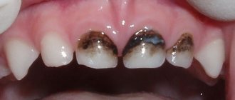
Photo. What does surgical drainage look like to remove pus?
Drainage after cholecystectomy for acute cholecystitis
A large prospective randomized study in 1991 and a meta-analysis of 1920 patients (ACA) summarized 10 similar studies. It was shown that when comparing patients with and without drainage in terms of mortality, reoperation or drainage due to bile accumulation, there were no differences. Wound infection more often accompanied patients with drainage (5). Thus, on the eve of the end of the era of BCI, routine drainage - the sacred cow of biliary surgery - was abandoned in many centers.
What is the trend in emergency LCE? In a recent study of Australian surgeons, drainage was left routinely in 1/3 of cases (6). Another small randomized trial comparing patients with and without drainage for LCE examined the effect of drainage on postoperative pain and nausea, in terms of gas removal, and found no difference (7). If routine drainage is pointless in ACE, why is it indicated in LCE? Therefore, Petrowsky et al. (3) drainage is not recommended for both ACE and LCE. In a prospective study of 100 patients who underwent LCE for acute cholecystitis, all of them underwent cholescintigraphy one day after surgery. Bile leakage was detected in 8, but all of them were asymptomatic (8). Most postoperative collections, be it bile, serous fluid or blood, remain asymptomatic, the fluid is absorbed by the peritoneum and this is well known from ultrasound studies since the time of AChE.
Drainage is much more effective at removing bile than stool or pus. Therefore, it is logical to leave a drain if the surgeon is concerned about possible bile leakage. For example, if a subtotal cholecystectomy is necessary, or when there are difficulties with sealing the cystic duct, or there is a suspicion of additional bile ducts in the area of the gallbladder bed, which manifests itself in the form of bile leakage from the surface of the bed.
Thus, although most patients do not require drainage, if the surgeon is concerned about possible bile leakage or excessive serous fluid, drainage is appropriate. In most cases, almost nothing is separated through such drainage. It is extremely rare that preventive drainage becomes therapeutic in the case of profuse and persistent bile leakage. In cases where the need for an existing drain is questionable, it is extremely important to remove it as soon as possible. “Dry” drainage for 24 hours indicates that it has served its role. Finally, Howard Kelly (1858-1943) said: “Drainage is the recognition of defective surgery.” Clinicians must be careful not to confirm this statement in practice: if it is safer to switch to an open procedure and carefully close the ultrashort cystic duct than to rely on questionable clip closure and safety drainage, then the choice is clear.
Drainage after gallbladder removal
After cholecystectomy, surgeons often install a gallbladder drain. Indications for cholecystectomy are:
- cholelithiasis;
- acute cholecystitis;
- gallbladder carcinoma.
The operation is performed laparotomy or laparoscopically. Drains from the abdominal cavity after open cholecystectomy are removed on the eighth day, and in weakened and oncological patients - on the twelfth day. To prevent the skin around the drainage from becoming inflamed, the bile is drained into a special vessel. The skin around the wound is lubricated with zinc ointment or Lassar paste. Drains are changed no earlier than the twelfth postoperative day. In this case, fistulography is performed through the drainage to ensure the free patency of the bile ducts. Removal of drains after cholecystectomy is not performed on the fourteenth day, and when draining the bile ducts, no earlier than the twenty-first day after removal of the gallbladder.
Make an appointment by calling. Doctors at the Yusupov Hospital use various methods of draining the bile ducts for obstructive jaundice. Medical staff care for the drainage of the gallbladder and bile ducts.
Endoscopic method
Using an endoscope, doctors perform nasobiliary drainage of the biliary tract. Indications for endoscopic drainage of the biliary tract are:
- obstructive jaundice caused by malignant and benign neoplasms;
- acute purulent cholangitis;
- external biliary fistulas;
- damage to the walls of the extrahepatic ducts, retroduodenal perforations;
- acute cholecystitis.
There are no contraindications to endoscopic drainage, except in cases where the tube for drainage of the biliary tract cannot be passed through the area of tumor narrowing. The endoscopic kit for drainage of the biliary tract through the nose includes:
- conductor wire;
- drainages of various shapes;
- a connecting tube for collecting bile and flushing the drainage;
- nasal tube, clamp and spatula.
The operation of endoscopic drainage of the biliary tract includes the following steps:
- cholangiography to determine the level and location of drainage;
- introduction of drainage with a metal guide-conductor;
- removal of the guidewire and endoscope;
- control cholangiography;
- assessment of drainage position;
- transferring the drainage from the mouth to the nose and fixing it on the head.
After using the endoscopic method of drainage of the bile ducts, complications do not develop. They may occur due to the progression of the disease.
Methods of drainage of the biliary tract
Oncologists prefer to perform external internal or, if technically feasible, external drainage of the bile ducts in patients with obstructive jaundice of tumor origin. Both methods are quite effective in preoperative preparation for radical surgery or as a final treatment method. Their advantage is:
- constant monitoring of bile flow;
- the possibility of active removal of blood, pus, and clots from the bile ducts;
- washing the ducts with aseptic solutions;
- dynamic x-ray monitoring of the location of the drainage tube.
Unlike external-internal drainage, bile completely flows out through the external drainage of the gallbladder. The disadvantage of external drainage of the bile ducts compared to external internal drainage is the complete flow of bile through the drainage to the outside. To compensate for the vital substances contained in bile, patients are forced to drink their own bile or medical personnel administer it through a nasogastric drainage. With external internal drainage, the distal end of the tube is located further than the narrowing site and most of the bile flows directly into the intestine. It remains possible to control the patency and flush the drainage and replace it with an internal transpapillary endoprosthesis.
Internal endoprosthetics of the bile ducts is performed after the elimination of jaundice. This is the final stage of treatment for inoperable patients. To successfully perform external or external internal cholangiostomy, oncologists use a set of special instruments: wire guides, special puncture needles, bougies and catheters.
Under local anesthesia using a Shiba needle, the surgeon tightly fills the bile ducts with a contrast agent. A long needle with a diameter of 1.5-1.7 mm performs a puncture of one of the segmental ducts. A conductor wire is then passed through it. The end of the conductor is passed beyond the narrowing, the narrowed area is widened with bougies, and a drainage tube is installed. It is fixed to the skin and the bile ducts are washed with sterile solutions.
This drainage method has disadvantages: there is a risk of bile and blood leaking into the abdominal cavity when the needle is removed, when passing a guidewire or bougienage the canal. Additionally, this complication may be encountered if the outer diameter of the needle is larger than the outer diameter of the guidewire. In order to reduce the number of complications associated with liver puncture, oncologists use the technique of installing a cholangiostomy using a stylet catheter.
How to remove the drainage system, why can it fall out?
The stripe is small, sometimes it interferes with eating and creates a lot of inconvenience for its owner. Many people are interested in how long it will take to use the system, and whether it can be delivered ahead of schedule. During the application process, the dentist warns that if the drainage falls out of the gums, it will be safer to seek help from a specialist than to remove it yourself.
If an edge falls out, carefully adjust it and try to put it back in place. If you remove it yourself, you will need to wash your hands, disinfect your mouth, stand in front of a mirror, pull the edge of the strip with your fingers, and pull it completely out. Doing procedures at home is not so much difficult as it is dangerous. It’s better not to take risks and seek professional help. Gently rinsing the treated area will help speed up the tissue healing process. If the tape falls out in the first days of treatment, it must be promptly replaced to avoid possible infection with sad complications.