What can inflammation of the salivary gland ?
Read the answer to this question in this article. The human body is a complex mechanism consisting of a huge number of organs and systems. Several structures are responsible for performing the same function, constantly complementing each other. For example, different organs take part in the digestion process.
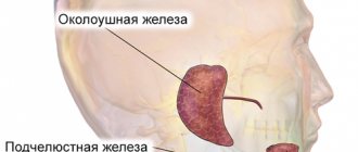
In the human body during normal development there are three pairs of salivary glands
The parotid gland is most often affected by various diseases. There are a number of diseases in which the sublingual and submandibular salivary glands become inflamed. If you do not start treatment on time or undergo inappropriate therapy, then serious complications may occur after such diseases, such as encephalitis, orchitis, meningitis, nephritis, neuritis and pancreatitis. However, do not worry, treatment often gives positive results. To avoid inflammation of the salivary glands, you just need to follow a few recommendations.
Anatomy and pathology of the salivary glands. Sialadenitis, sialosis
In the article, the authors consider the normal and pathological anatomy of the salivary glands. Sialadenitis and sialosis are treated. They are trying to consider the macroscopic and microscopic picture of these diseases.
Key words: salivary glands, sialadenitis, sialosis, pathological anatomy, dental diseases, parotid gland, sublingual gland, submandibular gland.
The salivary gland is an important part of the human digestive system. It performs exocrine, endocrine, filtration and excretory functions. The excretory ducts of the glands open into the oral cavity. There are large and small salivary glands.
The large salivary glands are located outside the oral cavity and include the parotid, sublingual and submandibular glands. The minor ones are located deep in the mucous membrane and are represented by the labial, buccal, palatine, molar and lingual glands.
Parotid gland, glandula parotidea
It is the largest gland. Located in the mandibular fossa, the excretory duct runs along the outer surface of the masticatory muscle and opens on the mucous membrane of the vestibule of the mouth in the area of the second upper molar [2].
In terms of its structure, it is complex alveolar, serous type, and has a lobular structure. The gland is covered with fascia, which encloses it in a capsule [4].
Submandibular gland, glandula submandibularis
Located in the submandibular space, the excretory duct opens on the sublingual papilla, like the duct of the sublingual gland. In structure it is a complex alveolar-tubular gland of a mixed nature, which also has a lobular structure. The submandibular gland secretes protein-mucosal saliva with a predominance of the protein component [4].
Sublingual gland, glandula sublingualis
It lies on the mylohyoid muscle, directly under the mucous membrane of the floor of the mouth, and has a thin connective tissue capsule. The excretory ducts of some lobules open independently into the oral cavity, and the main duct is located next to the duct of the submandibular gland [5].
The structure is a complex alveolar-tubular salivary gland, producing mixed saliva with a predominance of mucous secretion.
Diseases of the salivary glands can be either primary (independent) or secondary - a manifestation or complication of any systemic disease. Diseases can be caused by the development of infection, tumor, traumatic injury, as well as obstructive, tumor-like or autoimmune damage.
Sialadenitis
Inflammation of the salivary gland is called sialadenitis. Most often, the parotid gland is affected, less often - the submandibular gland, very rarely - the sublingual gland. Sialadenitis can be acute or chronic, and according to the etiology of the disease it can be viral, fungal or bacterial. The following routes of infection are possible: hematogenous, contact, lymphogenous and intraductal from the oral cavity of the body.
Acute sialadenitis
May occur due to exposure to local and general factors. Local factors - any surgical intervention, compression of the excretory ducts by a tumor, traumatic lesions. Common factors are diabetes mellitus, chronic gastrointestinal diseases, immunodeficiencies, cardiovascular diseases [1].
Acute sialadenitis is divided by morphology into gangrenous, serous and diffuse purulent. The salivary gland enlarges in this disease. Purulent sialadenitis is characterized by pronounced neutrophilic infiltration with an abundance of purulent bodies. With serous sialadenitis, swelling, neutrophilic infiltration, and plethora are observed.
Complications of acute sialadenitis include the occurrence of abscesses, sepsis and phlegmon of the neck tissue. Outcome: recovery or transition to chronic [3].
Chronic sialadenitis
Most often secondary, i.e. as a complication of traumatic injuries, infectious diseases, developmental defects. According to the degree of clinical manifestations, active and inactive sialadenitis are distinguished. Chronic sialadenitis often takes on a recurrent nature, so there are two stages: the remission stage and the exacerbation stage.
The salivary glands are enlarged, have a smooth surface and a densely elastic consistency. With exacerbation of chronic sialoadenitis, they acquire a doughy consistency
There are two morphological forms of chronic sialoadenitis: interstitial, ductal (Kussmaul sialodochitis).
Interstitial is characterized by focal or diffuse lymphohistiocytic infiltrates with macrophages, as well as the proliferation of connective tissue with atrophy of the epithelium of aciar structures. The preserved areas of the salivary gland in this case represent hypertrophied acini with hyperplastic cells of the intercalary and striated excretory ducts [3].
With ductal sialadenitis, between the acini and terminal ducts there are diffuse lymphohistiocytic infiltrates with an admixture of polynuclear leukocytes. The ducts are expanded and lined with multirow cubic epithelium. The cells of the epithelial layer of the interlobular excretory ducts are in a state of necrobiosis.
Outcome: with rare relapses, cirrhosis is possible, and with frequent relapses, the formation of large cysts with elements of purulent inflammation.
Full recovery usually does not occur. Preventive measures are aimed at preventing exacerbation of pathological processes and increasing the body's resistance.
Sialosis
It is characterized by hypertrophy of serous acinar cells, which contain mucoid substance, and is non-inflammatory in nature. It is based on dystrophic processes and changes. There is an enlargement of the glands, most often the parotid glands, painless or slightly painful swelling, and hyposalivation. Microscopically: narrowing of small and large ducts, as well as depletion of the gland parenchyma pattern due to hypertrophy and cell hyperplasia. There are three stages: the initial stage, characterized by hypersecretion, the clinically pronounced stage, which is characterized by depletion of secretion, dystrophic changes in the epithelium, and the late stage of lipomatosis and fibrosis [1]. Thus, the outcome is salivary gland lipomatosis.
Treatment is aimed mainly at eliminating the factor contributing to the development of sialosis, dystrophic changes in the salivary glands. In the late stage, there are indications for the use of surgical treatment methods: parotidectomy, extirpation of the excretory ducts of the salivary glands.
Pathology of the salivary glands is not so common, but still occurs, which is why it is important to know the normal and pathological anatomy of the salivary glands for the prevention, detection and treatment of pathological processes that occur in the oral cavity.
Literature:
- Afanasyev V.V., Abdusalamov M.R. Atlas of diseases and injuries of the salivary glands. - M.: VUNMC Roszdrav, 2008. - 192 p.
- Gaivoronsky I.V., Nichiporuk G.I. Anatomy of the digestive system: textbook. ELBI-SPb, 2006.-64 pp..
- Zavyalova M. V. Pathological anatomy of the head and neck: textbook / M. V. Zavyalova, S. V. Vtorushin, I. V. Stepanov; ed. V. M. Perelmuter; rec.: V. A. Shkurupiy, E. L. Kazachkov; Siberian State Medical University (Tomsk). - Electron. text data - Tomsk: Siberian State Medical University, 2013. - 167 p.
- Prives M. G., Lysenko N. K., Bushkovich V. I. Human anatomy. 9th ed. M.: Medicine, 1985. - 672 p. Textbook for students of medical institutes.
- Sapin, M. R. Atlas of human anatomy for dentists: atlas / M. R. Sapin, D. B. Nikityuk, L. M. Litvinenko. - Electron. text data - Moscow: GEOTAR-Media, 2013. - 600 p.
Three types of inflammation of the salivary gland
Depending on the disease, there are three types of inflammation, namely catarrhal, purulent and gangrenous. First of all, a swelling forms in the area of the salivary gland, which is often accompanied by pain. The inflamed area becomes red, and the skin there is tense and shiny. The exit site of the gland duct has a limited area of edema and inflammation.
In most cases, a specific liquid is released from it, similar to saliva or pus. Body temperature rises sharply to 39 degrees. Opening your mouth becomes more and more difficult and painful. If treatment is not started on time, the disease will develop into a more severe form with serious consequences.
Diseases of the salivary glands
Go to:
- Maxillofacial Surgery
The salivary glands produce secretions that keep your mouth moist, protect your teeth and mucous membranes, and help you digest food.
The major salivary glands include the paired parotid, submandibular (submandibular) and sublingual glands. The largest of them are the lunar glands.
Salivary gland problems
Many factors can interfere with the proper functioning of the salivary glands, preventing the production of sufficient secretions and preventing the flow of saliva into the oral cavity.
- Salivary stones (Sialolithiasis)
One of the most common causes of an enlarged gland is the presence of a stone in the gland or in its duct.
There are never too many promotions Read more
Sometimes salivary stones block the ducts, making it difficult for saliva to enter the mouth. Saliva accumulates in the gland, causing pain and swelling.
- Inflammation of the salivary gland (Sialadenitis)
A bacterial infection affects the gland, causing it to become inflamed, swollen, and its ducts blocked.
Sialadenitis is manifested by a painful enlargement of the gland and the appearance of purulent discharge from the ducts of the gland, opening into the oral cavity.
- Viral infections of the salivary glands
Viral infections such as mumps, influenza, Epstein-Barr virus, cytomegalovirus, coxsackievirus, and human immunodeficiency virus (HIV) can also lead to reactive swelling of the salivary glands. Swelling occurs in the parotid glands on both sides. Subsequently, after 48 hours, symptoms typical of a viral infection appear - headache and fever.
- Salivary gland cysts
A cyst can form in the salivary gland as a result of injury, infection, or the presence of a salivary stone. There are also congenital cysts of the parotid glands. The cyst is a slightly protruding round formation, soft in consistency, painless on palpation.
- Tumors of the salivary glands
There are malignant and benign tumors of the salivary glands.
The two main common tumors are pleomorphic adenoma and Warthin's tumor.
Pleomorphic adenoma usually occurs in the parotid glands. The tumor grows slowly and does not cause pain. Pleomorphic adenoma is a benign tumor.
Warthin's tumor is also benign and occurs in the parotid glands. It is usually observed on both sides more often in men.
-10% on all MRI and CT scans in the department on Narodnaya
More details
- Sjögren's syndrome, Mikulicz's disease
Systemic autoimmune diseases, in which the process mainly involves the salivary and lacrimal glands, which leads to dry mouth (xerostomia) and a feeling of sand in the eyes (xerophthalmia). Women get sick more often.
Treatment of diseases of the salivary glands
Treatment of diseases of the salivary glands depends on the cause and nature of the disease. When stones form in the duct, treatment usually begins with removing the stones, prescribing a salivary diet and physiotherapeutic procedures. If these measures do not produce the expected results, surgery may be required to remove the stone or affected gland.
Surgery is usually indicated for malignant and benign neoplasms of the salivary glands.
Other salivary gland problems can be treated with medication.
Bacterial infections can be eliminated with a course of antibiotics. If necessary, bougienage of the gland duct is carried out and massage is prescribed. You can make an appointment with a maxillofacial surgeon by phone or through an online appointment.
It is very difficult to diagnose this disease in the early stages.
In most cases, patients notice that something is wrong when the stone prevents the complete drainage of fluid. All this is accompanied by sharp pain, similar to salivary colic. At the site of inflammation, the tissues swell and become swollen. These symptoms are not constant, the pain either increases or disappears. During treatment, the stone is removed; if the situation is too advanced, sometimes the entire gland is removed.
The salivary glands under the tongue extremely rarely. But at the same time, the disease develops at a rapid pace and can have adverse consequences.
In order to maintain your health, first of all, you must adhere to the rules of hygiene and if you experience the slightest discomfort, immediately consult a doctor.
Inflammation of the salivary gland under the tongue
If the pain is associated with damage to the frenulum
The hyoid frenulum is a mucous membrane that connects the tongue to the base of the mouth. Inflammation can be caused by:
- Diseases occurring in the oral cavity - stomatitis, gingivitis, periodontitis;
- Using incorrect hygiene products: toothpaste, toothbrush, mouthwash;
- Allergies caused by taking medications.
Inflammation of the frenulum under the tongue can also occur as a result of injury to it during brushing teeth, eating (too hot or cold foods), talking loudly, sharply biting the tongue, or careless handling of cutlery.
The reason why it hurts under the tongue may also be a physiological feature. In cases where the frenulum is short from birth, the likelihood of damage increases significantly, and discomfort can occur even while eating.
What can you do
A visit to the dentist cannot be avoided if the frenulum under the tongue hurts. The doctor will examine the damage and prescribe treatment. At home, especially if you can’t immediately visit a specialist, in order to relieve pain, you can rinse your mouth:
- Soda solution (a teaspoon per glass of chilled boiling water);
- Chamomile or sage decoction;
- Preparations: Stomatofit, Rotokan, Chorophyllipt.
Also, if the frenulum under the tongue is inflamed, it is recommended to treat the affected area with Iodinol or place a cotton swab dipped in sea buckthorn oil at the damaged area. Such measures can only be used in cases where you know for sure that the cause of the pain under the tongue is an inflamed frenulum.