Chronic sialadenitis is an inflammation of the salivary glands, in which periods of exacerbation alternate with periods of remission. This type of disease manifests itself differently in men, women and children. During reduced activity of the pathological process, symptoms of the inflammatory process are practically absent. During an exacerbation, there is a painful swelling in the cervical spine, an unpleasant taste in the mouth, and difficult salivation. Treatment is aimed at containing changes in the structure of the salivary gland tissue and eliminating painful symptoms.
Classification of forms of chronic sialadenitis
Depending on the area of localization and similarity of symptoms, three main forms of sialadenitis are distinguished:
- Parenchymal sialadenitis . Parenchyma is the collection of functional cells that make up the salivary gland. The inflammatory process is based on changes in cells of this type. Women are more often susceptible to this form of the disease. Parenchymal sialadenitis affects the parotid salivary glands.
- Interstitial sialadenitis. This type of inflammation occurs in the interlobular space of the gland. Most often it affects men. Interstitial sialadenitis affects the submandibular salivary glands.
- Ductal sialadenitis Occurs due to congenital pathology of the salivary ducts or as a result of their injury. Most often it occurs in older people.
Symptoms of chronic sialadenitis
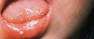
At the initial stage of parenchymal sialadenitis , symptoms are usually absent. The main complaints are related to a feeling of dry mouth. In the future, there may be a painful swelling in the neck, an unpleasant taste in the mouth, and nagging pain. Without treatment, the surface of the salivary gland becomes lumpy, and saliva separation is difficult.
With interstitial sialadenitis, a separate area of the inflamed gland may bother you. Complaints of hearing loss are rarely observed. With increasing inflammation, the salivary gland increases in size; upon palpation, the unevenness of its surface is felt, and the secretion of saliva is impaired.
Ductal sialadenitis affects people of advanced age or with weakened immune systems. Inflammation is accompanied by an obsessive salty taste in the mouth. When pressing on the mouth of the duct, cloudy saliva is released. The mouth of the excretory duct itself is noticeably thickened.
Sialadenitis - symptoms and treatment
Diagnostic measures for diseases of the salivary glands can be divided into clinical, laboratory and instrumental.
Clinical methods include the collection of complaints, anamnesis (medical history), as well as direct examination of the patient.
During the questioning, the patient’s complaints are clarified, the time of their occurrence, the nature, intensity of pain, the impact of these symptoms on the quality of life, the presence of relapses and remissions, and their duration are specified. Separately, it is worth dwelling on questions about the presence or absence of somatic and infectious diseases - sometimes they can be a cause or an aggravating factor in the course of sialadenitis. It is worth clarifying whether parents and relatives had similar conditions.
During the clinical examination, the doctor pays attention to the presence of swelling and asymmetry of the face, the size, consistency, shape and relief of the affected and healthy salivary gland. These data largely depend on the primary nature of the disease, the presence of relapses and the nature of the treatment performed or its absence. The more relapses there were, the more sclerotic the gland was, which negatively affected its functioning. In the oral cavity, you should pay attention to the mouth of the excretory duct, and also examine the excretory duct itself (if possible) for the presence of salivary gland stones and other pathological changes. It is important to determine whether there is salivation. To do this, massage the gland tissue, after which the amount of saliva, its color and consistency are assessed.
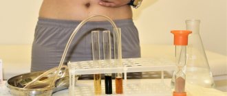
Laboratory methods are required if sialadenitis is suspected. Blood, urine and saliva itself are examined. A general blood test can detect leukocytosis (increased levels of white blood cells) - the primary sign of inflammation. In blood biochemistry, special attention should be paid to blood glucose levels, and in urine - to the amount of salts. In saliva you can find a large number of leukocytes, impurities and pus, possibly the presence of bacteria and sand. Physicochemical parameters of saliva are given special attention [8].
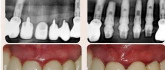
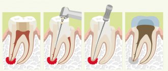
One of the first hardware diagnostic methods for diseases of the salivary glands includes an x-ray of the gland - sialography . It allows you to determine the presence of stones in the thickness and excretory duct of the gland. sialography with contrast appeared , with the help of which it is possible to detect not only stones, but also to identify a narrowing of the lumen of the excretory duct, the presence of cysts and other neoplasms that impede the normal functioning of the gland.
The most modern sialogram method is digital dynamic sialography , through which it is possible to eliminate the overlap of the bone components of the jaws, visualize the soft tissue component of the gland and evaluate the flow of saliva through the duct. Among other things, it significantly reduces the radiation dose to the patient.
Computer and magnetic resonance sialotomography of small foreign bodies (stones) in the excretory duct and the gland itself.
The sialosonography method gives a fairly complete picture of the structure of the gland. With its help, you can easily identify sclerotic changes in tissues, foreign bodies, estimate their number, density, size, and also exclude the presence of neoplasms.
Thermosialography makes it possible to study the dynamics of changes in the temperature of the gland. This allows you to evaluate the effectiveness of the treatment.
Ultrasound-guided salivary gland biopsy is a fairly common diagnostic method. It is especially effective in the presence of cavities (cysts) in the thickness of the glandular tissue [9].
Currently, both throughout the world and in Russia, the most minimally invasive and informative method for diagnosing lesions of the salivary glands is widespread - sialoendoscopy . Modern devices help to visualize not only the excretory and intraglandular ducts, but also the ductal system, up to ducts of 2-3 orders, sometimes it is even possible to examine ducts of 4-5 orders.
The presence of a second working channel in the endoscope body allows not only visualization, but also washing, expansion of the duct, and, if necessary, collection of biopsy material. Using an endoscope, you can evaluate the color of the walls of the excretory duct, their elasticity, and detect the reasons that impede the normal flow of saliva through the ducts - stones, mucus plugs, polyps, neoplasms, areas of narrowing of the lumen of the duct. Also, thanks to endoscopic support during surgery, you can not only get a complete picture of the problem, but also eliminate it with minimal intervention.
Diagnosis of sialadenitis
Diagnosis involves a thorough analysis of the patient’s complaints, as well as palpation of the salivary gland. To clarify the diagnosis, cytological studies of salivary secretions are possible. , radiography with a contrast agent may be prescribed . The purpose of the study is to evaluate the gland tissue and the patency of the salivary ducts. X-rays are performed only after acute inflammation has been eliminated. The goal of diagnosis is to differentiate chronic sialadenitis from other diseases with similar symptoms:
- acute bacterial or viral sialadenitis;
- benign neoplasms;
- Herzenberg's pseudomumps;
- sialadenosis.
1.General information
Sialolithiasis, or calculous sialadenitis, is the formation of calculi (stones) in the salivary gland, which is reflected in the Russian term “salivary stone disease.” This disease was known back in the time of Hippocrates, but to this day many of its aspects remain virtually unstudied.
Thus, thanks to special medical and statistical studies, the true epidemiological picture of sialolithiasis, which 2-3 decades ago was considered a rare pathology and of interest mainly to specialists, began to become clearer. In fact, it turned out that it is sialolithiasis that underlies a significant proportion of clinical syndromes and symptom complexes that sooner or later require the intervention of a maxillofacial surgeon. Frequency estimates in different sources still differ widely (the specific share of sialolithiasis in the total volume of diseases of the salivary glands is today estimated in the range from 30% to 80%), but it is already obvious that the problem is much more pressing than previously seen.
Data have been published showing that salivary stone disease is more common in men. Other authors do not find a reliable statistical dependence on gender, however, in the studied samples of patients, they reveal a three-fold predominance of city dwellers over rural residents. Even greater discord is observed in estimates of age-related incidence trends: data is provided that sialolithiasis is most often found in children of early school age, but the results of other studies refute this, and the age of primary primary diagnosis of salivary stone disease is determined in the range of 30-40 years.
Statistics of predominant localization, on the contrary, are distinguished by uniformity of estimates: in the vast majority of cases (more than 90%), sialolithiasis affects the submandibular gland, much less often detected in the parotid gland, and extremely rarely in all other salivary glands - sublingual or small (palatal, molar, buccal, etc.) .d.).
A must read! Help with treatment and hospitalization!
Treatment of chronic sialadenitis
Treatment of chronic sialadenitis is aimed at preventing changes in gland tissue, normalizing saliva secretion and eliminating the inflammatory process.
During an exacerbation, the dentist prescribes a course of broad-spectrum antibiotics. In the absence of a bacterial infection, therapy may be limited to antiseptic rinses and the administration of a vitamin complex to support immunity. In complex therapy, it is possible to carry out physiotherapeutic procedures, such as electrophoresis or magnetic laser therapy. In rare cases, if conservative treatment is ineffective and the gland loses function, surgical intervention is possible.
2. Reasons
The etiopathogenesis of calculous sialadenitis is currently being actively studied and clarified. Apparently, the disease should be considered polyetiological and multifactorial, i.e. One or more of many possible causes may lead to its development, making prediction and prevention extremely difficult.
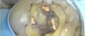
The most significant is considered to be the role of congenital anomalies in the structure of the salivary gland, in which the outflow of saliva and the leaching of solid organic and mineral microparticles are hampered. Amino acids predominate in the composition of stones.
The forming sialolith (sizes in different cases vary from fractions of a millimeter to several centimeters), in turn, expands the duct, creating abnormal mechanical pressure. Stagnation in the salivary gland, as in any other organ, creates direct preconditions for infection and inflammation; sooner or later an infection occurs, which forms a characteristic clinical picture.
Risk factors include an imbalance in the circulation of calcium and phosphorus compounds in the body, vitamin A deficiency, etc.
Visit our General Surgery page
4.Treatment
With small sizes and significant mobility of stones, it is sometimes possible to induce their spontaneous evacuation (heating and means of stimulating salivation are used). However, the effectiveness of conservative methods is low and unpredictable. More often the question is raised about surgical removal of the stone or the entire gland as a whole. Considering the abundant vasculature and innervation, as well as the development of irreversible histological changes in the most advanced and advanced cases, such an intervention is always very complex and risky in terms of consequences.
Therefore, you should consult a doctor at the first appearance of the discomfort or pain described above.
Recently, the possibilities of clinical application of alternative high-tech methods (for example, extracorporeal lithotripsy) have been studied, but the methodology is still at the stage of clinical development and is not widely used (at least in Russia). Minimally invasive sialoscopy, a method based on the use of ultra-thin endoscopes with built-in operating manipulators, should apparently be considered the most promising.