Publication date: 04/02/2021
Dental exostosis is a benign formation on the tissues of the jaw bone or on the palatal surfaces. Exostosis is manifested by the formation of hard – bone or osteochondral growths – osteophytes.
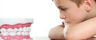
Thickening and overgrowth of bone tissue often occurs in children and adolescents. In this case, you need to contact a pediatric dentist. Exostosis can also develop in patients who have suffered a fracture of the jaw bone (then exostosis is localized at the site of fusion of bone fragments) or trauma.
Causes of osteophytes
There is no absolute understanding of the etiology of the process of the appearance of osteophytes. But it can be noted that:
- The risk of developing exostosis increases if there have been inflammatory or tumor processes in the bone tissue.
- Exostosis may appear after tooth extraction. Especially when it comes to the problematic removal of impacted or dystopic wisdom teeth. This is explained by the high probability of bone trauma during this procedure.
- A long and advanced process of periodontal disease can provoke bone growths.
General overview
Classifications of edentulous jaws play an important role in dental science.
They allow specialists to adhere to uniform terminology and features of determining existing anomalies in the structure of the dentofacial rows. Thanks to generally accepted classification criteria developed by renowned scientists and doctors of medical sciences, orthopedic specialists are able to accurately plan further treatment and determine in advance what problems may be encountered during therapeutic measures.
Symptoms of exostoses of the jaw
In most cases, exostosis does not cause pain or discomfort. Almost the only way to recognize pathology at an early stage is an x-ray, which the doctor sends to a patient who comes in for caries, pulpitis or other problems. However, its peculiarity is the constant increase in size of osteophytes. This means that at a certain stage the patient can feel their presence in the oral cavity.
- The tongue feels a hard formation under the mucous tissue.
- If osteophytes are located on the jaw bones and have grown greatly, discomfort or even pain may occur due to pressure on the surrounding nerve endings.
- Visually, exostosis is visible as an area of a lighter shade than the surrounding mucous tissue on the gum or palate.
- In serious cases, large and advanced exostosis can cause a lisp and other speech defects.
- If pathological bone growths are localized in the “eight” area, they can provoke problems with wide mouth opening.
- The oval of the face changes.
How to get rid of bone growth in your mouth
Before removing the growth, an x-ray of the jaw is taken.
Bone spines in the mouth on the right or left are often discovered by chance during a dental examination. The main method of differential diagnosis is x-ray and study of the medical history.
Exostosis of the jaw resolves on its own in 50% of cases. This happens when you change your diet, eliminate vitamin deficiency and disrupt salt metabolism in the body.
If the growth on the jaw grows, begins to hurt, and interferes with chewing and talking, it is surgically removed in the clinic. Excision of the spine is also carried out before prosthetics. Contraindications – disorders of the endocrine system, adrenal glands, diabetes, poor blood clotting. There are no medications or folk remedies for removing osteophytes.
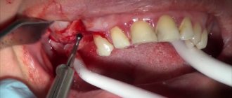
The operation is simple and is performed under local anesthesia. After treating the oral cavity with an antiseptic, a small incision is made on the gum, the base of the growth is cut off with a laser or bitten with dental forceps.
Using a special attachment on a drill, smooth out all sharp corners on the bone. Stitches and wound healing ointment are applied.
Treatment at home
After removal of exostosis, it is necessary to maintain oral hygiene.
Further treatment takes place at home. Before the wound heals completely, you need to provide the oral cavity with proper care. Every day, rinse your mouth with Miramistin, Chlorhexidine or soda solution. Procedures cannot be performed on the first day after surgery. After a day, passive rinsing is indicated - take the solution into your mouth, hold for 5-7 minutes without performing active actions. Hydrogen peroxide and potassium permanganate should not be used for mouth rinsing.
Cotton pads soaked in Levomekol or Solcoseryl ointment should be applied to the incision. Sometimes doctors prescribe antibiotics to prevent bacterial infections from developing. If the immune system is functioning normally, the recovery period is 7 days.
You can also rinse your mouth with folk remedies based on herbs with antiseptic and anti-inflammatory effects:
- a collection of equal parts of sage and mint;
- a mixture of lemon balm and oak bark;
- oregano and chamomile;
- nettle and birch;
- Golden mustache;
- eucalyptus;
- soda and salt - dissolve 1 tsp each in 250 ml of warm water.
To prepare any herbal infusion, pour 2 tsp. raw materials 200 ml of water at room temperature. Simmer in a water bath for 20 minutes. Strain and use warm.
To prevent suture dehiscence, you can only eat soft pureed food, soups and broths for 2 weeks after surgery. Avoid hard and viscous foods, hot and cold foods, smoking and alcohol.
Online consultation with a doctor
If you are bothered by a growth on the jaw bone that you feel with your tongue, it bothers you, or causes discomfort, then it is best to undergo an examination and seek advice from a dentist. Because these may be symptoms not just of exostosis, but of a more global disease, which, if neglected, will lead to the necessary more complex and expensive treatment: cytology testing and surgical intervention. The specialist will listen carefully, visually assess the condition and color of the fabric and give appropriate recommendations. With this information, you can safely begin treatment.
Classification of toothless jaws
The classification to a certain extent determines the treatment plan, facilitates the relationship between doctors and facilitates recording in the medical history; the doctor clearly understands what typical difficulties he may encounter. None of the known classifications claims to be an exhaustive description of toothless jaws, since there are transitional forms between their extreme types.
Schroeder (1927) distinguished three types of upper edentulous jaws.
The first type is characterized by a well-preserved alveolar process, well-defined cusps and a high palatal vault. The transitional fold, the places of attachment of muscles, folds of the mucous membrane are located relatively high.
This type of toothless upper jaw is most favorable for prosthetics, since there are well-defined points of anatomical retention (high arch of the palate, pronounced alveolar process and tubercles of the upper jaw, high points of attachment of muscles and folds of the mucous membrane that do not interfere with the fixation of the prosthesis).
In the second type, an average degree of atrophy of the alveolar process is observed. The latter and the cusps of the upper jaw are still preserved, the palatine vault is clearly defined. The transitional fold is located somewhat closer to the apex of the alveolar process than in the first type. With a sharp contraction of the facial muscles, the fixation of the prosthesis may be disrupted.
The third type of toothless upper jaw is characterized by significant atrophy: alveolar processes and tubercles are absent, the palate is flat. The transitional fold is located in the same horizontal plane with the hard palate.
When prosthetizing such a toothless jaw, great difficulties are created, since in the absence of the alveolar process and tubercles of the upper jaw, the prosthesis gains freedom for anterior and lateral movements when chewing food, and the low attachment of the frenulum and transitional fold contributes to the shedding of the prosthesis.
A.I. Doynikov expanded Schroeder’s classification by adding to it:
The fourth type is a well-defined alveolar process in the frontal region and significant atrophy in the lateral regions
Fifth type - Pronounced alveolar process in the lateral sections and significant atrophy in the frontal section.
Keller distinguished four types of edentulous mandibles.
In the first type, the alveolar parts are slightly and uniformly atrophied. The evenly rounded alveolar ridge is a convenient base for the prosthesis and limits its freedom of movement when moving forward and to the side.
The attachment points of the muscles and folds of the mucous membrane are located at the base of the alveolar part. This type of jaw occurs when teeth are removed at the same time and alveolar ridge atrophy occurs slowly.
It is most convenient for prosthetics, although it is observed relatively rarely.
The second type is characterized by pronounced but uniform atrophy of the alveolar part.
In this case, the alveolar ridge rises above the bottom of the cavity, representing in the anterior section a narrow, sometimes even sharp, like a knife, formation, unsuitable as a base for a prosthesis. The muscle attachment sites are located almost at the level of the ridge.
This type of edentulous lower jaw presents great difficulties for prosthetics and obtaining a stable functional result, since there are no conditions for anatomical retention, and the high location of muscle attachment points during their contraction leads to displacement of the prosthesis. Using a prosthesis is often painful due to the sharp edge of the maxillary-hyoid line, and prosthetics in some cases are successful only after smoothing it.
The third type is characterized by pronounced atrophy of the alveolar part in the lateral sections with relatively preserved alveolar ridge in the anterior section. Such a toothless jaw is formed with the early removal of chewing teeth.
This type is relatively favorable for prosthetics, since in the lateral sections between the oblique and mylohyoid lines there are flat, almost concave surfaces, free from muscle attachment points, and the presence of a preserved alveolar part in the anterior part of the jaw protects the prosthesis from displacement in the anteroposterior direction.
In the fourth type, atrophy of the alveolar part is most pronounced in the front, with its relative preservation in the lateral sections. As a result, the prosthesis loses support in the anterior region and slides forward.
I.M. Oksman (1967) proposed a unified classification for the upper and lower edentulous jaws.
According to his classification, there are four types of upper edentulous jaws.
In the first type , a high alveolar part, high tubercles of the upper jaw, a pronounced arch of the palate and a high location of the transitional fold and points of attachment of the frenulum are observed.
The second type is characterized by moderate atrophy of the alveolar ridge and cusps of the upper jaw, a shallower palate and lower attachment of the mobile mucous membrane.
The third type is characterized by significant but uniform atrophy of the alveolar edge, tubercles, and flattening of the palatine vault. The mobile mucous membrane is attached at the level of the apex of the alveolar part.
The fourth type is characterized by uneven atrophy of the alveolar ridge, i.e. combines various features of the first, second and third types.
The first type of toothless lower jaw is characterized by a high alveolar ridge, low location of the transitional fold and points of attachment of the frenulum.
In the second type, moderately expressed uniform atrophy of the alveolar part is observed.
This is interesting: Is it necessary to remove dentures at night: types of dentures, material, rules of use and storage, oral hygiene and dental advice
The third type is characterized by the absence of the alveolar edge; sometimes it is present, but poorly. Atrophy of the jaw body is possible.
In the fourth type, uneven atrophy of the alveolar part is noted, which is a consequence of the removal of teeth at different times.
V.Yu. Kurlyandsky proposed his classification of types of lower jaw.
This classification takes into account both the atrophy of the alveolar process and the topography and sites of muscle attachment:
The first type - the alveolar process protrudes above the level of muscle attachments on the inner and outer sides.
The second type - the alveolar process and the body of the jaw are atrophied to the level of the muscle attachment points on the inner and outer sides.
The third type is atrophy of the jaw body below the level of muscle attachment.
The fourth type is more pronounced atrophy in the area of the chewing teeth.
The fifth type is more pronounced atrophy in the area of the front teeth.
Anatomical
It is removed using standard impression trays and a large amount of dental plaster. Has high edges.
Functional tests are not used in this case, as a result of which the condition of the tissues bordering the prosthetic bed is not taken into account.
Functional
To make this type of impression, a personal spoon and special functional tests are used, with the help of which the mobility of the folds of the mucous membrane is reflected.
The edges of the impression are slightly lower than those of the previous type, and the border of the manufactured prosthesis covers the mucous membrane by no more than 2 mm.
Based on pressure on the oral mucosa, functional impressions are divided into three types:
- unloading – removed using a plaster mass using minimal pressure on the mucous membrane;
- compression - used when the mucous membrane is highly pliable, and is performed under pressure using silicone, gypsum or thermoplastic mass;
- combined - allow you to compress areas of the mucosa with high compliance, without overloading areas with low compliance.
Unloading impression
With its help you can minimize pressure on the mucous membrane. Relief impressions are made using plaster without pressure.
There are 2-3 holes on the palatal side of the individual spoon. When pressed, excess plaster flows out through them. This minimizes pressure on the palate.
Compression impression
It is used for pliability of the mucous membrane. It is removed using thermoplastic, silicone and alginate materials. They are inserted into the mouth under pressure. In some cases, gypsum can also be used. However, in this case the pressure must be continuous. There should be no holes in the spoon.
Functional suction impression
It is also removed with an individual spoon. However, the boundaries of such a print should be slightly larger and overlap the neutral area by 1-2 mm. The oral edge of the upper part should be 1-2 mm behind the “A” line.
Diagnostics
If there are visual symptoms of exostosis, the doctor prescribes diagnostic measures to the patient to confirm the disease and differentiate benign bone growth from malignant processes. For this purpose, a person is sent to:
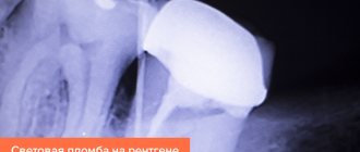
- X-ray – after receiving the image, the dentist sees exactly the size of the formation, its location, as well as the presence of contact with the roots of neighboring teeth, which can lead to damage.
- Biopsy and tissue cytology - based on the research, one can make an unambiguous conclusion about whether the bone growths are osteophytes and the patient has exostosis, or whether it is a tumor process.
Diagnosis of exostosis after tooth extraction or injury
In order to make a correct diagnosis, CELT dentists:
- listen to the patient’s complaints and collect anamnesis;
- conduct a dental examination;
- The patient is prescribed an x-ray.
Examination reveals the presence of a dense formation that is not fused to the soft tissues. The mucous membrane over it is thinned and can be damaged by the sharp edges of the destroyed units. The x-ray image clearly shows a protrusion with clear boundaries. Jaw osteophytes must be differentiated from bone tumors of benign or malignant etiology.
Pros and cons of prints
Some experts are against the use of unloading impressions. This position is based on the fact that all chewing pressure falls on the alveolar process. In this regard, its atrophy begins.
Dentures made using compression impressions rest on the tissues of the buffer areas, like cushions. In this case, the alveolar process remains unloaded. When chewing under pressure, the vessels of the buffer area are emptied of blood. The prosthesis puts pressure on both the buffer zones and the process. As a result, the latter does not atrophy.
Sources:
- https://spas-dent.ru/drugoe/klassifikatsiya-po-oksmanu-ortopediya.html
- https://estetikaplus29.ru/drugoe/klassifikatsii-bezzubyh-chelyustej-po-shrederu-kelleru-oksmanu-kurlyandskomu-i-dojnikovu.html
- https://www.vash-dentist.ru/protezirovanie/semnyie-p/klassifikatsiy-bezzubyih-chelyustey.html
- https://zubodont.ru/klassifikacija-bezzubyh-cheljustej/
- https://FB.ru/article/332400/osnovnyie-klassifikatsii-bezzubyih-chelyustey
- https://rsdent.ru/shredera-klassifikaciya
Changes in the mucosa
Atrophy of the bone bed is accompanied by changes in the mucosa, which should also be taken into account when making prosthetics. The nature of the mucous membrane is different. There are several types of mucous membrane, based on its mobility and pliability.
Mobility is influenced by the connection between the mucosa and the muscles. In the cheek area, the mucous membrane is located on the muscles and makes movements at the moment of muscle contraction; it is called actively mobile.
In the area where submucosal tissue is found under the mucosa, and under it there is adipose tissue and glands, the mucosa is called sedentary and good pliability under pressure is noted. Fused with the periosteum, the mucosa is considered immobile.
Types of mucous membrane of the prosthetic bed
It is characterized by the degree of pliability, sensitivity and mobility. There are three types of mucosa:
- Normal. It is characterized by moderate pliability and good moisture. The mucous membrane has a pale pink color. It is considered the most favorable for installing a prosthesis.
- Hypertrophied. When palpated, the mucous membrane is loose, with a high content of intermediate substance. Features good hydration. With this type of mucosa, it is not difficult to create a valve, but the prosthesis will be mobile due to the pliability of the membrane.
- Atrophied. This mucous membrane is distinguished by its high density and whitish color. The shell is dry. It is considered the most unfavorable for the installation of a prosthesis. The mucous membrane covering the maxillary alveolar process is immovably connected to the periosteum. Almost along its entire length it consists of its own layer and flat multilayered epithelium. On the latter, in the area of the process, there is a stratum corneum.