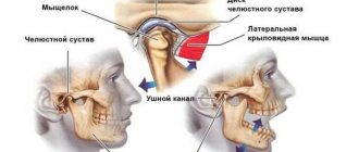
Treatment of
jaw
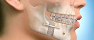
fractures is a characteristic of conservative and surgical treatment methods. Treatment of any injury must begin with first aid. For fractures of the maxilla, it is necessary to restore the prominence and height of the face and recreate the pre-traumatic occlusion. The jawbone is often stable, so treatment is carried out by fixing the upper and lower teeth together. Various options for osteosynthesis of facial bones with titanium mini-plates. When the upper jaw is fractured, its fragments are displaced downward, disrupting the usual relationship of the teeth of the upper and lower jaw and somewhat lengthening the face.
PROVIDING ASSISTANCE FOR FRACTURES OF THE UPPER AND LOWER JAWS
Qualified assistance is provided before admission to a specialized hospital.
When providing qualified surgical care, the surgeon must:
1) anesthetize the fracture site;
2) inject antibiotics into the wound and administer antibiotics internally;
3) carry out simple transport immobilization, for example, apply a standard transport bandage;
4) make sure there is no bleeding from the wound, asphyxia or its threat during transportation;
5) carry out anti-tetanus measures according to the instructions;
6) ensure proper transportation to a specialized medical institution, accompanied by medical personnel (determine the type of transport, the position of the patient);
7) clearly indicate in the accompanying documents everything that was done to the patient.
Patients with complex and complicated facial injuries should be referred to a specialized department if there is a need for primary plastic surgery of soft tissues and the use of the latest methods for treating facial bone fractures, including primary bone grafting.
Temporary (transport) immobilization of fractures of the lower and upper jaw. It is carried out outside a specialized medical institution or at the scene of an incident by paramedics, doctors of other specialties, sometimes in the form of mutual assistance.
For temporary immobilization use:
1) Circular bandage parietal-chin bandage
2) Standard transport bandage
3) Soft chin sling Pomerantseva-Urbanskaya
4) Intermaxillary ligature fastening
Circular bandage parietal-chin bandage.
Circular tours of the bandage, passing through the chin of the lower jaw and parietal bones, do not allow fragments to move during transportation of the victim. For this purpose, you can use a mesh elastic bandage.
Standard transport bandage.
A standard transport bandage consists of a rigid chin sling and a support cap (dimensionless). The latter has 3 pairs of loops for fixing rubber rings, which tightly press the sling to the chin area. Under the loops there are fabric pockets for cotton pads, which allow you to remove the rubber rings from the swollen soft tissues of the face and prevent their injury. The cap is placed in such a way that it tightly covers the occipital protuberance, and its straps are tied on the forehead. A rigid chin sling is made with a cotton-gauze liner so that it covers the edges of the sling along its entire perimeter. This prevents direct contact of the rigid structure with swollen soft tissues, and can also serve as a protective bandage in case of damage to the skin of the chin area. Depending on the number of pairs of rubber rings used in the cast, the sling can hold the fragments without pressure or apply pressure to them. For fractures of the lower jaw behind the dentition or for a fracture of the upper jaw, a standard bandage can be applied using 3 pairs of rubber rings (as a pressure bandage)
In case of fractures of the lower jaw within the dentition, it should be applied only to support the fragments. Excessive pressure on displaced fragments will lead to even greater displacement with the risk of developing asphyxia. However, such a differential approach is only possible in a specialized department where there is a dental surgeon. Non-specialists should be advised to apply a standard transport dressing as support.
Soft chin sling by Pomerantseva-Urbanskaya.
Its chin part is made of several layers of canvas or calico. The intermediate section is represented by two wide elastic bands (haberdashery), which go into the peripheral section of the bandage, made of the same material as the chin part. The latter has lacing, which allows you to adjust the degree of tension of the rubber strips of the sling. This bandage is comfortable for patients, easy to use and provides good fixation of fragments
Therapeutic immobilization of jaw fragments using laboratory-made splints
Laboratory-made splints are classified as orthopedic immobilization methods. They perform both an independent immobilization function and can be an additional device for various surgical methods of fastening fragments. Removable orthopedic structures include supragingival splints (simple or inclined plane Weber supragingival splint, Vankevich splint, Vankevich-Stepanov splint) and Porta supragingival splint. Non-removable orthopedic structures include dental splints with fixing elements of various modifications. Indications for the use of laboratory-made splints : - severe injuries of the jaws with significant defects of bone tissue, in which bone grafting of the jaw is not performed; — the presence of concomitant diseases in the victim (diabetes mellitus, stroke, etc.), in which the use of surgical immobilization methods is contraindicated; — the patient’s refusal to surgically fix the fragments; — the need for additional fixation of fragments simultaneously with the use of wire splints. To produce laboratory splints, the following conditions are required: a dental laboratory, special materials. Dental work is carried out by dental technicians.
Simple Weber dentogingival splint.
Can be used independently or as one of the main elements when using the surrounding suture method for fractures of the lower jaw. The Weber splint is used for significant defects of the lower jaw as a result of traumatic osteomyelitis or after resection of the lower jaw for a tumor. In these cases, long-term wearing of the splint (for 2-3 months) can lead to the elimination of pronounced lateral displacement of the lower jaw after removal of the splint. The Weber splint is prepared in the laboratory, after taking impressions of jaw fragments. To prevent lateral displacement of fragments, an inclined plane is made on it in the area of the molars. You can make a splint directly in the patient’s mouth from quick-hardening plastic.
Bus Vankevich and bus Vankevich-Stepanova.
They are dental splints supported on the alveolar process of the upper jaw and the hard palate. In the lateral sections it has two inclined planes facing downwards, which abut against the anterior edges of the branches or the alveolar part of the lateral sections of the body of the lower jaw, mainly from the lingual side and do not allow fragments of the lower jaw to move forward, upward and inward. The Vankevich splint is used to fix and prevent lateral and rotational displacement of fragments of the lower jaw, especially with significant defects, due to the emphasis of the inclined planes on the anterior edges of the branches of the jaw. The Vankevich splint as modified by Stepanov differs in that instead of a maxillary base there is a metal arch, like a clasp prosthesis. The Port splint is used in case of a fracture of the edentulous lower jaw without displacement of the fragments and the patient does not have removable dentures and teeth in the upper jaw. The splint consists of two base plates for each jaw, similar to complete removable dentures, rigidly connected to each other in the position of central occlusion. There is a hole in the front section of the tire for food intake. The Porta splint is used in combination with wearing a chin sling bandage.
Dental splints with fixing elements.
Used for immobilization of fragments of the lower jaw in the presence of a bone tissue defect within the dentition, when the fragments have a sufficient number of stable supporting teeth. These splints consist of metal caps fitted to the teeth of the lower jaw. The caps are soldered together and fixed on the teeth of each fragment. With the help of various locks (pins, levers, etc.), after their reposition, the fragments are secured for the period necessary for consolidation. The teeth used for splinting are not prepared.
INTERDENTAL AND INTERMAXILLARY LIGATURE BINDING:
Requirements for using the method:
1) on each fragment there are at least two adjacent stable teeth and two antagonist teeth;
2) the bandage should not include teeth located in the fracture line, with signs of periodontitis and pulpitis, and having pathological mobility.
Contraindications to the application of intermaxillary ligature bonding:
1) concussion;
2) the possibility of bleeding in the oral cavity;
3) danger of vomiting;
4) transportation of the patient by water or air.
Among the many types of intermaxillary ligature fastening, the following are most often used: simple, figure eight, according to Ivey.
a) simple ligature binding.
With a simple intermaxillary ligature fastening, the end of a ligature wire 5-6 cm long is passed into the interdental space, covers one of the teeth included in the bandage from the lingual side and returns it through the other interdental space to the vestibule of the mouth. On the vestibular side, both ends of the wire are twisted together. The twisted wire tightly wraps around the neck of the tooth. The second ligature is fixed in the same way on the adjacent tooth. These two wires are then twisted together, combining the two teeth into one bandage. A similar bandage is applied to the teeth of the second fragment, then to the antagonist teeth. Having reduced the fragments, they are brought into contact with the teeth of the upper jaw and fixed in this position by twisting the wire extending from the teeth of the lower and upper jaws with each other on each side in turn. The ends of the wire are cut with scissors for cutting metal and bent so that they do not injure the mucous membrane of the cheek and gums.
b) figure eight ligature binding.
When fastening in the form of a figure eight, both ends of a ligature wire 6-8 cm long are passed into the interdental spaces from the vestibular side to the oral side so that the wire covers two teeth included in the bandage at once. Then both ends of the wire are returned to the vestibular side, passing them through the gap between the teeth included in the bandage. In this case, one end is passed over the wire covering the teeth from the vestibular side, and the other - under it. On the vestibular surface, the ends of the wire are twisted together. Then the same bandage is applied to the teeth of the second fragment and the antagonist teeth. As in the previous case, the wire fixed to the teeth of the upper and lower jaws is twisted together. The excess is cut off with scissors.
c) ligature binding according to Ivey
When fastening according to Ivy, a wire 10 cm long is first bent into the form of a hairpin, leaving one end 1-1.5 cm longer than the other. At the end of the “hairpin” a loop with a diameter of about 0.2 mm is formed. To do this, you can use a small piece of aluminum wire, crampon tongs, or tweezers. Both ends of the wire are passed from the vestibular side to the oral side between the teeth included in the bandage. The long end of the wire is returned to the vestibular surface through the interdental space located posterior to the loop and passed through it. The short end is brought out to the vestibular side through the interdental space located anterior to the loop and twisted with the long end. The excess wire is cut off by bending the remaining end about 0.5 cm long so that it does not injure the mucous membrane of the cheek. The same bandage is applied to the teeth of the second fragment, the antagonist teeth. The fragments are reduced and fixed to the teeth of the upper jaw with wire passed through the loops of the ligature bandage on each side.
This method has some advantages over the simple one: it is less traumatic, it allows you to examine the oral cavity without removing the entire structure, but only by cutting off the ligatures connecting the teeth.
Therapeutic (permanent) immobilization using non-laboratory dental splints
Individual dental wire splints by Tigerstedt. Types of Tigerstedt dental splints: - smooth splint-bracket; — bus-bracket with a spacer bend; - tire with hook loops.
The splints are made of aluminum wire d=1.8-2.0 mm and 12-15 cm long. They are tied to the teeth using bronze-aluminum wire d=0.5-0.6 mm. The splint is bent individually for each patient using crampon forceps. General rules for applying dental splints . 0.5 ml of a 0.1% atropine solution is injected subcutaneously to reduce salivation, splinting is carried out under local anesthesia, it is necessary to remove tartar to freely pass the ligature into the interdental space, bend the splint from the side of the fracture, try it on the teeth in the mouth, and bend it outside the oral cavity, the splint should be adjacent to the neck of each tooth at least at one point, the splint is tied to each tooth with a ligature wire, which is twisted clockwise. The production of the splint begins with bending a large hook that clasps the first tooth, or a hook spike inserted into the interdental space. To try on a splint, it is applied to the teeth in the mouth.
Smooth splint.
It is used to treat fractures of the lower jaw, provided that the larger fragment contains at least four, and the smaller fragment contains at least two stable teeth.
Smooth splint
Indications for use : linear fractures of the lower jaw, located within the dentition, without displacement or with easily reducible fragments, fractures of the alveolar process, fractures and dislocations of teeth, tooth mobility in acute odontogenic osteomyelitis and periodontitis, fractures of the upper jaw (Adams and Dingman methods), to prevent pathological fracture of the lower jaw. After treatment, before removing the splint, loosen the ligatures and check the lack of mobility of the fragments by rocking them. The splint is removed after 4-5 weeks. The patient needs to take liquid food. The doctor should regularly examine the patient 2-3 times a week. In this case, it is necessary to monitor the state of the bite, the strength of fixation of the fragments, the condition of the tissues and teeth in the fracture gap. When the fixation of the splint on the teeth becomes loose, it is necessary to tighten the ligatures by twisting them. If the ligature bursts, it is replaced with a new one. The patient is taught hygienic measures to prevent the development of gingivitis. For this purpose, the patient should brush his teeth and splint with toothpaste and a brush 2 times a day, after each meal, remove food debris with a toothpick, and rinse his mouth with antiseptic solutions 3-5 times a day.
Busbar with spacer bend.
The spacer bend prevents lateral displacement of fragments.
Busbar with spacer bend
Indications for use : fracture of the lower jaw within the dentition and the presence of a bone tissue defect of no more than 2-4 cm, fracture of the lower jaw without displacement or with easily reducible fragments, if the fracture gap passes through the alveolar part, devoid of teeth.
Tire with hook loops.
The splint is most often used to treat jaw fractures. Two splints with hooking loops are made for the teeth of the upper and lower jaw.
Tire with hook loops
Indications for use : fractures of the lower jaw outside the dentition, within the dentition - in the absence of four stable teeth on the larger fragment and two stable teeth on the smaller one, fractures of the lower jaw with difficult-to-reduce fragments requiring traction, bilateral, double and multiple fractures of the lower jaw, fracture of the upper jaw (with the obligatory use of a chin sling), simultaneous fractures of the upper and lower jaw. When making a splint, its hook loop should be at an angle of 45° relative to the gum. The towing loops are bent on the tire so that they are located in the area of the 6th, 4th and 2nd teeth. If the patient does not have these teeth, then hooking loops are made in the area of other teeth that have antagonists. Usually, 3-4 hook loops are bent on the splint adjacent to the teeth of the larger fragment, and 2-3 hook loops on the smaller one. The base of the loop must be within the crown of the tooth. If the displacement of the fragments is large and it is difficult to bend one splint onto both fragments, splints can be made and secured to each of the fragments. After their reposition, rubber rings are put on the hook loops at an angle so that they create compression of the fragments, which significantly prevents their movement. Periodically (2-3 times a week) the patient is examined, ligatures are tightened, rubber rings are changed, the vestibule of the mouth is treated with antiseptic solutions, and the condition of the bite is monitored. 10-25 days after applying the splint, an X-ray examination is performed to monitor the position of the fragments. After fusion of the fragments, before removing the splints, it is necessary to remove the rubber rings and allow the patient to walk for 1-2 days without fixation, eating soft food. If the fragments do not move, the splints are removed. If there is a slight change in the bite, the rubber traction is retained for another 10-15 days.
Splinting using the A.P. method Vikhrov and M.A. Slepchenko.
The authors proposed using a polyamide thread to strengthen the attachment of the splint to the teeth. To do this, take a bronze-aluminum wire ligature, fold it in the form of a hairpin and insert both of its ends into one interdental space from the mouth towards the vestibule of the mouth. The ligature is tightened so that a small loop is formed on the lingual surface of the interdental spaces. A similar procedure is performed in the area of all interdental spaces. Take a polyamide thread with a diameter of 1 mm and pass it through all the loops on the lingual side, the ends of the thread are brought out into the vestibule of the mouth behind the last teeth on both sides. Next, a previously made splint is placed on the teeth so that it is located between the two ends of the same previously made bronze-aluminum ligatures, which are then twisted. According to the authors, the advantages of their method are the following: stronger fastening of fragments, reduction of splint fastening time, and absence of trauma to the gum mucosa.
Splinting using the Vikhrov-Slepchenko method
Tooth standard splints.
Good manual skills are required to make custom wire splints. Their production requires a lot of time and frequent fitting to the dental arch. It is especially difficult to bend them in case of malocclusion, dental dystopia, etc. Taking into account the above, standard splints were proposed, which are manufactured in the factory, do not require bending of the hook loops and simplify splinting. In Russia, standard belt tires were proposed by V.S. Vasilyev. The tire is made of a thin flat metal strip 2.3 mm wide and 134 mm long, which has 14 hooking loops. The tire bends easily in the horizontal plane, but does not bend in the vertical plane. The Vasiliev splint is cut to the required size, bent along the dental arch so that it touches each tooth at least at one point, and tied to the teeth with ligature wire. The advantage of the splint is the speed of its application . The disadvantage is the impossibility of bending it in a vertical plane, which does not allow to avoid injury to the mucous membrane in the lateral parts of the jaws due to the discrepancy between the splint and the curve of Spee. This splint is not suitable for single-jaw splinting due to its low strength. Abroad, there are various designs of standard tires made of steel wire (Winter tires) and polyamide materials that can be bent in any plane. Tires are manufactured with pre-made hooks.