Jaw cancer is a malignant neoplasm that affects bone tissue. The disease has no age restrictions; it is characterized by rapid growth, metastasis, and pronounced pain. The structural features of the maxillofacial region, proximity to important vessels and nerve centers further complicate treatment.
Anatomical structure
The oral cavity is the initial section of the digestive tract, in which food is chewed and saliva is produced to digest food. It is involved in the process of breathing, swallowing, articulation and speech.
The composition of the oral cavity includes:
- vestibule (lips, front side of teeth, inner surface of cheeks);
- gums;
- the bottom on which the tongue lies;
- two thirds of the tongue;
- teeth;
- retromolar triangle - the space on the lower jaw behind the third molar;
- hard and soft palate.
Classification
Oral cancer is divided into three types:
- papillary. The nodule in the mucous membrane increases in size and hangs into the oral cavity. The neoplasm progresses slowly;
- infiltrative. The seal on the pinkish mucosa is distinguished by a whitish color, clear contours and shape, and thinning of the membrane around it. On palpation from the side of the cheek, a dense infiltrate is felt. The tumor tends to grow rapidly. The patient complains of unbearable pain;
- ulcerative The most common form of the disease. Ulcers on the mucous membrane do not heal, they grow, and the border around them turns red. The outline is torn and its edges are bleeding.
Tumor metastases appear quickly. Malignant cells grow into the mental, submandibular, and deep jugular lymph nodes. This process is influenced by the thickness and depth of the tumor. Thus, when the tumor deepens by 4-5 mm, metastases occur in 98% of cases. At the T1 stage of oncology, metastasis is detected in half of the cases, and when the T4 stage is reached, distant spread of cancer cells is observed in 85% of cases.
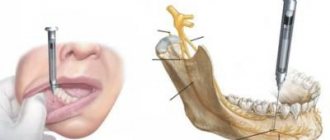
Painkillers
The symptom that most clearly indicates the development of a malignant tumor is pain. An actively growing tumor most often causes pain. Pain can be caused by damage to the nervous tissue, the development of an inflammatory process in the tumor. To alleviate the patient's condition, pain medication is administered. It is prescribed individually, taking into account the patient’s condition, and can significantly reduce the intensity of pain. Pain relief for stage 4 cancer:
- If mild pain occurs, non-steroidal anti-inflammatory drugs are prescribed.
- pain of moderate intensity is treated with combination drugs: ketorol and other potent drugs.
- severe, debilitating pain is treated with the help of strong drugs containing narcotics - fentanyl, morphine, promedol.
Causes
The prevalence of oral cancer is growing and is currently diagnosed in 2% of patients among the total number of cases. Since 2009, the incidence has increased by 25%, with mostly squamous cell carcinoma being detected and only in isolated cases adenocarcinoma.
Most foci of oncology are observed in the tongue. Slightly less malignant formations on the floor of the mouth. Cancer of the soft and hard palate, gums and cheeks is detected in 20% of cases. Much less frequently diagnosed is damage to the alveoli of the lower jaw - 4%, the arches of the palate, retromolar region and vestibule - 3%.
Based on practice, men are more susceptible to oral cancer than women. This is due to bad habits, for example, the abuse of cigarettes or chewing tonic mixtures increases the production of saliva, which washes away beneficial elements from the mucous membrane. The risk group includes patients with HPV, elderly people, workers in hazardous industries, patients with lichen planus, people whose oral mucosa is systematically injured by fillings, prostheses, and metal objects.
Forecast criteria
The survival rate of patients with maxillary sinus cancer averages about 40% over 5 years.
At an early stage, when treated in European oncology clinics, tumors have a cure rate of up to 80%.
Patients with unresectable tumors treated with radiation had a survival rate of less than 20%. Survival rates have now improved slightly due to advances in skull base surgery.
In patients with squamous cell carcinoma of the nasal cavity or maxillary sinus, better survival outcomes are achieved when patients receive adjuvant radiation therapy, adjuvant chemoradiotherapy, or neoadjuvant therapy in addition to surgery.
In patients with sinonasal adenocarcinoma, better disease-free survival was associated with surgery followed by radiation therapy compared with surgery alone, for all tumor stages (T1-T4).
Symptoms
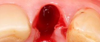
A malignant ulcer from ordinary stomatitis in the mouth can be identified by swelling and swelling of the cheeks, pain and constant discomfort even at rest. You should be wary of prolonged non-healing of the wound and its bleeding.
As the disease progresses, the symptoms intensify:
- swelling increases and spreads to the neck;
- the red or white spot on the oral mucosa intensifies;
- discomfort when chewing and swallowing;
- difficulty speaking due to friction of the mucous membrane on the teeth when moving the jaw;
- the appearance of bad breath;
- feeling of a foreign object in the throat;
- anemia of the mouth.
In the late stages of cancer, teeth fall out and body weight rapidly decreases.
Detection of pathology
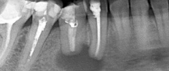
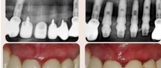
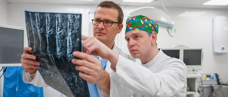
Detection of tumors of the lower jaw is based on a thorough study of the course of the disease, as well as morphological and radiological data. The diagnosis is made by x-ray, since x-rays can detect destructive changes in the bone early.
In the initial stages of sarcoma or jaw cancer, x-rays show bone loss. Its changed area does not have clearly defined boundaries; they are blurred. If jaw cancer is localized in the area of the alveoli, then the cortical plates of its walls are destroyed, and an extensive zone of destruction of the spongy substance is visible around the circumference. If the process has spread, then the radiograph shows complete destruction of a certain part of the bone.
Histological analysis of the tissues of the surface of the extracted tooth during its mobility is informative. It is always necessary to strive to establish the cause of loose teeth. If the mucous membranes of the mouth have ulcerations and the area is clearly visible, a cytological analysis of the mucosa is performed to confirm the diagnosis.
Diagnostics
At the initial consultation, the doctor examines the oral cavity, examines ulcers, erosions, damage to the mucous membrane, and then takes a smear for examination. To confirm the inflammatory process, the patient is sent for a general and biochemical blood test.
The diagnosis is confirmed by the results of the examination:
- MRI and ultrasound of soft tissues of the neck. The images reveal the localization of the pathology, the depth of germination and the structure of the tumor, compaction from blood and lymph, decomposition of the cortical layer of the bone;
- if metastases are suspected, a fine needle aspiration biopsy of the lymph nodes under the chin, under the jaw and in the upper third of the neck is performed;
- positron emission tomography. Shows the depth of the tumor, as well as early metastases;
- osteoscintigraphy. Skeletal bones are examined to look for displaced cancer cells;
- CT scan of facial bones with contrast. The images show the tumor growing into the neck vessels, jaw or base of the skull.
Metastasis
The process of metastasis of pathology occurs through the lymphogenous route. Most often, metastases are observed in the submandibular zone and fuse with the lower jaw quite early, after which they infiltrate the skin.
If jaw cancer is in an advanced form, then metastasis to the spine, liver and other distant organs is noted. But often metastasis to the cervical lymph nodes and distant organs is not present. In the case of sarcoma of the lower jaw, metastasis to distant organs rarely occurs, and they, as a rule, do not form in regional lymph nodes.
Secondary (metastatic) tumor formations are detected much less frequently compared to primary ones, and they are more often observed in women. Pathological metastases occur in lung, breast, thyroid, and stomach cancer.
In case of metastases, they are resected together with excision of the cervical tissue: if there is one metastasis in the submandibular region, then an upper fascial-sheath excision of the neck tissue is performed on one side; if metastases are present at the site of the branching of the common carotid artery, then a Krail operation is performed, if necessary, fascial-sheath excision cervical tissue.
Treatment
The choice of treatment tactics depends on the stage and extent of the tumor. When the tumor grows rapidly, treatment methods are combined.
Operation
The doctor determines the principle of surgical intervention after determining the stage of the tumor and its spread. If cancer cells have penetrated the periosteum and surrounding tissues, a wedge-shaped, planar or sagittal resection of the jaw is performed. If the examination reveals the growth of cancer cells directly into the bone or the defect is noticed during surgery, segmental resection of the lower jaw is performed. The doctor assesses the lesion on site and determines the thickness of the excised layer.
The next stage of the operation is partial or complete excision of the cervical lymph nodes to prevent metastases if the thickness of the tumor is more than 4 mm or the location of the tumor in the floor of the mouth or on the tongue. If the tumor is located in the midline, then the cervical lymph nodes are excised on both sides. The operation ends with the immediate replacement of damaged tissue.
After removal, the tumor is sent for histological examination. Its size, thickness, depth, edges are assessed. Further treatment is affected by cell growth beyond the boundaries of the capsule of the removed lymph node, and the spread of cancer cells to neighboring organs.
Radiation therapy
Radiation after surgery is prescribed when diagnosing T3, T4, N2, T3 stages of the disease no later than six weeks after tumor removal. The need for radiation therapy increases with perineural invasion of the lymphatic vessels. The total focal dose for all sessions is 60 g, and the single focal dose for one session is 2 g. When metastases are detected on the neck, the SOD increases to 66 g, and if there is no risk of metastasis, the SOD decreases to 50 g.
As the main treatment, radiation therapy is used in a total focal dose of 60-70 g. The procedure is performed five days a week and is combined with chemotherapy. Every three weeks, 100 mg of cisplatin is administered.
Chemotherapy
Anticancer drugs are prescribed before surgery or along with radiation therapy to reduce the size of the tumor. Sometimes therapy is prescribed simultaneously with surgery.
Treatment involves the use of a 5-fluoroacyl regimen together with cisplatin or other agents - carboplatin, methotrexate, bleomycin. They cause a number of side effects, for example, vomiting or nausea, hair loss, decreased appetite, and increased bleeding. Symptoms disappear after treatment, but permanent hearing loss is sometimes observed after taking cisplatin.
The prognosis of oral cancer depends on the stage at which the disease is detected. If treatment is started at stage zero, the disease will stop. It is worth noting that smoking provokes relapse or degeneration of the tumor, so repeated surgery or radiation may be required. Surgery at the first stage increases survival rate to 80-85%, and the combination of radiation therapy with surgery at the second stage by 60-80%. Already at subsequent stages of cancer development, the survival rate is no more than 50%, and all three treatment methods are used simultaneously.
Dispensary observation
Since the tumor can recur and metastasize, after completing the course of treatment the patient is registered with the oncology clinic. The first year you should visit a doctor every month, the second year a preventive examination is carried out every 4-6 months, and then once a year or in case of any ailments. The examination involves an examination - ultrasound and contrast MRI of the soft tissues of the neck, PET, osteoscintigraphy. Consultation with an otolaryngologist, dentist and oncologist is required. The doctor may shorten the period of medical examination if there is a high risk of relapse.
List of references on the topic:
- Gantsev Sh.H. Oncology – M, 2012 – P.204-205.
- Golovin D.I. Errors and difficulties in diagnosing tumors, D.: Medicine. Leningr. department, 2015 305 pp.
- Selected lectures on clinical oncology/Ed. IN AND. Chissova, S.L. Daryalova. – M., 2010
- Matyakin E.G., Alferov V.S. Chemotherapy of head and neck tumors // Mat. 2nd Ros. oncol. conf. “Current trends in the development of drug therapy for tumors” December 8–10, 2016 – M., 256 p.
- Tumors of the head and neck: hands/ A.I. Paches. - 5th ed., add. And revised - M.: Practical Medicine, 2013. -478 p.
- Shine A.A. Oncology. M – 2014 365 pp.
- Encyclopedia of Clinical Oncology/Ed. M.I. Davydova. – M., 2014 –P.140-179.
- Bityutsky P.G., Kitsmanyuk Z.D., Trofimov E.I. Diagnosis and treatment of cancer of the oral mucosa // Medical consultations. - 2014. - No. 1. - P. 23-27.
- Byakhov M. Yu. Options for combined and complex treatment of locally advanced cancer of the oral mucosa and oropharynx: Dis. Dr. med. Sci. - M., 2013.
Rehabilitation period
- Adjusting the obturator to a comfortable state.
- Classes with a speech therapist to restore speech.
- A month after the operation, when the surgical wound has healed, a course of radiotherapy and chemotherapy is carried out (as indicated).
- Manufacturing and installation of permanent jaw prosthesis.
- Periodic observation by the attending physician.
Oncology doctors can confidently say that with timely treatment, accurate diagnosis and correct radical treatment of jaw cancer, the patient’s life prognosis will be favorable, and subsequent jaw prosthetics will completely eliminate the cosmetic defect and facilitate the patient’s adaptation to society after returning to his homeland.
If you or your loved ones have a similar problem, then you simply must contact specialists in this field as soon as possible. You can make a request using the feedback form attached below (to ensure that your application is processed as quickly as possible, fill out all fields). Our consultants will contact you at the specified phone number and answer all your questions.