Painful lumps that appear behind the ear or in the nose area not only cause discomfort, but in some cases can pose a serious danger. It is strictly not recommended to leave the phenomenon without attention and treatment, since these disorders may turn out to be a malignant neoplasm or a serious abscess.
A lump behind the ear, in the earlobe or in the nose area can appear at any age. Gender, like age, does not affect the likelihood of developing pathology. If the lump does not go away within 5 days, or if it grows rapidly and there are symptoms of general intoxication, such as fever and general weakness, you should urgently visit a doctor.
For help, you can initially contact an ENT doctor or therapist. After the examination, if necessary, the patient will be referred to an appointment with a surgeon or oncologist. In most cases, treatment is carried out by an otolaryngologist.
Causes
The causes of lumps in the jaw area are different. But if they appear outside, on the right or left side (closer to the ear) and under the chin, most often this indicates inflammation of the lymph nodes.
When pathogenic bacteria enter the body, the immune system activates the production of lymph. The external manifestation of this process is an increase in the size of the submandibular lymph nodes.
One of the reasons for the development of lymphadenitis is diseases of the oral cavity:
- Pulpitis is inflammation of the dental pulp caused by streptococcal or staphylococcal infection.
- Alveolitis is an acute inflammation of the walls of the socket at the site of tooth extraction.
- Periodontitis is an inflammation of the tissues surrounding the root of the tooth. Its main symptoms are: pain in the area of the affected tooth, swelling of the gums and swelling of the cheek. Can lead to the formation of jaw cysts and osteomyelitis.
- Stomatitis is an inflammation of the oral mucosa.
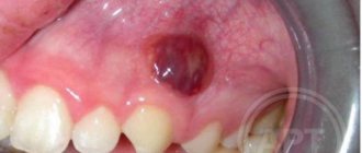
- An abscess is an inflammation of the root of a tooth, accompanied by an accumulation of pus.
When signs of lymphadenitis appear, dental pathologies are first excluded. Enlarged lymph nodes are a secondary symptom: it occurs when a visit to the dentist is delayed or after the removal of a tooth affected by a purulent infection. The primary signs of dental disease are gum inflammation and pain.
In 15% of cases, the reasons for the formation of seals in the maxillofacial area are dental. In addition to lymphadenitis, processes occurring in the oral cavity lead to the appearance of bumps on the gums.
They may be a sign of the growth of a permanent (in young children - baby) tooth or pathology of an existing dentition. The following appear as bumps on the gums near the teeth:
- Exostosis is a jaw abnormality characterized by the appearance of bony protrusions on the gums.
- Epulis is a pink or red mushroom-shaped tumor. Appears when the gums are mechanically damaged by a filling or tartar. Other causes of its occurrence: bruises and burns, congenital malocclusion, the use of low-quality dentures, hormone imbalance.
- A fistula is a small seal on the gum with a hole. When an infection occurs, pus flows out of it. The main reason for the formation of fistulas is the granulating form of periodontitis.
On this topic
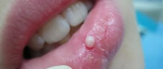
All about the ball on the frenulum under the tongue
- Olga Alexandrovna Novikova
- September 30, 2022
Benign tumors of bone and soft tissues of the oral cavity have a similar manifestation. The most common are:
- Ameloblastoma - mainly affects the lower jaw. It develops intraosseously and sometimes grows into soft tissues. Diagnosed at the age of 20-40 years. At first, the course of the formation is asymptomatic, but over time it increases. This leads to jaw deformation, swelling, bumps on the face on the affected side, and displacement of teeth.
- Osteoma is a tumor growing from bone tissue. It can form both on the outside of the jaw and on the inside (most often in the area of the upper dentition). With the active growth of the seal, pain appears, jaw mobility is impaired, and facial asymmetry occurs.
- Fibroma is a pathological formation that develops from the connective tissue of the tooth germ. Clinical signs of the tumor are mild: sometimes there is aching pain and inflammation at the site of its location.
- Cementoma is localized, as a rule, on the lower jaw: in the area of the tooth root. When it reaches large sizes, it causes facial deformation.
Malignant tumors of the oral cavity (carcinoma, sarcoma) are rarely diagnosed. But if a lump appears on your gums, you should definitely see a doctor: a dentist and, if necessary, an oncologist.
Non-dental reasons for formation
If the patient does not have pathologies of the teeth, soft and bone tissues of the oral cavity, one should look for another reason for the formation of lumps on the jaw. Inflammation and an increase in the size of the submandibular lymph nodes provoke diseases.
- systemic lupus;
- rheumatoid arthritis;
- leukemia.
Traumatic injuries to the lower jaw, head and hearing organs are no less rare causes of inflammation of the lymph nodes and the formation of lumps near the ear, in the chin area. At the site of the bruise, inflammation, swelling or compaction with a dense structure appears.
In the jaw area there are not only lymph nodes, but also blood vessels, salivary and sebaceous glands, bone and soft tissues. Therefore, when lumps appear on the outside of the jaw, the presence of cancer should not be ruled out. Sometimes growths under the skin of the face are benign or malignant neoplasms.
It occurs due to lipid metabolism disorders, clogging of the sebaceous gland, accumulation of toxins in the body
There are many reasons for the formation of lumps on the jaw. It is difficult to independently understand why the lumps appeared: you need to consult a doctor.
Dental causes of inflammation of the lymph nodes on the chin
Since very often in their work dentists are faced with huge lymph nodes in patients, let us dwell in more detail on the diseases that cause this symptom:
- Periodontitis. It can occur in acute or chronic form. Leads to tooth loosening and severe pain. If you do not receive qualified dental care in time, there will be a need to remove the affected unit.
- Stomatitis. Causes painful ulcers to appear on the mucous membranes of the mouth. It is the result of injuries, low immunity, vitamin deficiency. In advanced cases, the entire oral cavity becomes covered with wounds. Then active production of lymphocytes is observed.
- Pulpitis. Inflammatory lesion of pulp tissues. A very insidious disease. Through the pulp, pathogenic microorganisms penetrate into the lymph nodes and other tissues. It is important to clean the canals and seal them as soon as possible.
- Advanced caries. Despite the fact that many people do not take caries seriously, it is a rather insidious infectious process. If the deep tissues of the tooth are damaged, the lymph nodes come to the aid of the body - they try to neutralize pathological agents by producing lymphocytes. This can be understood by their increased size.
- Periodontal disease. Causes hypertrophy of periodontal tissues, leading to local hypoxia. Periodontal disease does not cause pain, so most often the patient learns about it by chance during a medical examination for some other condition. With periodontal disease, the gums become lighter, the interdental papillae atrophy, and the dental necks become exposed. If treatment is not started in time, the teeth will begin to loosen and fall out.
- Periodontitis. A disease that develops due to metabolic disorders, neurosomatic abnormalities, poor oral hygiene, and deficiency of vitamins and minerals. It manifests itself as bleeding gums, bad breath, and loose teeth.
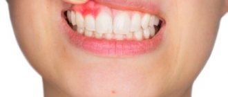
- Gingivitis. Inflammatory reaction in the gums. Appears against the background of gastrointestinal pathologies, allergies, infections. The patient experiences bleeding gums even with minor mechanical stress. A burning sensation in the mouth bothers me. Ulcerative-necrotic areas may form.
These are not all dental conditions in which the lymph nodes under the chin become inflamed. In medical practice there are many more such diagnoses. This once again proves that the patient himself will not be able to understand why his submandibular area is swollen.
Diagnostics
To find out the reason for the appearance of a lump on the jaw, a comprehensive examination is prescribed. First, the doctor examines and collects the patient’s complaints. If a patient has lymphadenitis, at the initial stage of the disease the following symptoms appear:
- slight enlargement of lymph nodes, their mobility;
- pain of the nodes on palpation, turning the neck (pain can radiate to the ear or other parts of the body nearby);
- general weakness;
- low-grade temperature ;
- decreased sleep quality;
On this topic
What could be a growth on the gum?
- Olga Alexandrovna Novikova
- September 3, 2022
With inflammation of the lymph nodes, accompanied by purulent processes, the patient has complaints of:
- increased pain when moving the jaw;
- redness of the skin in the area where pus accumulates;
- severe swelling of the lymph nodes, their pain even without pressure;
- temperature rise to 38 ºC or more.
In the last stages of lymphadenitis, the patient’s well-being deteriorates significantly: inflammatory processes spread to the neck and shoulders, fever appears, and bluish skin is observed at the site of inflammation.
Since, in addition to inflammation of the lymph nodes, there are other reasons for the appearance of lumps on the jaw, it is impossible to make a correct diagnosis based on the clinical manifestation of the pathology. Therefore, laboratory and instrumental diagnostic methods are used.
- blood test (general, advanced);
- bacteriological examination of mucus from the tonsils and upper respiratory tract;
- radiography of the jaw (in different projections);
- ultrasound examination of pathological formation;
- CT or MRI of the head;
- biopsy (if necessary).
Regardless of the mechanism of occurrence of a lump on the jaw, it is important to begin taking therapeutic measures in a timely manner. The earlier treatment is started, the lower the risk of complications.
How to understand that the problem is in the lymph nodes
Among the symptoms indicating damage to the lymph nodes:
- pain in the area where they are located;
- migraine;
- weakness;
- increased body temperature;
- discomfort under the chin;
- unpleasant pulsation in the lower jaw;
- difficulty chewing food;
- swelling noticeable to the naked eye.
If a purulent process develops, the skin in the area of inflammation becomes red and becomes very hot to the touch. Additionally, symptoms characteristic of the disease that provoked the disorder may appear.
Types of bumps on the face
Subcutaneous formations can be relatively harmless, appear, for example, in the form of acne, or indicate more serious diseases, including cancer. If we consider the main types of pathological processes in connection with which subcutaneous bumps can form on the face, the following stand out:
- Lipoma. Sometimes such seals are called wen, since the formation contains fatty and connective tissue. It is small in size, does not cause pain, and is a benign neoplasm. It forms relatively deep under the skin, a kind of capsule is noted, which can be removed surgically. Squeezing, for example, like a pimple is not only difficult, but also dangerous, as there is a risk of infection.
- Hemangioma. It is most often diagnosed in infants, but is sometimes observed in adults due to injuries and improper skin care. The lump differs in color from the skin, as it consists of accumulations of capillaries that can be compressed and injured. Subcutaneous bumps on the face do not hurt and can go away on their own. If treatment is necessary, physiotherapeutic, medicinal or surgical measures are prescribed.
- Purulent formations, foci of abscess. Such seals are quite painful, especially when pressed. They are formed due to the entry of bacteria and various types of infections into the layers of the epidermis; they can appear in connection with inflammatory processes already occurring within the body.
- Subcutaneous cyst. Refers to benign formations consisting of keratinized epithelium, connective, adipose tissue, and cavity. If the cyst grows rapidly, removal is recommended. A subcutaneous cyst or atheroma can develop together with the inflammatory process and suppuration. Traditional methods for treatment are not used.
- Pimples, boils. The seals have ulcers, inflammation and redness of the skin are even visually noticeable. It is extremely undesirable to squeeze out such formations, although many try to do it on their own. Such actions increase the risk of infection of other tissues, and other complications are possible.
- Oncological formations. It is impossible to distinguish cancerous tumors without appropriate diagnostic measures. Visually, they are similar to other types of cones.
In addition to the above diseases that cause various types of subcutaneous formations, there are others that are less common. In any case, the final diagnosis should be determined solely by the doctor, as well as the choice of treatment methods.
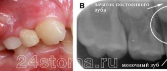
Lump on a child's gum
This problem doesn't only affect adults. A lump on the gum can also appear in children. Here, to the above reasons, another, quite logical, is added. If a hard lump on the gum hurts when pressed, this may be a sign that a molar is about to appear.
To protect your child, it is best to visit your doctor regularly. This way you will prevent possible problems and eliminate risks in a timely manner. For example, if the doctor finds a lump indicating the imminent appearance of a new tooth, you just need to wait a little. But if after some time the tooth does not appear and the lump remains, you will have to take action.
In addition, a lump on a child’s gum can occur due to infection and injury, just like in an adult. The treatment will be similar - for each diagnosis there is a separate sequence of procedures. Therefore, first of all, it is necessary to establish the cause of the appearance and nature of the tubercle.
Causes
There is an opinion that dermatological diseases occur mainly due to insufficient or improper skin care, but this is not entirely true. This reason is one of the common ones, but there are others. These include:
- Hormonal disorders, diseases of the endocrine system.
- Lifestyle, nutrition, bad habits.
- Use of low-quality cosmetics and care products.
- Metabolic disorders, including chronic diseases, such as diabetes.
- Infectious, viral diseases.
- Injuries. Subcutaneous bumps on the face after a blow can occur not only in the form of bruises, but also as other structural seals. These can be infectious formations, cystic, and so on.
Treatment
Conservative or surgical methods are used to treat subcutaneous lumps. Defining
factors are the size of the compaction and the reason for its development. If the formation is benign, small in size and does not cause any physical or emotional discomfort, then you can only observe it. If the lump grows, symptoms of inflammation and other signs of activity, measures are taken to eliminate the problem area.
Conservative therapy is used only if there is a possibility that the lump may disappear over time. To do this, antibiotics or antivirals are selected, external agents are prescribed, the action of which is aimed at the problem area. If necessary, antihistamines, antiallergic drugs, and hormonal drugs are taken.
For each patient, depending on the clinical picture, an individual treatment regimen and surgical options are determined.
Types of seals
Seals that appear in the tissues behind the ear, in the earlobe or in the nose area are divided into several types. Depending on which of them the diagnosed compaction belongs to, the necessary therapy is prescribed. An error at this stage of diagnosis will result in ineffective treatment or even worsening of the patient’s condition.
Whatever type of lump it is, it must be shown to a doctor. Painful sensations can be eliminated only after the necessary therapy. The seals themselves pass extremely rarely, and you should not count on this.
What kind of education is this
A lump is a lump on the face in the form of a ball under the skin. They can be found on the forehead, chin, and cheeks. Such lumps are often found in the area of the corners of the eyes and lips. There are many reasons for lumps to appear. Depending on them, all such formations are divided into groups. It is necessary to consult a doctor so that, after conducting a diagnosis, he can determine why the lump appeared.
Important! To accurately understand the cause of the lump under the skin on the face, you need to undergo a short examination, take a blood test and take a smear of the dermis. After receiving the results, it will be possible to clearly say what caused the disease.
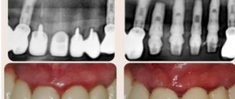
Periodontitis
Description
A hard lump on the gum, an abscess formed at the base of the tooth root. In the absence of therapy, it transforms into a benign tumor.
Treatment
If such compaction of the gums under the tooth appears, contact your dentist. The teeth are unfilled and root canals are cleaned. After removing the exudate, oral baths with medicinal herbs or soda solution are prescribed. Treatment for periodontitis may require multiple visits to the doctor.
Types of formations and reasons for their appearance
An abscess is a lump under the skin that forms after infection. The formation in this case is very dense, since there is a clot of pus in the lesion. This lump is red around its entire perimeter, and very often patients experience an increase in temperature. It will not be possible to surgically remove or simply squeeze out such a lump without consequences; it will form in the same place again. This will continue until the infection is eliminated.
An intradermal cyst is a solid formation, which is accompanied by regular discharge from it onto the surface of the skin. Such a compaction can occur as a result of disturbances in the body and the secretion of the sebaceous glands of the dermis: around the ear, in the auricle, on the nose, on the neck, and also on the face. This disease is treated with anti-inflammatory drugs, which are prescribed by the doctor in accordance with the examination results.
Dense lump under the skin of the face
Rheumatoid nodes are the result of an inflammatory process in the lymph nodes. If the disease is not treated correctly, deformation may occur in the joints, which stand out on the face in the form of lumps.
Lipomas are spherical lumps that can move under the skin if you press on them with your finger. They can often be seen in the area near the eyes, as well as on the cheeks. By themselves, if they are small in size, they are painless, but in cases where lipomas grow, discomfort sets in, and they can even ache a little when pressed. They can damage other tissues, as well as organs if they are located near the formation.
The reasons for the appearance of such wen can be varied:
- swipe;
- smoking;
- alcohol;
- disorders of the nervous system;
- disorders of the secretion of the sebaceous glands.
Cancerous lump - in shape it can be similar to any formation described above. At the beginning of the development of the disease, no symptoms may appear; in many cases, the lumps are difficult to detect visually. When the formation grows inside the dermis, damaging a fair amount of tissue, it may appear on its surface in the form of a small reddish pimple. Subsequently, it can grow very quickly or, conversely, slowly, and the formation is accompanied by unpleasant sensations and sometimes pain.
Wen on the body: reasons, how to get rid of it
A small growth on the skin, a pea-sized “nodule”, can cause anxiety and concern. Concerns increase if the formation begins to gradually increase in size. The specialist’s diagnosis of lipoma only adds fuel to the fire of worry, since it is very similar to the name of many dangerous malignant tumors.
In fact, there is no real reason for concern, since wen extremely rarely (almost never) degenerates into a malignant neoplasm. This is a benign tumor that is characterized by slow growth or stable size over a long period of time. Why wen appears on the body and how to deal with them, you will learn from the publication presented to your attention.
What is a wen?
Wen is an unofficial, “folk” name for a subcutaneous formation formed by adipose tissue. In official medicine, the scientific term “lipoma” is used, which reflects the histological composition of the tumor.
Lipoma is a benign tumor that remains as such throughout its existence. A wen on the body is not characterized by malignant degeneration. From this point of view, it does not pose a health threat. The problem is mainly of an aesthetic nature, especially if the tumor is located on an open area of the body - on the face, on the neck, on the skin of the distal parts of the arms.
A distinctive feature of any benign tumor is that it is formed from cells surrounded by a fibrous capsule. In this way, wen on the body differs from atheroma - another neoplasm, the appearance of which is caused by fat. Atheroma is an accumulation of sebum secreted by the sebaceous glands. Atheroma is formed due to blockage of the ducts of the sebaceous glands. The secretion of the gland cannot come out, accumulates under the skin and manifests itself in the form of a small or medium-sized formation that rises above the skin.
Lipoma, unlike atheroma, does not consist of the secretion of the sebaceous glands, but of fat cells surrounded by a capsule of connective tissue. A skin formation appears as a result of abnormally active division and growth of adipocytes. All cells, regardless of the size of the wen, remain differentiated, that is, they retain their typical characteristics, which makes it easy to distinguish a lipoma from a malignant tumor. A cancer tumor is characterized by impaired differentiation and atypical cell structure. In the case of lipoma, these signs of malignancy are not present.
Lipoma is a benign tumor of adipose tissue, which consists of differentiated adipocytes surrounded by a fibrous capsule. Malignant degeneration is not typical for wen on the body; they do not pose a danger to health or life.
Reasons for the appearance of wen
Numerous studies devoted to the problem discussed in the publication have not made it possible to establish a single cause of lipomas. Currently, it is generally accepted that a wen on the body appears as a result of the combined action of a number of predisposing factors, among which endogenous causes predominate.
Among the risk factors, the most important role is played by metabolic disorders associated with enzymatic deficiency. Particularly dangerous in this regard are disorders affecting the biochemical mechanisms of lipid and carbohydrate metabolism. The cause of lipoma can be hormonal disorders not related to the pathology of the endocrine system. In particular, in women, a neoplasm can appear during pregnancy, lactation or after the end of the lactation period.
Endocrine diseases are also among the risk factors. If a person has wen on the body, the reasons should be sought in the functioning of the hormonal system. Dysfunction of the pituitary, adrenal, thyroid (hypothyroidism) or pancreas (diabetes) may be the root of the problem. A risk factor is an imbalance in the secretion of sex hormones, which is important for women taking oral contraceptives and athletes using pharmacological agents to accelerate results.
A fatty deposit on the body can occur even against the background of complete well-being, that is, in the absence of any endocrine disorders, hormonal changes or metabolic problems. Doctors explain this by genetic predisposition: people whose relatives have encountered a similar problem have a higher risk of developing lipoma. Hereditary, or familial, lipomatosis is characterized by the appearance of multiple wen at a young age.
Among exogenous factors, nutrition is of greatest importance. The risk of lipoma is higher if a person's diet is dominated by simple carbohydrates (sugar, confectionery, flour), as well as saturated and hydrogenated fats. A risk factor is systematic alcohol consumption. All these substances interfere with the natural course of metabolic processes, which is fraught with the formation of a benign tumor of adipose tissue.
Another possible cause of the appearance of a wen on the body is mechanical trauma, which leads to neurotrophic disorders in the integumentary tissues. They, in turn, can provoke the proliferation of subcutaneous fat cells. It is possible that exposure to ultraviolet radiation on the skin plays a certain role, but the connection between this factor and the appearance of lipomas has not been proven.
Localization, appearance, symptoms
The concept of “typical localization” does not apply to wen on the body. The tumor can appear on any part of the body: on the back of the foot or hand, on the shoulder or forearm, on the back, on the chest, on the face or on the abdomen. In short, lipoma does not have a “favorite” location; the tumor can appear anywhere.
As for the symptoms, that is, the appearance of the tumor, there is more certainty. Externally, the lipoma looks like a pea; it is characterized by a rounded shape with regular and even edges. On palpation, the adipose tissue tumor is soft or moderately dense. The surface is smooth. The color can be flesh-colored, reddish or brownish, depending on how abundantly blood vessels grow into the tumor.
An important diagnostic criterion is the mobility of the tumor. The subcutaneous wen on the body is always mobile; it is not fused to deeper tissues and moves freely with the skin. This symptom makes it easy to distinguish a lipoma from a malignant tumor. The latter grows into deep tissues, which is why it becomes immobile.
Size is difficult to use as a diagnostic criterion because it can vary over a wide range. Typically, a fatty tissue tumor has a diameter of 0.5 to 1 cm, but there may be exceptions to this rule. Wen on the body can reach 2-3 cm in diameter. There are cases when, as a result of growth, a lipoma reached gigantic sizes - more than 10 cm in diameter.
The last symptom - pain - is not typical for wen. As a rule, education does not remind us of itself. The exception is situations when a person mechanically injures him. When struck, scratched or pinched, a sharp pain may occur, which is not too intense and passes quickly. However, if a tumor of adipose tissue puts pressure on superficial vessels and nerves, the clinical picture may include moderate or even severe pain.
Are wen dangerous?
As already mentioned, the prognosis for this benign tumor is extremely favorable. The neoplasm is not characterized by malignant degeneration, so nothing threatens life. However, this does not mean that you can forget about lipoma, since certain health risks are still present.
In case of mechanical damage (cut, pinching, blow) inflammatory processes may begin in the depths of the formation. If the integrity of the skin is damaged, there is a risk of infection in the wound. The result of this may be the development of a necrotic or purulent-inflammatory process, after treatment of which visible scars will remain on the skin.
Scars can also form after removal of a large tumor. A small lipoma can be removed without leaving a trace, so that there are no noticeable scars left on the skin. When removing large and giant wen, it is difficult to avoid scar changes. Meanwhile, large wen must be removed, since sooner or later they will begin to put pressure on the nerves and blood vessels. It follows from this that it is better to get rid of the wen at an early stage, before it grows to a large size, the only question is how to do this?
How to get rid of wen?
Many people, in an attempt to get rid of the tumor, resort to traditional medicine recipes. Numerous publications devoted to this problem offer various remedies: compresses made from onions, garlic or sprouted wheat, applications of celandine and other medicinal herbs. Unfortunately, such an approach is doomed to failure; it is not possible to get rid of education using folk remedies. Traditional recipes are unlikely to provoke the growth of a lipoma, but they certainly will not lead to a reduction in its size.
Official medicine offers two options for solving the problem. You can get rid of small lipomas using a minimally invasive procedure, which involves injecting a special solution deep into the formation that destroys adipocytes. The disadvantage of this method is that there is a high risk of relapse, the capsule is not removed, and the method can only be used for small (up to 2-3 cm) tumor sizes.
Surgical removal of a wen on the body is an effective and universal treatment method. The lipoma is removed along with the surrounding capsule. Minor surgery is performed using a traditional, radio wave or laser scalpel. Accordingly, traditional surgical, radio wave and laser removal are distinguished.
All methods of surgical treatment are effective, but from an aesthetic point of view they are not equivalent. After traditional surgical removal of a tumor, small scars often remain, even if the doctor removes a small tumor. Radio wave surgery allows you to minimize the severity of scars, and the laser method makes it possible to perform the operation absolutely without leaving a trace.
Laser lipoma removal has many benefits. The laser knife seals the vessels immediately after they are damaged, reduces bleeding, and injures the skin to a lesser extent. Since laser pulses have a detrimental effect on pathogenic and opportunistic microflora, there is no risk of infectious complications. In addition, the laser stimulates regeneration, which shortens the recovery period, accelerates skin healing and avoids scar formation.
The problem is that the laser method is used only for medium-sized and small-sized wen; large and gigantic ones (more than 10 cm in diameter) are forced to be removed by the doctor using the traditional surgical method. Given their size, it is almost impossible to avoid scar formation.
Since the aesthetic component of the operation is a concern for all patients, doctors recommend removing lipomas in the early stages. The deciding factor is the size of the education. A tumor on the face can be removed without a trace if its diameter does not exceed 1 centimeter. Wen on the body are removed without scarring if their size is no more than 2.5-3 cm.
Laser removal of wen is the most effective and safe method. It is better to remove lipomas in the early stages, when their size does not exceed 1 cm if localized on the face or 3 cm if localized on the body.
The operation is performed on an outpatient basis under local anesthesia; only giant lipomas require the use of general anesthesia (anesthesia). The patient returns home on the day of surgery. During the recovery period, it is necessary to protect the skin from ultraviolet rays and avoid exposure to high or low temperatures. It is advisable not to wet the wound with water and regularly treat it with antiseptic solutions.
Removal of fatty tissues on the body: price
Surgeries to remove fatty tissue are carried out in aesthetic medicine and plastic surgery clinics. It is advisable to contact a medical institution equipped with modern equipment for laser manipulation.
The price of removing wen with a laser is low.
The exact cost depends on the size of the education. The cost of removing a small lipoma (up to 3 cm) in Moscow clinics is about 5 thousand rubles. The cost of treating a tumor up to 10 cm in size increases to 8-9 thousand rubles. Treatment of large and giant lipomas costs more - up to 15 thousand rubles, depending on the size of the formation and the category of complexity of the operation. You will receive more detailed information about lipoma removal methods during a consultation with a doctor at the Galaktika aesthetic medicine clinic (Moscow)
Common Causes of Bumps
Most formations on the face are a consequence of an unhealthy lifestyle
Most of the reasons that lead to the formation of a tubercle are a consequence of an unhealthy lifestyle, regular stress, and bad habits. Some of the most common causes of bumps:
- hormonal disbalance;
- violation of diet and sleep patterns;
- poor nutrition;
- allergy;
- bad habits;
- injuries;
- disruption of the endocrine system;
- malfunctions of the nervous system.
- infections.
- incorrect selection of cosmetics and skin care.
About the subcutaneous boil
A boil is a purulent inflammation of the skin. There are many reasons for its appearance:
- obesity;
- diabetes;
- metabolic disorders;
- intestinal disorder;
- abrasions and cuts.
Boils are painful subcutaneous pimples on the face that are accompanied by pain at the slightest touch. At the base of this formation there is a purulent rod, and at its tip there is a head filled with purulent fluid. Often they try to remove it to remove the pus, but this only leads to worse consequences: the boil begins to increase in size, damaging more and more tissue. Many, not understanding the danger, try to squeeze out the unfortunate pimple, enduring extreme pain when pressed. But the inflammatory process can only deepen into the skin with such actions.
Under no circumstances should you apply compresses; they only create excess moisture, which contributes to the development and enlargement of acne. To reduce the size of the formation, it is necessary to dry the skin; even heating helps to reduce the inflammatory process and its faster maturation. An abscess will gradually form, which should be lubricated with a special ointment with the addition of an antibiotic; you must first consult a doctor. He will analyze the condition of the pimple and give recommendations for its removal.
Important! Subcutaneous boils on the face are treated comprehensively. It is mandatory to prescribe a course of medications for external and internal use, as well as a complex of vitamins to cleanse the blood. A preliminary examination is carried out, which is aimed at finding out the cause of the disease.
Benign formations
Relatively safe are:
- Lipoma (wen) is a formation consisting of adipose tissue covered with a fibrous membrane. It does not pose a threat to health if it is not inflamed, and is more of a cosmetic defect. Occurs due to disruption of lipolysis processes in adipose tissue.
- A furuncle is an inflammation of the hair follicle, which is a cavity in the skin filled with pus. The causative agent of this inflammation is staphylococcus. Untimely surgical treatment can lead to blood poisoning.
- Fibroma is a benign formation of connective tissue that forms at the site of an injury or scratch. Fibroma is completely painless and does not degenerate into malignant, but nevertheless, it must be kept under control.
- Inflamed lymph nodes are a constant companion of infectious diseases. As you recover, the seals decrease and return to normal.
- An enlarged thyroid gland is a consequence of impaired hormone production. The thyroid gland changes in size due to iodine deficiency or in case of genetic predisposition. The danger of nodular or diffuse goiter is that it can develop into a malignant tumor.
- Inflammation of the submandibular salivary glands can occur if it is blocked by a stone. As a result, saliva accumulates in the gland, causing pain and swelling. Infection may also be the cause of the increase.
Treatment options
Many small formations can appear during a cold, so there is no need to make hasty conclusions and resort to treating them. The body itself will cope with the underlying disease, and then begin to fight the pimples; they will gradually begin to dissolve on their own. They are most often flesh-colored and can appear in the forehead area, along the jaw. The patient may experience discomfort, as such pimples may peel and itch.
Flesh-colored bumps may be the result of a cold
Other types of bumps on the face need to be shown to doctors so that they can prescribe treatment. It will not be possible to get rid of them by squeezing out - this will only lead to damage to the cover, and the compacted lesion itself will not be removed. This may lead to the need to resort to surgery, after which it will be necessary to treat the skin for a long time from the consequences of the patient’s incorrect actions.
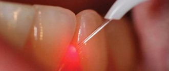
Laser removal of cones allows you to painlessly remove them from the surface of the dermis, while the beam penetrates to the depth of the entire formation and reduces it, pushing the pus out. The use of injections and ointments can help get rid of acne. Most often, this method is used in combination with surgery.
Important! It is worth remembering that after each treatment procedure, redness and induration may persist on the face for another 6-8 days. This is why many people think that the course is not suitable for them or there is no result.
What formations hide danger?
The following types of neoplasms require urgent medical intervention:
- Atheroma is similar to a lipoma, but unlike it, it often becomes infected and turns red, and can also increase in size. It is this moment that poses a health hazard, since there is a danger of fistula formation and pus leaking into nearby tissues.
- A median cyst is a congenital benign round formation located on the front of the neck. Up to a certain point, it may not bother the child, but in 60% of cases it becomes inflamed with the formation of a fistula, causing pain and discomfort when swallowing. In addition, the cyst is prone to degeneration into cancer.
- Neurogenic tumors are formed at the ends of nerve trunks and represent a wide range of neoplasms. They can be safe, but malignant formations can also occur. Under no circumstances should they be left without the attention of a doctor.
- Lymphogranulomatosis (Hodgkin's lymphoma) is cancer of the lymph nodes. The first symptom of this cancer is expressed in enlarged lymph nodes, but they do not hurt and slowly enlarge.
The otolaryngologist makes conclusions about the safety of formations only after conducting a comprehensive examination. It is impossible to do this on your own.
Further skin care
Before resorting to your usual skin care, you should visit a cosmetologist. He will select gentle care that will help restore the skin, and gradually the ulcers will stop hurting. For the first week after treatment, you should care for your skin only with water and a cotton pad; you should avoid all kinds of cosmetics, as they can cause acne to reappear.
After treating subcutaneous diseases, it is worth visiting a cosmetologist
Over the next couple of weeks, you need to use products that will protect your skin from exposure to sunlight. They will also gradually relieve itchy skin after bump treatments.
Preventive measures
In diseases that lead to the formation of acne, improper care, frequent bruises and untreated wounds lead to the appearance of knobby formations. To avoid health problems, you should adhere to preventive measures:
- remember the rules of personal hygiene and follow them. Change your razor in a timely manner, use decorative cosmetics as little as possible, which leads to clogging of pores;
- monitor your diet. It is worth minimizing the consumption of alcohol, sweets, marinades, preservatives, sausages and sauces;
- undergo a complete examination by doctors once a year.
Getting rid of bumps under the skin on your face is difficult on your own. This is best done in beauty salons. Specialists will be able to do this practically without damaging the surface of the dermis.