Cheek cancer is a malignant neoplasm of the oral cavity.
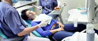
Examination of patients with symptoms of cheek cancer at the Yusupov Hospital is carried out using modern diagnostic equipment from leading companies in the USA and European countries. Oncologists provide comprehensive treatment for cancer of the buccal mucosa. The medical staff is attentive to all the wishes of the patient. Chefs provide special dietary meals. Professors and doctors of the highest category develop tactics for managing patients with cheek cancer at a meeting of the expert council. Early diagnosis of the disease and multidisciplinary therapy improve the prognosis and five-year survival of patients after treatment.
Causes of cheek cancer
Cancer of the buccal mucosa develops under the influence of the following provoking factors:
- Use of tobacco in any form (cigarettes, cigars, pipes, chewing tobacco);
- Alcohol abuse (the risk of developing cancer increases when the use of alcohol and tobacco is combined);
- Infection with carcinogenic forms of human papillomavirus.
One risk factor is exposure to sunlight. Both family history and genetic predisposition, as well as exposure to mutagenic environmental factors, play a role in the development of cheek cancer. The formation of a malignant tumor occurs in several stages. The most important is the disruption in the functioning of oncogenes and genes that inhibit tumor growth. The development of malignant neoplasms of the cheek is associated with inactivation of the p16 gene, mutations in the p53 gene, and the introduction of the human papillomavirus.
Why do jowls appear?
Our face consists of 57 muscles and fat, which is concentrated mainly in the cheek area. This cheek fat, in honor of the French anatomist Marie François Bichat, has its own name - Bichat's fat pads, and reaches a very impressive size.
With age, any person experiences drooping of facial tissues. The lower jaw to the left and right of the chin is devoid of muscle support (this can be easily seen in the figure), and nothing prevents sagging in this area.
As a result, characteristic sagging is formed along the jawline - “bulldog cheeks”, which make the facial contour wavy.
In thin people, shaving is usually due to excess skin and only skin. In all other cases, sagging is enhanced by subcutaneous fat or descending lumps of Bisha
It is believed that Bisha's structures contribute not only to the “blurring” of the contour of the lower jaw, but also cause nasolabial folds and form sufas on the face.
Most often, sagging skin in the lower jaw area is the visible result of a number of invisible changes that have occurred to our face over the years:
- Low collagen and elastin content. Collagen inside our skin is like the reinforcing mesh in buildings made of monolithic concrete. Only with age does the old collagen “break down”, and the new one is not synthesized actively enough. This causes the skin to lose support and elasticity.
- Weakening of the facial muscles, which cannot hold the skin and subcutaneous fat high in the same place. Thus, as a result of weakening of the cervical platysma muscle, a “goiter” and a double chin are formed in the neck area. The same mechanism underlies sagging cheeks.
- Due to a lack of certain elements and hormonal changes, the bones become thinner and the face becomes flattened. Cheekbones lose their “sculpted” volume. Deprived of this important support, all facial structures, like on a sled, literally “slide” down.
- An age-related decrease in the amount of melanin pigment in the skin makes it vulnerable to sunlight and accelerates aging.
A face with jowls is not just a problem of age. There are other reasons too. Individual facial characteristics, stress, constant exposure to wind and cold, smoking, alcohol, coffee, poor nutrition, certain medications, endocrine diseases, rapid weight loss, a poorly developed chin and even the force of gravity all contribute.
The risk of developing early jowls is especially high in heavy individuals with a large amount of cheek fat. These are overweight people and those who, at normal weight, inherited chubby cheeks.
The same factors apply to men. But due to gender characteristics, they have less facial fat. Their skin is not subject to hormonal fluctuations during pregnancy and menopause; it remains dense for a long time. Therefore, men's jowls appear 10-15 years later.
Expert comment:
“The appearance of jowls in youth is usually a consequence of being overweight. In all other cases, this is an obvious sign of age.
. But if jowls appear after 40 years, there is no need to urgently remember the forgotten cosmetologist.
Don't get me wrong, you definitely need to continue to take care of yourself. But creams, face masks, peelings and mesotherapy do not work on jowls. And do not rush (no matter what they tell you) to invest money in fillers and 3D mesothreads - they do not work against sagging of the lower jaw, and you will simply waste your money.
To get rid of jowls on your face after 40, you need to work with the causes, not the consequences.
.»
Andrey Iskornev, plastic surgeon.
The mechanism for the formation of jowls on the face is the drooping of intrabuccal fat.
The mechanism of development of cheek cancer
Malignant tumors of the cheek occur against the background of precancerous changes in the epithelial and subepithelial layers (leukoplakia or erythroplakia). The risk of leukoplakia degenerating into invasive cheek cancer is about 4-6%, with erythroplakia it reaches 30%. Subsequently, dysplasia transforms into “cancer in situ”, which penetrates into surrounding tissues and metastasizes to local and regional lymph nodes.
In some patients, even those cells that do not initially raise suspicion of dysplasia upon initial microscopy may gradually become malignant. As the tumor process progresses, distant metastases occur in the bones, lungs, and liver. Buccal cancer can grow through the skin.
Doctors' advice and recovery period
According to the advice of experts, after excision of the affected area (in the event that the internal bump on the cheek was removed surgically), the patient should be very careful and take his condition seriously. For example, you should completely review your diet, eliminating roughage from it.
At first, if a bandage has been applied to the patient, you must be extremely careful not to touch the seam; it is important to allow it to tighten efficiently and painlessly. In addition, in case of excision of areas on the cheeks, the risk of scar formation remains.
We must not forget that the operation itself is additional stress for the body; after it, it is very important to strengthen all your strength from the inside. To do this, you will need to eat right, in addition, good sleep is important along with giving up bad habits, and if possible, you need to avoid any worries and stress. You should not self-medicate; seeking medical help will help you quickly cope with the disease.
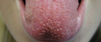
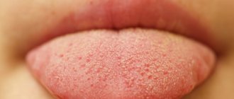
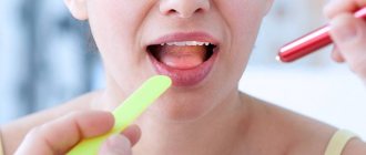
Symptoms of cheek cancer
Cheek cancer in the initial stages of the tumor process is asymptomatic. Any ulcer that is not prone to rapid healing and any area of hyperkeratosis should be regarded as an early stage of cancer. In the early stages of cheek malignancy, there is little or no pain. As the size of the cancer tumor increases, the following symptoms appear:
- Pain;
- Induration and infiltration of underlying tissues;
- Enlargement of regional lymph nodes, including damage to the lymph nodes of the submandibular triangle, jugular-digastric group and deep cervical lymph nodes.
What does cheek cancer look like? Any non-healing oral mucosal ulcer should be assessed as a potentially malignant tumor and consultation with an oncologist should be sought.
Home therapy
Treatment can be started at home in the case of a single manifestation, when the tendency for the lump to grow in size does not persist. Here are a few recipes that can help you cope with this disease on your own without seeking medical help:
- If the manifestation is completely fresh, then you should try using raw chicken egg white several times a day.
- You should take a small piece of cotton wool and soak it in castor oil, then apply it to the bump in the cheek area.
- The procedure should be repeated twice a day.
- It is necessary to cut a clove of garlic into slices, and then lubricate the growths with them three times.
- You need to prepare a tincture, for which you will need the peel along with walnut leaves. You should pour some alcohol into the dry ingredient and let it sit for two weeks, and then lubricate the formation with it once or twice daily.
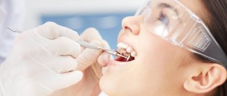
Diagnosis of cheek cancer
If a tumor of the mucous membrane of the cheek is suspected, oncologists at the Yusupov Hospital conduct an examination using a mirror, palpation of the tumor and lymph nodes. If a long-term non-healing ulcer is detected, a biopsy is performed. If a negative result of histological examination of the material obtained during the biopsy is obtained, the suspicion of the malignant nature of the tumor remains, the biopsy is performed again.
Oncologists clarify the stage of the tumor process, assess the extent of spread of the malignant tumor from the buccal mucosa to adjacent tissues, and find out whether there are metastases to regional lymph nodes and distant organs. If the presence of metastases is suspected, computer and magnetic resonance imaging, ultrasound, and scintigraphy are used. Distant metastases are found in 20% of patients at the time of diagnosis.
Computed tomography and magnetic resonance imaging make it possible to assess the condition of the deeper anatomical structures of the oropharynx and surrounding tissues. If there is a suspicion of metastases to the lymph nodes or tumor infiltration of the floor of the mouth, a cytological examination of the aspirate obtained under ultrasound guidance is performed. To exclude distant metastases, a chest x-ray in two projections and an ultrasound examination of the abdominal organs are done.
Considering that the prognosis for cheek cancer is serious, a tomography of the neck, chest and upper abdominal cavity is performed. Using bone scintigraphy, I exclude bone metastases. Positron emission tomography allows one to identify the source of metastases in cases of undetected primary tumors.
The danger of atheroma on the face
Sebum and keratin masses accumulated in the atheroma capsule have a cheesy consistency. This is a favorable environment for the development of bacteria. An attempt to squeeze out the atheroma can lead to infection getting inside the capsule and causing suppuration. The main symptoms of its onset are:
- redness of the skin around the atheroma;
- pain in the affected area;
- Migraine-like headaches;
- temperature increase.
Inflamed atheroma can lead to phlegmon - inflammation of adjacent tissues without precise boundaries.
Rice. 3 Self-squeezing of atheroma usually leads to inflammation of facial tissues
Treatment for cheek cancer
Malignant tumors are successfully cured using radical radiation therapy while preserving the function of the oral cavity. Radiologists implant radiation sources because it is possible to irradiate a small volume of tissue at a high dose. Radioactive isotopes of cesium, gold, radium, iridium, tantalum, which have the same efficiency, are used as sources.
For small tumors, the size of which does not exceed 1 cm, they are limited to implantation of a radiation source, without resorting to additional external influence. In cases of slightly larger tumors that are not suitable in size for implantation, external irradiation is used along with source implantation.
Traditionally, large cheek tumors are treated with external beam radiation. Recently, oncologists have been using a combination of radiation and chemotherapy. Mobile lymph nodes are radically excised. During prophylactic removal of lymph nodes without signs of damage, in a significant number of cases, foci of micrometastasis are found in them. For this reason, radiologists prefer to perform prophylactic irradiation of the neck in patients without evidence of lymph node involvement by the external beam, sometimes in combination with surgical treatment.
After surgery for cheek cancer, a cosmetic defect is formed. The results of surgical intervention are improved using the technique of microvascular free skin grafting. If a patient develops dry mouth after radiation therapy, he is prescribed oral pilocarpine. The drug increases salivation, which is usually accompanied by minor side effects - sweating and increased urination. For primary or secondary treatment of tumors of the buccal mucosa, patients are prescribed chemotherapy drugs.
Early diagnosis of a cheek tumor allows for effective treatment. If the tumor process is at a late stage, the prognosis worsens. If you experience unpleasant sensations in the oral cavity or identify ulcers of the buccal mucosa, undergo an examination at the clinic by calling the Yusupov Hospital.
What should you do if you have an illness?
After a biopsy, such patients may be prescribed the following types of therapy:
- Carrying out surgical treatment of a bump on the cheek under the skin. For this, electronic scissors are used, and immediately after removing the tumor, the affected area is cauterized. Therapy is continued with a course of antibiotics.
- Injection treatment. It involves special injections containing alcohol, which are given directly to the area of the granuloma.
- Performing laser treatment.
- Local injections using aletretin gel.
So, a bump appeared on my cheek. Regardless of the size of the pathological growth, one should not delay going to a specialist, this way a person can quickly feel comfortable and avoid all sorts of complications.
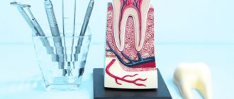
How to treat a lump after tooth extraction
The technique of dealing with a dental disorder depends on the factors that caused it. First, the doctor studies the dominant symptoms and carries out various diagnostic measures. Based on the data obtained, he makes a decision on an effective treatment technique. She may be:
- conservative;
- surgical.
In the first case, we are talking about cleansing tissues from purulent masses and carrying out drainage measures. The patient is prescribed medications of different pharmacological groups. These can be antibiotics, anti-inflammatory, antiseptics, agents that suppress the activity of pathogenic flora, regeneration stimulants, pain relievers.
Medicines can be taken orally. They are also applied to the affected area. The rinses have proven themselves to be excellent. With their help, it is possible to speed up the evacuation of the contents of a purulent lesion.
Surgical therapy is indicated if there is a dense sac on the gum containing dead tissue. Then the surgeon makes a small incision and installs a drainage tube into the formed cavity. The exudate leaves the pathological focus along it. When the lump subsides and all the pus is removed, the doctor stitches it up. To avoid complications, the patient is prescribed antibacterial agents.