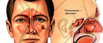
Liquororrhea (from Latin liquor
- liquid and Greek
rhoia
- outflow) - outflow of cerebrospinal fluid. Depending on the location of the outflow of cerebrospinal fluid from the cranial cavity, several types of spontaneous liquorrhea are distinguished: nasal, auricular and orbital.
Spontaneous nasal liquorrhea (SNL) - the leakage of cerebrospinal fluid from congenital or resulting from various non-traumatic causes of defects in the bones of the skull and dura mater - is a rare disease that accounts for 3-4% of all cases of nasal liquorrhea [1, 2]. It can be constant or periodic, drip or jet. Intensification of liquorrhea is possible when changing the position of the head or straining. In some cases, SNL is hidden, and cerebrospinal fluid flows into the nasopharynx. SNL is also divided into primary (idiopathic), the obvious cause of which cannot be determined, and secondary, when a pathological process in the paranasal sinuses and bones of the base of the skull leads to the formation of a fistula (defect).
There are a small number of reports in the literature summarizing the experience of monitoring patients with SNL. Thus, a number of foreign scientists consider SNL a symptom of idiopathic intracranial hypertension [3], other researchers consider defects of the ethmoid bone and the walls of the sphenoid sinus as the cause of this pathology [4]. However, it is generally accepted that the reliable cause of SNL cannot be established.
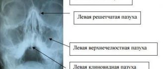
Computed tomography (CT) as a method of non-invasive radiation diagnostics has been used since the 70s of the last century. It allows, even before surgery, to create a visual model of the nasal cavity and its paranasal sinuses - a kind of “visual reality”, taking into account which the surgeon can plan adequate treatment measures [5]. The advantages of K.T. are the ability to eliminate the summative effect inherent in a conventional radiograph, visualize organs and tissues separately, as well as assess their density characteristics.
The purpose of the study is to develop a diagnostic algorithm for examining patients with SNL using CT.
The authors conducted a detailed analysis of the CT results of patients with SNL, assessed the anatomical features of the ethmoid bone structure, their possible impact on the development of this disease and the condition of the bone structures of the paranasal sinuses and pathological changes in them.
How do we treat sinusitis?
Sinusitis (sinusitis) is inflammation of the maxillary sinuses. Microorganisms (bacteria or viruses) enter the maxillary sinus through the nose or through the bloodstream and cause inflammation of its mucous membrane, often accompanied by the accumulation of pus in the sinus. To drain pus, “punctures” are used - punctures of the maxillary sinus. Unfortunately, many patients, fearing such “punctures,” delay seeing a doctor for sinusitis, thereby harming themselves: creating the threat of developing severe complications of sinusitis associated with the possible spread of infection, as well as conditions for the formation of chronic sinusitis.
Is it possible to treat sinusitis without a puncture?
Very often - yes. Subject to timely contact with an ENT doctor. At the initial stage of sinusitis, it is quite possible to do without a puncture. A “puncture-free” method of treating sinusitis is nasal lavage using the fluid displacement method (“cuckoo”) in combination with laser therapy. The rinsing procedure clears the nasal passages and nasal cavities of pus and mucus, and the laser relieves inflammation. The course is designed for 5-7 procedures. Already after the 1st procedure, patients see a significant improvement in their condition. Another way to avoid a puncture for sinusitis is to use a device that creates negative pressure in the nasal cavity, and thereby helps remove secretions from the maxillary sinuses. A modern method of ensuring outflow from the inflamed sinus and the possibility of carrying out therapeutic actions in it is balloon sinuplasty, performed in our center. It is possible to cure sinusitis without a puncture only if there is nasal discharge, indicating that there is an outflow of the contents of the maxillary sinus. Even the anatomical features of the structure of the ENT organs are important. That is, to determine the optimal treatment tactics for sinusitis, it is necessary to contact an otolaryngologist as early as possible. In our clinic, puncture-free treatment of sinusitis is possible!
- Without pain and blood
- Without piercing the maxillary sinuses
- In just an hour and a half
Healing sinusitis is a huge relief for your body.
Causes of sinus perforation during dental treatment
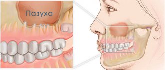
The maxillary sinuses or maxillary sinuses are located inside the upper jaw. They are separated from the oral cavity by a septum and the alveolar process, which contains the roots of the teeth. Treatment of the canals of such teeth is complicated by the risk of sinus perforation for a number of reasons:
- Anatomically thin septum Sometimes the height is only 1 mm. It happens that the teeth penetrate the cavity with their roots and only the sinus mucosa separates.
- Treatment of teeth with inflammation on the roots If there is inflammation on the roots of the teeth (periodontitis, cyst), the surrounding bone melts and becomes thinner.
- Excessive efforts by the doctor If the dentist does not calculate the efforts when passing the canals, the canal may rupture, damage the root, or perforate the bottom of the sinus.
Without a preliminary diagnosis, the endodontist, having no idea about the size of the sinus septum and the location of the roots, may not calculate the effort and damage the tooth and the thin bone layer that lies between the maxillary sinus and the root. As a result, infection penetrates into the sinus cavity, and fragments of instruments and filling material may fail. In our practice, there are cases when a foreign body in the sinus attracts secondary infections with the development of neoplasms - fungal colonies, cysts, polyps.
A fragment of an endodontic instrument in a tooth canal
Causes and development of sinusitis
- rhinitis (runny nose) - infectious or allergic,
- deviated nasal septum (congenital or acquired)
- difficulty in nasal breathing due to hypertrophy of the nasal turbinates and adenoids (in children);
- the existence of foci of chronic infection in the body (for example, staphylococcus);
- weakening of the immune system.
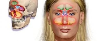
ENT medical doctors have all the technical capabilities necessary to diagnose and treat sinusitis. Extensive experience and attentive attitude of doctors to your problem and your concerns will ensure timely and correct treatment of sinusitis at the modern level and prevent the development of complications. With a runny nose, inflammation of the nasal mucosa can also affect the mucous membrane of the maxillary sinus. There, mucus production, reproduction and accumulation of pathogenic microorganisms increase. The outflow of the contents of the nasal sinus is difficult due to the swollen nasal mucosa, which blocks the opening connecting the nasal cavity and the maxillary sinus. This is how sinusitis develops. The accumulation and stagnation of mucus leads to increased pressure on the walls, which is felt as pain. The formation of pus and the absorption of toxins produced by microbes cause symptoms of intoxication - fever, malaise, headache.
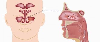
Cysts, polyps of the paranasal sinuses. Odontogenic sinusitis: symptoms, causes, treatment
The formation of a cyst occurs due to impaired ventilation of the paranasal sinuses and the outflow of mucus, i.e. There is no physiological gas exchange and removal of mucous membrane secretions from the sinus. This leads to dysfunction of the secretory cells of the mucous membrane, which begins to grow, the gland duct is blocked and a cyst is formed in the form of a thin-walled bubble with liquid inside. The causes of the appearance of cysts are chronic inflammation of the nasal cavity, the polypous process, as well as inflammatory processes in the roots of the teeth located in close proximity to the sinuses.
Polyps of the maxillary sinus are growths of the nasal mucosa, which appear due to the presence of chronic inflammation with insufficient treatment of colds or allergic diseases, as well as in the presence of immunological changes.
Odontogenic sinusitis develops due to the penetration of infection into the maxillary sinus from an inflammatory focus in the roots of the teeth located in close proximity to the maxillary sinus. As medical practice shows, patients very often turn to an otorhinolaryngologist with symptoms of odontogenic sinusitis after dental surgery. In some patients, inflammation of the maxillary sinus was caused by an infection remaining in the tooth socket, in others, the infection penetrated into the sinus after surgical removal of the tooth, and still others complained of sinusitis due to the filling substance getting into the nasal appendages.
Symptoms of odontogenic sinusitis
Symptoms of odontogenic sinusitis, cysts and polyps
- frequent colds
- migraine-like headache
- temporary or permanent nasal congestion and difficulty breathing
- unpleasant odor from the nose and mouth
- accumulation of mucus and phlegm in the throat
- mucous, light or cloudy purulent discharge from the nose
- dysfunction of the sense of smell
Treatment
At the Federal State Budgetary Institution National Medical Research Center of Otorhinolaryngology, Federal Medical and Biological Agency of Russia, the treatment of sinusitis, cysts and polyps is carried out by specialists from the scientific and clinical department of diseases of the nose and pharynx under the guidance of a talented doctor, Ph.D. V.M. Averbukh. The Center's modern diagnostic equipment, extensive experience of doctors and the successful use of the latest developments and techniques in the field of treatment of sinusitis help not only to correctly diagnose the disease, but also to prescribe effective treatment.
The Center's specialists believe that not all types of cysts and polyps require surgical removal. As practice shows, in most cases, acute inflammation in the maxillary sinus responds well to conservative treatment - the right combination of anti-inflammatory, antibacterial and antiallergic drugs. Patients should be aware that the cyst does not resolve on its own and only in very rare cases is it evacuated from the sinus spontaneously. Only a specialist can determine the type and nature of the cyst and prescribe the correct solution to the problem. If the cyst is large and its volume puts pressure on the walls of the sinus, surgical intervention is indicated.
But before prescribing an individual course of treatment, it is necessary to conduct a thorough diagnostic examination of the patient.
Diagnostics
In the scientific and clinical department of diseases of the nose and pharynx, various modern types of diagnostics are used:
- radiography
- orthopantogram (panoramic X-ray of the jaw)
— CT scan of the paranasal sinuses
— Dental CT
— Videoendoscopy of the nasal sinuses
— MRI of the paranasal sinuses
The Center’s specialists have at their disposal various techniques developed not only by the best domestic and foreign clinics, but also their own. Endoscopic surgery on the maxillary sinus, which is performed both under local anesthesia and general anesthesia, is one of the most modern techniques used in the Center. This operation requires high skill and extensive experience of the surgeon. Through the natural anastomosis of the maxillary sinus, the surgeon carefully performs endoscopic removal of the cyst. The instrument is inserted through the patient's nose without incisions or punctures. The department’s surgeons believe that this technique has no contraindications for use, does not lead to complications, and does not require a long hospital stay. However, if it was not possible to completely remove the cyst using the mentioned method, the surgeon uses a trocar to make a 4mm hole under the upper lip in the vestibule of the oral cavity.
When a foreign body or cyst is located in the bottom of the sinus, the most modern surgical technique is used - minimally invasive removal of pathological formations using infraturbinal access (through the lower nasal passage), without forming a permanent connection between the sinus and the nasal cavity and preserving the mucous membrane. The choice of the most effective surgical intervention and the method of its implementation depend on the nature, localization of the pathological process and the experience of the surgeon.
For each patient of the Center, specialists develop an individual treatment program, and the attention and caring attitude of the staff is guaranteed to everyone!
Symptoms and main complaints of sinusitis
- Runny nose, nasal discharge (if the drainage is not blocked), often due to yellowish-greenish pus
- Difficulty in nasal breathing
- Pain in the nose and inflamed maxillary sinus (right or left of the nose)
- General symptoms: fever, headache, weakness.
Sinusitis is confirmed by the results of radiography or computed tomography. As soon as the diagnosis of sinusitis is confirmed, full-fledged qualified treatment is required under the guidance of an ENT doctor (in no case self-medication!).
Material and methods
The article analyzes the CT results of 45 patients with SNL who were treated in the department of otorhinolaryngology of the Kursk Regional Clinical Hospital from 2000 to 2014. Among the patients there were 39 women aged 37 to 66 years and 6 men aged 34 to 66 years . The studies were carried out in axial and coronal projections with a slice thickness of 0.625-3.0 mm on a computed tomograph, followed by evaluation of diagnostic images on a workstation. All patients underwent biochemical analysis of nasal discharge.
Possible complications of sinusitis
If adequate treatment of sinusitis is not carried out, then pus and inflammatory effusion from the maxillary sinuses can penetrate into the surrounding tissues. First of all, the tissues of the eye react, since in the area of the eye sockets there is a lot of loose fiber that easily absorbs liquid. Swelling and redness of the eyelids appears, and exophthalmos may even develop - protrusion of the eyeball forward. If pus destroys the wall of the maxillary sinus, there is a danger of developing osteomyelitis - inflammation of the bone tissue, in this case the upper jaw. With proper and timely treatment, the risk of such complications is low.
Why you should entrust your treatment to the ENT Department of Dentistry
ENT dentistry is a comprehensive approach to the treatment of complications in the maxillary sinuses after dental treatment
The symbiosis of two areas - dentistry and otolaryngology - makes it possible to identify the cause, assess the situation, and select competent treatment tactics.
The ENT department of the Doctor Levin Center has been providing assistance for many years when problems arise after treatment and removal of teeth located on the border with the maxillary sinus. Surgical treatment is carried out by candidates of medical sciences, maxillofacial surgeons with otolaryngological training .
Patients come to the Center after a painful search for a solution to the problem. Repeated treatment by an ENT doctor in the hospital does not bring results. But a thinking patient should understand that if there is concern on the side of the sinuses where endodontic treatment once took place, you need to contact an oral and maxillofacial surgeon with ENT training. Only in this case can a comprehensive assessment of the situation be made and an adequate treatment plan drawn up.
Treatment of sinusitis
The earlier treatment for sinusitis is started, the better the prognosis. If you consult a doctor before the accumulation of pus in the maxillary sinus (in the first days of the onset of symptoms of sinusitis), the treatment effect is the best (without puncture). Early use of vasoconstrictor drops and sprays will help relieve swelling of the nasal mucosa and restore the outflow of secretions from the maxillary sinuses. Antibacterial therapy and antiallergic treatment are mandatory. Local rinsing of the nose with antiseptic solutions, physical treatment - UHF, UV irradiation of the nasal cavity.
How is the treatment carried out?
We respect the patient’s personal time and strive to carry out all activities comprehensively, in one day :
- Professional hygiene Preparation of the oral cavity to ensure sterility during surgery to avoid secondary sinus infection
- Surgery to eliminate inflammation Performed while you sleep; the method of access to the sinus is selected depending on the location of the foreign body and the presence of tumors
- Temporary prosthetics If the causative tooth had to be removed, a temporary crown or immediate prosthesis is installed to mask the defect
Surgical operations in our Center are performed under sedation. The patient does not feel anything, fears and worries are excluded. Sedatives put you into a controlled drug-induced sleep without falling into unconsciousness - this is not general anesthesia! They do not contain toxic components, act gently, preserving reflexes. The artificial lung ventilation device is not connected, hospitalization is not required.
The surgical intervention is completed by a mandatory CT examination , which is necessary to assess the quality of the work performed.
After 10-14 days, the patient is invited to the clinic for suture removal and a control CT image. A schedule of professional inspections is drawn up.
Puncture of the maxillary sinus
If sinusitis is advanced and there is thick pus in the sinuses, then a puncture is necessary. After removing the pus through a needle, the doctor rinses the maxillary sinuses with antibacterial and anti-inflammatory drugs. After the puncture, catheters are inserted into the sinus through the hole - thin tubes, which allow the sinuses to be washed every day in the future without additional punctures. Relief occurs immediately upon completion of the procedure. And recovery is going very quickly. If the doctor insists on performing a puncture, it means that it is necessary for your health.