- Author:
Naumovich Yulia Yakovlevna - Specialty:
Dentist-orthodontist - Category:
Doctor of the highest qualification category
Learn more about the doctor Get a consultation
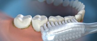
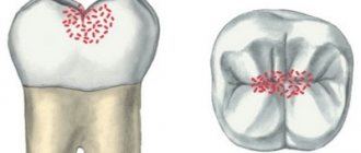
Plaque is a layer of bacteria that accumulates on the surface of teeth and forms immediately after brushing. Such a film on the teeth is considered a normal physiological phenomenon. Its benefit lies in the fact that it protects the enamel from excessively acidic foods, and also prevents the development of pathogenic bacteria. However, if the amount of plaque is excessive, it can cause serious diseases such as caries and gingivitis. Therefore, during examinations, the dentist takes into account the thickness of plaque and draws up a treatment plan in such a way as to achieve an optimal balance: to preserve the protective functions of the teeth without allowing complications to arise.
Symptoms, how to determine the presence of deposits
The formation of dental plaque cannot be tracked, since this process occurs at the cellular level. But its quantity must be controlled and not allowed to turn into hard stone, since this is fraught with the emergence of many problems. The following are symptoms that will help detect the presence of deposits in the oral cavity:
- yellow or white plaque that is located on the surface of the teeth, between them, as well as along the entire length of the gingival margin,
- teeth become rough if you run your tongue over them,
- there is an unpleasant odor from the mouth,
- As plaque turns into stone, bleeding gums appear, mucous membranes can move away from the surface of the teeth, change their color to bright scarlet or even bluish.
There are two types of dental plaque in terms of their localization, i.e. locations:
- superficial, that is, supragingival: visible to the naked eye, has a white, yellow or even brown color,
- subgingival: can be detected during an examination by a dentist, because is located under the mucous membranes and often the patient does not see or feel it (especially if the amount is small). It has a darker shade, even brown. It is impossible to remove it at home.
Kinds
There are two types of tartar, depending on its location - subgingival and supragingival.
Subgingival tartar can only be detected by a dentist using a special probe, since it is hidden from view by the edge of the gum. It is located in the periodontal pocket and fits tightly to the root. The color of this stone is usually dark brown or greenish-black, it is extremely hard and consists mainly of calcium phosphate, magnesium phosphate and calcium carbonate. If subgingival stone is not removed in a timely manner, it can provoke an inflammatory process in the periodontium and cause periodontitis.
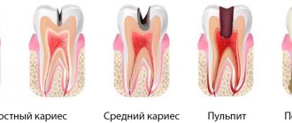
Supragingival tartar can be diagnosed independently; it stands out on the surface of the enamel with its yellowish or brownish color and is easily removed using a dental excavator. It is usually located above the crest of the gingival margin, so it is easy to recognize. In terms of density, supragingival stone is much softer than subgingival stone, since it is a salivary type formation and consists mainly of epithelial cells, bacteria, mucus and food debris. If it is not removed in a timely manner, over time the hardened plaque turns into caries.
Causes of plaque and tartar
The main reason for the appearance of plaque and tartar is associated with the accumulation of pathogenic microflora in the oral cavity. In general, this is a normal process, but deposits must be removed independently and several times a day. If this is not done, their number will increase significantly, which will lead to various complications - diseases of the teeth and gums.
In addition to poor hygiene, there may also be other factors that cause increased plaque formation on teeth. These include:
- smoking and drinking alcohol,
- unhealthy diet - eating a large amount of carbonated drinks and sweets, focusing on soft foods and avoiding hard foods, which have a cleansing effect on the enamel (especially a diet without vegetables and fruits),
- taking certain antibiotics and medications that disrupt the composition and quantity of saliva,
- diabetes mellitus, hormonal disorders.
Causes
If you have stones, the reason for their occurrence may be the following:
- lack of systematic oral hygiene;
- use of low-quality accessories for brushing teeth;
- the presence of a large amount of dental plaque;
- violation of salt metabolism;
- increased viscosity of saliva, as a result of which it loses its cleansing properties and does not wash away plaque;
- insufficient consumption of solid foods, which promote self-cleaning;
- unilateral chewing of food (only on the left or only on the right side of the jaw) as a result of the presence of diseased or missing teeth in the patient’s mouth;
- increased roughness of the enamel surface as a result of filling or orthodontic treatment;
- smoking.
Possible complications
Plaque is the #1 cause of gum inflammation (gingivitis and periodontitis). Therefore, in the long term, this problem can even lead to tooth loss. In addition, deposits are localized on the teeth, and the bacteria that make them up lead to the destruction of enamel, the appearance of caries and other dental diseases.
The aesthetics of a smile are also disturbed, an unpleasant and even putrid odor from the mouth occurs, which affects a person’s psychological comfort. Another consequence is disruption of the digestive tract, since plaque is bacteria and, along with food, they also enter the stomach.
Diagnostic features
If supragingival stones are easy to see on your own, then those located under the gum can only be seen with the help of special tools from a dentist. Diagnosis is carried out as follows: the doctor immerses the probe into the periodontal pocket, which makes it possible to verify the presence of hard deposits, as well as determine the depth of the pocket. This will allow you to choose how to remove the stones.
It is possible to diagnose supragingival calculus even in the early stages of its appearance. There are special tablets on sale that, when bitten, stain areas of the tooth with stone.
Methods of prevention and treatment
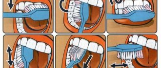
Both prevention and treatment come down to careful and thorough oral care. It is necessary to strictly follow the rules in order to protect yourself from the formation of pathological plaque:
- Brushing your teeth twice a day – morning and evening, for at least 2 minutes, and using properly selected products. It is important to use a brush with medium-hard bristles (even if gum inflammation is present); one that is too soft will not cope with deposits,
- using dental floss and irrigator after every meal,
- using interdental brushes to help clean narrow spaces between teeth,
- Visit your dentist every six months for a checkup.
But you should understand that even with careful hygiene, plaque will still accumulate. For example, under the gums. And even if you missed cleaning at least once or were unable to use the product after meals, especially against the background of reduced immunity or hormonal changes, plaque will again accumulate in greater quantities. Therefore, it is very important to periodically seek help from a dentist for professional oral hygiene.
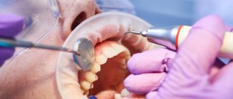
Professional oral hygiene The procedure allows you to quickly and effectively remove plaque and tartar, as well as get rid of subgingival deposits. For this, an integrated approach is used: ultrasound, Air Flow air abrasive device, manual cleaning, chemical destruction of deposits (if necessary), as well as thorough polishing of the enamel and application of a strengthening composition. This is an excellent method of preventing many oral diseases. It is recommended to carry out an average of 1-2 times a year.
Price:
3000 rubles more details about the solution
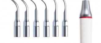
Treatment with the Vector and Varius apparatus The use of apparatus treatment is advisable in the presence of pathological gingival pockets in which plaque and stone accumulate, and there is also pronounced inflammation of the mucous membranes. These devices allow you to remove stone located deep under the gums due to targeted vertical action along the tooth, unlike conventional ultrasonic scalers.
Price:
10,000 rubles more about the solution
Preventive actions
In addition to regularly brushing your teeth, follow these rules:
- Eat right. The diet should contain hard foods.
- After eating, eat a sweet and sour apple. Its juice will dissolve some deposits.
- Use floss and a special rinse after every meal.
Visit your dentist regularly. All of the listed preventive measures are relevant when there is a small amount of supragingival hard deposits. What is happening in the area of the roots of the teeth can only be seen on x-rays. As practice shows, in approximately 50% of cases, tartar is deposited under the gums. This means that only a doctor can remove it.
In what cases does stone plaque form?
The reason for the formation of tartar is poor quality dental care, in which soft plaque is not completely removed. High mineral content causes plaque to harden. Using a too soft toothbrush, lack of attention to the interdental spaces and gum margins, irregular oral hygiene, etc. are the main sources of the problem.
But there are factors that increase the likelihood of hard deposits forming on teeth:
- diseases of the digestive system and endocrine system, in which the chemical composition of saliva changes;
- malocclusions and dental defects that form particularly difficult areas to clean;
- wearing dentures, braces, brackets, plates and other structures that make it difficult to properly clean teeth using a traditional brush and paste;
- a large amount of carbohydrate foods in the diet, which facilitates the gluing of soft plaque particles.
What should be the prevention of dental plaque [4]
To reduce the rate of plaque formation, you should pay due attention to hygiene, give up bad habits, review your diet, and pay attention to the quality of water.
Regular use of fluoride-containing toothpastes plays a leading role in the prevention of dental caries at the individual and community levels.
According to the conclusion of WHO experts and due to the presence of strictly proven facts about their anti-caries effectiveness, it is considered unethical to recommend toothpastes that do not contain fluoride to the population. Daily use of fluoride-containing toothpaste is recommended for every person, regardless of the presence of any other source of fluoride, systemic (fluoridation of water, salt or milk) or local (gels and varnishes with a high fluoride content, rinses, glass ionomers, etc.) Official position of the Russian Dental Association [6].
First of all, it is necessary to reduce the consumption of sweet foods and drinks, and also take into account that the formation of dental plaque depends not only on the composition, but also on the consistency of the food. Soft crushed food leaves more plaque on the teeth than coarser food. Therefore, for example, gnawing on whole raw carrots is healthier than eating carrot salad. After eating, it is advisable to rinse your mouth with mouthwash or water to remove food debris and reduce acidity levels.
Who is contraindicated for dental cleaning?
Manual removal of tartar or soft plaque has practically no contraindications, which cannot be said about the use of devices. Such removal of deposits is contraindicated in the following conditions and pathologies:
- for heart diseases;
- if there are implants in the oral cavity;
- bronchial asthma and bronchitis;
- presence of hepatitis and tuberculosis;
- diabetes;
- if there is increased sensitivity, which can provoke a gag reflex;
- during pregnancy and while breastfeeding, it is better to refrain from such procedures;
- Such methods are also contraindicated for children and adolescents.
When visiting a dentist, you must inform him about the presence of chronic pathologies or existing acute diseases.
Plan Introduction.
Classification of dental plaque. 2. Surface formations on the teeth: cuticle. Supragingival dental plaque: pellicle, green plaque, smoker's plaque, dental plaque, soft plaque, food debris, tartar (salivary). Subgingival hard dental deposits: tartar (whey) Dynamics of formation of dental deposits and their role in the development of dental diseases: Cariesogenic properties of dental plaque and their role in the development of dental caries The influence of dental plaque on the permeability and solubility of enamel The role of dental plaque microorganisms in the etiology and pathogenesis of diseases periodontal To date, there is no uniform terminology in the literature that objectively characterizes dental plaque. The same name often refers to different structural formations. The most popular term at present is “plaque” and its translations into Russian are “plaque” or “dental plaque”. The cuticle (Nasmitian membrane) is an embryonic epithelium that is lost after tooth eruption. The enamel surface is exposed to saliva and microorganisms. As a result of erosive demineralization on the surface of the enamel or the dissolution of its proteins, ultramicroscopic tubules are formed that penetrate the enamel to a depth of 1-3 microns. Subsequently, the tubules are filled with an insoluble protein substance. Since the cuticle is lost before or shortly after tooth eruption, it does not subsequently play a significant role in the physiology of the tooth. A study of the ultrastructure of the enamel surface showed that the cuticle is present only in its subsurface layer, sometimes reaching the surface in the form of a microscopic film. There are various divisions of dental plaque: I. Groshikov M.I., 1973. Soft plaque (white plaque, smoker’s plaque, green plaque). Hard dental deposits (tartar). II. Pakhomov G.N., 1982. Non-mineralized dental plaque: Pellicle Dental plaque White matter (soft plaque) Food debris Mineralized dental plaque: Supragingival tartar Subgingival tartar However, it is not entirely appropriate to classify food debris as dental plaque; rather, they should be considered as a favorable environment for the growth and maturation of dental plaque. All dental deposits have varying degrees of fusion with the surface on which they were formed, while food residues do not have real fusion; only certain types of food may have short-term adhesion to the surface due to their viscosity, but over time their adhesion quickly decreases. III. An expanded classification of dental deposits should be considered the classification of Ulitovsky S.B., 1999: Division Dental deposits are divided according to the following characteristics: By degree of density By degree of mineralization By localization By degree of expression By place of deposition By stages of growth. By degree of density: Soft Mixed Semi-dense (compacting) Hard. According to the degree of mineralization: Plaque (0th stage of inception) Non-mineralized (1st - soft phase of spread and accumulation) Mineralizing (2nd - transition phase, compaction occurs) Mineralized (3rd - final phase of formation of tartar). By localization: Supragingival: by density (soft, hard, mixed) by distribution (towards the periodontal sulcus, cutting edge, deep into the fissures, stable) Mixed Hybrid Subgingival: by density (hard) by growth (in the cervical region, the upper third of the root, up to mid-length of the root, to the root apex) by localization (one root surface, two, three, root bifurcation area, total). By degree of severity: Dental plaque Soft plaque Soft plaque with minor supragingival tartar Pronounced supragingival tartar without destruction of the periodontal sulcus Minor supragingival tartar with destruction of the periodontal sulcus Pronounced supragingival tartar with destruction of the periodontal sulcus Pronounced subgingival tartar growing along the root of the tooth. By stages of growth: Growth of dental plaque from outside Growth inside Mixed growth Stable growth (in some cases can be considered as a transitional phase) By place of deposit: On teeth On fillings On crowns On fixed dentures On removable dentures On dental calculus Some foreign authors (Doc. MUDr . Ivo Drizhal, CSc., 2001) offer an extremely simplified division of dental deposits: Supragingival dental plaque Subgingival dental plaque Fissural dental plaque SUPRAGINGIVAL DENTAL POLITUS TOOTH PELLICULE The tooth pellicle is an acquired thin (from 1.5 to 50 microns) transparent organic film that came to replace the Nasmith shell. It is a structureless, microbial-free coating formation on the surface of tooth enamel, resulting from spontaneous deposition (adsorption, polymerization and denaturation) of the protein-carbohydrate components of saliva: mucin, glycoproteins, sialoproteins, and is not related to collagen, keratin, or hemoglobin-like substance. The pellicle is difficult to identify with the naked eye; bacteria quickly colonize on its surface and dental plaque forms. To detect pellicles in clinical studies, dyes are usually used, in particular, Bismarck brown, Lugol's solution, fluorescent sodium solution, 0.75% and 6% solutions of basic fuchsin, 4-5% alcohol solution of erythrosine, erythrosine tablets (6-10 mg), Schiller-Pisarev solution, 2% aqueous solution of methylene blue. If the tooth is thoroughly cleaned, removing the plaque, and stained again, the colored film will be visible; if this film (pellicle) is also removed with pumice, the tooth will not be stained. Quite often, colored pellicles are found in the clinic, which can arise under the influence of chromogenic bacteria, tar and tar (from smoking), coloring food components, blood pigments, medications, etc. The pellicle is quickly restored: if the pellicle is removed from the surface of the teeth with abrasives, then within 20-30 minutes their surface is again covered with pellicle, provided that the tooth is in contact with saliva. The thickness of the daily pellicle is 2-4 mlm. The dental pellicle plays an important role in the processes of diffusion and permeability in the subsurface layer of enamel, in protecting teeth from the effects of dissolving agents. It gives the enamel selective permeability. As mentioned above, after eruption, as the tooth loses its embryonic formations, ultramicroscopic tubules are formed on the surface of the enamel, which are filled with a protein substance, where sialic acid, alanine, glutamic acids, amino sugars - derivatives of the bacterial membrane predominate. Precipitation of salivary mucoproteins, sedimentation, growth, and then destruction of microorganisms on the tooth surface lead to the formation of a thicker organic layer of pellicle, consisting of a mixture of salivary proteins and lysed bacteria. Due to local conditions, microbes invade these structures and multiply, causing the onset of dental plaque development. Electron microscopic studies have proven that the pellicle consists of several layers. The subsurface component is located in the thickness of the enamel and has many processes that fill its pores or demineralized areas. The middle part of the pellicle is closely connected to the tooth enamel and has a uniform thickness, usually less than 1 micron. The thickness of the surface layer of the pellicle reaches 10 microns; it is located under the plaque or in areas of the tooth that are rarely cleaned. This layer can be formed from adherent salivary glycoproteins, excretory products of bacteria, or degenerated epithelial cells. The pellicle is free from bacteria, but it gives rise to the formation of dental (microbial) plaque, sorbing microbial associations in its niches. Adsorption Str. sanguis to the enamel is significantly accelerated if the enamel is covered with a pellicle. The pellicle thus creates a situation for the initial phase of microbial colonization to occur on the tooth surface. The processes of diffusion and permeability in the surface layer of enamel and solubility in an acidic environment depend on the state of the pellicle. Changes in the composition and properties of the pellicle may favor the development of caries. The acquired pellicle, like most protein adsorbed layers, is a membrane that gives the enamel selective permeability. Because of this, the diffusion of various solutions from saliva into the tooth and from the tooth into saliva can be significantly altered. Solutions must either penetrate the pores of this membrane or dissociate into ions that easily cross the barrier. This barrier is a factor influencing the rate of caries and tooth remineralization. Pakhomov G.N. (1982) noted that the pellicle can reduce the initial flow of fluoride into the tooth, at the same time, it can delay the reverse diffusion of the resulting fluoride enamel products from the tooth. An important factor is the reaction of fluoride with enamel at high and low concentrations. Low concentrations of fluoride when applied topically promote the formation of fluorapatite, but it is quickly washed out from the tooth surface. If the pellicle reduces the initial flow of fluoride into the tooth, then a higher ratio of fluorapatite to calcium fluoride is likely to be formed. GREEN POLICY Green plaque was previously called greenstone or pristley mass. According to Priestley, the cause of staining of the pellicle and/or dental plaque is the development of the fungus Lichen dentalis, which produces chlorophyll. In this regard, such deposits are observed to a greater extent on the front teeth, which are exposed to light. But its deposition on molars cannot be ruled out. Characteristically, only the labial and buccal surfaces of permanent and temporary teeth are affected in the form of a border or individual stripes at the neck of the tooth (mainly in children). Deposits are most often observed on the teeth of the upper jaw and very rarely on the teeth of the lower jaw. The most intense coloring was noted in the area of the gingival margin. This plaque cannot be easily removed with a brush and is difficult to remove instrumentally, since it is very tightly fixed to the enamel surface. Under a microscope, the enamel of such teeth reveals numerous thin holes densely populated with bacteria. There is an idea that green plaque is the result of the formation of sulfmethemoglobin, which is released when gums bleed, and is also observed among workers in some industries when copper dust enters the oral cavity. 'S PLATE Smoker's plaque is the result of the deposition of tobacco pigment mainly on those surfaces of the teeth that are not involved in the act of chewing and are poorly cleaned when brushing the teeth. These are the lateral and cervical surfaces of the teeth, the lingual and palatal sides. The color of the plaque ranges from brown to black. The layer is small, but difficult to remove. DENSE PLAQUE, OR DENTAL (MICROBIAL) PLAQUE Dental plaque is located above the pellicle of the tooth. To detect it, staining solutions are used. But upon careful examination of the teeth in the cervical part of the crown and on the lateral surfaces after removing the white matter with a toothbrush (especially in people who do not regularly care for the oral cavity), a formation with a rough surface can be detected. It has been established that plaque on the teeth begins to accumulate within 2 hours after brushing the teeth, and after 3-4 days it covers half or even most of the tooth crown. Dental plaque is a soft amorphous granular deposit with a specific histological structure. It sticks tightly to the pellicle located underneath it, is not washed off, and is not always removed when brushing your teeth; rinsing and air or water jets do not completely remove it; it is scraped off with an excavator or trowel. Only after careful removal of dental plaque does the shiny surface of the enamel appear. Often, under the plaque, an area of demineralized enamel with a changed dull color (white, gray) is found. Dense dental plaque is a product of microbial growth, where their active life activity occurs, accompanied by acid formation, enzymatic activity and other metabolic processes. Colonization of body surfaces or organs by saprophytes is a common phenomenon; the body is protected from excessive proliferation of microorganisms by regularly separating the surface layers of the epithelium, which are carriers of biofilm microbes. It's about self-purification. Dental plaque has a completely different status than other biofilms in the body. It cannot be removed by self-cleaning, only mechanically. In small quantities, the plaque is not visible unless it is pigmented (green plaque, smoker's plaque, etc.). When it accumulates in large quantities, it becomes a visible mass of gray or yellow-gray color. The plaque occurs above the gum, most often on the gingival third of the teeth, and under the gum. It is formed equally on the upper and lower jaws, more on the molars and on the lateral surfaces of the teeth, in smaller quantities on the buccal and labial surfaces, and least of all on the lingual surface. Plaque formation begins with the attachment of a monolayer of bacteria to the acquired pellicle or tooth surface. Microorganisms “attach” to the tooth using a sticky interbacterial matrix, with carbohydrates playing a significant role, which are partially converted into dextrans and levans during the process of microbial decomposition. Of these, dextran is the most important component in the adhesion process. It has high adhesive properties, as well as relatively low solubility and high resistance to bacteria. Dextran is produced from sucrose by streptococci. The formation of an immature plaque is regulated by the degree of cleaning of the oral cavity - the worse the hygiene, the faster the plaque matures. Studies of dental plaque using an electron microscope have shown that young mature dental plaque has a certain structure. It becomes similar to tissue - it contains cells (epithelial, lymphocytes, macrophages), intercellular substance and tissue fluid. A few days after ripening, it can be divided into cellular and acellular components. Organic and inorganic components make up ¼ of the mass of the plaque and represent an intermicrobial jelly-like substance, which also contains water, mainly inside bacterial cells, which does not have a strong connection with protein, and an intercellular matrix. Bacteria make up approximately 75% of the solid residue (1 mg wet mass of plaque - 300 bacterial strains). The matrix consists of a complex of polysaccharides and proteins, in which the main components are hydrocarbons and proteins (30% each), as well as about 15% lipids. The protein component is formed due to the precipitation of sialic acids from saliva under the influence of enzymes produced by microorganisms. The remaining components are represented by extracellular metabolic products of plaque bacteria, remnants of their cytoplasm and cell membrane. Carbohydrates, present in small quantities in the matrix, are represented by dextrans, mutons, fructans - polysaccharides, their derivatives and bacteria, which constitute approximately 9.5% of the total mass of the solid plaque. Other matrix hydrocarbons include levan polysaccharide bacterial product (4%), galactose (2.6%) and methylpentose in the form of ramanose. The main inorganic components of the plaque matrix are calcium and phosphorus. Magnesium, potassium and sodium are contained in small quantities. They are associated with organic components of the matrix. There are more inorganic components in the plaque matrix of the anterior teeth of the lower jaw, mainly on the lingual surfaces. The plaque contains fluoride, its concentration increases in people whose teeth are treated with fluoride preparations or who consume water with an optimal fluoride content. Dental plaque contains proteolytic (spirochetes, neisseria, veillonella) and acidophilic microorganisms (actinomycetes, lactobacilli, leptotrichia, streptococci - Str. mutans, mitis, sanguis, salivarius). In the group of acidophilic bacteria, acidogenic bacteria are isolated separately, and acidogenic bacteria, which synthesize lactic and sometimes acetic acid from sucrose, are isolated separately. The first group is characterized by anaerobic fermentation; the substrate is carbohydrates, food proteins and amino acids. All of them are capable of breaking down collagen. In addition to lactic acid, dental plaque contains formic, butyric, propionic and other organic acids. Insufficient neutralization of acid formed in dental plaque is explained by limited diffusion of neutralized salivary compounds, for example, calcium, into plaque, and limited diffusion of acidic products from it. As the plaque grows, its microbial flora changes from a predominance of cocci to rod-shaped and filamentous microorganisms. Initially, the plaque consists of characteristic cocci. Streptococci make up approximately 50% of the bacterial flora with a predominance of Str. mutans and Str. sanguis. As the plaque thickens, anaerobic conditions are created inside it, and the flora changes. Superficial microorganisms, as a rule, take power from the oral medium, deeper also use metabolic products of other bacteria of plaques and components of the plaques matrix. This leads to the fact that gram-negative cocci and sticks (2/3 of the total number of microorganisms) appear on 2-3 days, of which 15% make up anaerobic sticks: leptotrichia, bacteroids, campilobacteria. On 4-5 days, fuzobacteria, actinomycetes, Weilonella appear. After 7 days, spirella and spirochetes appear, the number of fibrous microorganisms increases. From 28 to 90 days, the amount of streptococci decreases from 50%to 40-30%, and the number of spindle and threaded microorganisms, moving sticks and spirochet increases to 40%. Mature plaques contains about 325 types of microorganisms and is represented by 4 layers: 1) acquired by pellikula 2) polysadomously located fibrous microorganisms fixed on pellikul 3) dense network of fibrous bacteria 4) with a surface layer of cocci while 1.2.3 layers are a structured part And 4 layer - freely located clusters of microorganisms. A significant role in the formation of plaques is played by saliva. It contains a mixture of glycoproteins, called mucin. All salivary glycoproteins consist of proteins in conjunction with various carbohydrates: sialic acid, fucosa, galactose, glucose, mannose and two hexosamine - acetylactosamine and acetylhlucosamine. Enzymes of glycosidase of microorganisms split carbohydrates that they use as nutrients. Depending on the symbiosis of individual groups of microorganisms and food residues, alkaline and sour dental plaques are formed. So, with metabolism of a protein substrate and a small amount of carbohydrates, an alkaline plaque is formed. With metabolism of protein substrate and a large number of enzymatic carbohydrates - acidic. In addition, Dexstra serves as a barrier that prevents the diffusion of saliva and the manifestation of its neutralizing action. Therefore, the pH of a plaque can range from 4.0 to 8.0. The consistency of food affects the rate of plaque formation. Plush quickly occurs when drinking soft food, adding sucrose to food accelerates the formation of plaques. During sleep, the plaque forms faster than after eating, because The mechanical effect of food and mechanical salivation during chewing can delay the formation of plaques. Soft plaque
Mechanical removal of deposits
Removal of dental plaque in this way is carried out manually or using special equipment. You can combine different cleaning methods to achieve the perfect result. At the end of the procedure, the dentist uses brushes and special pastes to polish the enamel, and the patient leaves the office with a snow-white smile.
- The manual method is carried out using special curettes. It is often combined with other methods. Personal manual cleaning is used only when the patient is contraindicated to use the devices. This happens in the presence of epilepsy, diabetes and some other pathologies.
- Ultrasound removal of dental plaque. This is an effective method of removal using a modern device. The method allows you to remove even large stones without damaging tooth enamel.