The dentofacial system is an important component of the human masticatory apparatus. It meets morphological and functional characteristics, such as number, size, shape, color, structure of dental units. There are a number of genetically determined or external factors that lead to disruption of these characteristics. Dental anomalies can have various consequences: some lead to periodontal pathologies, various forms of caries, others seriously limit chewing function, cause injury to the mucous membrane, and others cause aesthetic problems.
Improper growth of teeth results in speech and bite defects and breathing problems. As a result, the patient’s general health seriously suffers, and psychological problems arise.
Reasons causing anomalies
Anomalies of the dental system are very diverse and include both anomalies of dental units and incorrect formation of the dentition, as well as abnormalities in the development of the jaws. The most frequently diagnosed abnormalities of dental units. They can be caused by a wide variety of reasons, including both exogenous (caused by external factors) and endogenous (formed as a result of the physiological state of the patient).
Endogenous causes
Endogenous anomalies are formed as a result of genetic and endocrine disorders.
- Many structural features of the dental system, leading to improper development of teeth, are genetically determined. This can occur through direct inheritance (abnormal number and shape of teeth, edentia, diastema), through inheritance of a mismatch in the size of the jaw bones, through inheritance of a mismatch in the size of the jaw and teeth (crowding of teeth or sparse arrangement of teeth). Dental anomalies of this type include disorders associated with hereditary and congenital pathologies (clefts of the soft and hard palate, cleft lip, Down syndrome, Vaanderburg syndrome, Seckel syndrome, Shershevsky-Turner syndrome).
- Another part of the endogenous causes of dental anomalies are problems of the endocrine system, such as hypothyroidism (in later stages), hyperparathyroidism, hypocortisolism. Their frequent symptoms may be delays in the eruption and replacement of teeth, and disturbances in the formation of the enamel layer.
Exogenous causes
Exogenous (external) causes of dental anomalies are, for the most part, external unfavorable factors that affect the baby’s body during the formation of tooth germs. There are prenatal (prenatal), postnatal (postpartum) and intranatal (associated with childbirth) periods.
- An example of a prenatal factor is a pregnant woman living in poor environmental conditions. Various developmental disorders of the fetus can also occur as a result of the mother’s poor lifestyle.
- Factors in the intranatal period may include complicated labor, birth injuries, consequences of asphyxia and oligohydramnios.
- In the postnatal period, dental anomalies develop as a complication of pathologies in early childhood. These include childhood infections, rickets, hypovitaminosis, and micronutrient deficiency.
All these reasons are of a common nature. Local factors that affect dental development include everything related to improper oral care, feeding errors or bad habits. Among them are feeding too soft food, prolonged sucking of a pacifier or finger in early childhood.
Childhood injuries and caries with complications lead to dental anomalies.
The death of tooth germs or the development of supernumerary teeth can occur due to osteomyelitis.
Etiology of the pathological phenomenon
Tortoanomaly is a polyetiological problem. Various factors, both acquired and congenital and/or genetically determined, can lead to its development.
The most common reasons include the following:
- Lack of space in a row due to narrowing of the jaw and/or macrodentia.
- Pressure on the causal unit from neighboring supernumerary or displaced units.
- Incorrect follicle formation.
- Too early replacement of “milk jugs” with permanent units or, on the contrary, their persistence (delay in eruption).
- Injuries in the area of the anterior teeth.
- Genetic predisposition.
- Malignant and benign neoplasms that put pressure on the problematic tooth.
Other factors cannot be excluded as the cause of tortoanomalies. In particular, bad habits, disorders in the prenatal period, improper treatment of other anomalies, etc.
What is coronoradicular teeth separation, in what cases is it performed, what does it give.
Come here if you are interested in the dangers of a split tooth.
At this address https://orto-info.ru/zubocheliustnye-anomalii/zubov/razmera/nazyivayut-korotkimi.html we will tell you how short teeth are corrected.
Anomalies in the number of teeth
All anomalies in the number of teeth are divided into three types:
- hyperodontia (excess of dental units above normal);
- edentia (complete absence of teeth);
- hypodontia (insufficient number of teeth).
Hyperodontia
Hyperodontia is a disorder of the formation of the dentition, which is expressed in the presence of supernumerary teeth (over 32 dental units in an adult). The cause of the disease may be a violation of the mechanism of formation of tooth germs in childhood or infancy. Hyperodontia mainly affects the incisors of the upper jaw. Other teeth are involved in the pathological process less frequently. Extra dental units grow small and have a cone shape.
The disease can be true or false.
- With true hyperodontia, the formation of extra dental units occurs in the human jaw.
- False hyperodontia occurs when the loss of primary teeth is delayed, which interferes with the normal growth of permanent teeth.
With polyodontia, teeth may erupt away from the dentition or next to a correctly positioned dental unit, which leads to its gradual displacement. The dentition can be seriously deformed, which leads to serious diction problems.
If supernumerary teeth interfere with the development of normal teeth, they must be removed. Broken teeth are corrected by installing braces.
In those rare cases when a row violation does not occur, the supernumerary tooth is preserved. Prosthetics are used to correct the shape.
Hypodentia
Hypodentia is a deficiency of dental units. This disorder occurs most often due to the death of tooth germs in the fetus. A small number of teeth causes a shift in the midline, the formation of wide gaps between teeth (diastema and three), and shortening of the dentition. All this adversely affects the bite. Possible ways of correction are prosthetics.
Edentia
Adentia is a complete or partial absence of teeth. With edentia, the continuity of the dentition is disrupted, which leads to difficulties in chewing and speaking. The reason is a violation of the development or death of tooth germs under the influence of genetic factors or under the influence of unfavorable external factors during the formation of the dental plate in the fetus (the formation of the germs of permanent teeth in the fetus occurs after the 17th week of intrauterine development). The disorder is corrected using prosthetics or dental implantation.
Methods for correction and treatment of anomalies
Treatment depends on the clinical situation and is determined by the doctor after a comprehensive dental examination. Correction of dental abnormalities may include:
- surgical removal of wisdom teeth or supernumerary units;
- prosthetics: for adentia, hypodentia;
- orthopedic correction with a crown, veneer, lumineer: for size anomalies;
- restoration with crowns, composite materials, veneers: for non-standard shapes;
- if the structure of enamel and dentin is damaged, complex therapeutic, orthodontic, orthopedic treatment, enamel mineralization, and drug therapy are prescribed;
- eruption disorders: physiotherapy, massage. Sometimes orthopedic treatment (plates, aligners), removal of a baby tooth (if a permanent tooth has formed underneath it), surgical care (for impacted segments), or orthodontic preparation (system braces) may be required in order to create conditions for stretching the segment retained in the gum.
Malocclusions are most often corrected with fixed orthodontic structures (braces, aligners) in adults. Children are mostly fitted with removable devices.
Anomalies of magnitude
There are two forms of this type of deviation, including macrodentia (increase in the size of dental units) and microdentia (small teeth).
Macrodentia
With macrodentia, the size of dental crowns is significantly increased. The cause of the disorder is, in most cases, dysfunction of the endocrine system, which is characterized by the fusion of several rudiments together. The disease is localized mainly in the area of the upper incisors. Large teeth interfere with the process of eruption and growth of neighboring dental units, which leads to their abnormal arrangement and crowding. Giant teeth are found on or outside the dentition line. This is a serious cosmetic defect that causes serious functional impairment and psychological disorders. Pathologically enlarged teeth are removed, and the growth of adjacent teeth is corrected using prosthetics or braces.
Microdentia
Microdentia – teeth that are too small. The disorder mainly affects the upper lateral incisors, but may affect the incisors of the lower or both jaws. The cause of the anomaly has not been studied, but it is reliably known that it develops in genetically predisposed patients. With the problem of small teeth, patients develop too large interdental spaces, which significantly disrupts the aesthetics of the dentition. To correct the pathology, incorrectly growing teeth are removed or covered with crowns.
Causes of polyodontia
Medicine has not yet found an exact answer to the question of what are the causes of supernumerary teeth. Scientists put forward several hypotheses:
- Atavism. Supernumerary teeth are explained by the fact that the dental system strives to return to the original number of elements laid down by nature. There is evidence that our ancestors had 6 incisors on both the lower and upper jaws. As a result, many doctors consider atavism to be the cause of the development of polyodontia in humans.
- Splitting of the tooth germ. Even in the embryonic period, the activity of the dental plate is disrupted in the child, as a result of which hyperdontia is formed. Violations can be caused by viruses, poor ecology, drugs, medications prohibited during pregnancy, alcohol and other factors. This hypothesis is increasingly supported today, because recently the disease has been rapidly progressing due to bad habits and poor ecology.
The causes of hyperdontia continue to be researched. Scientists cannot give an exact explanation for this anomaly, but most of them are inclined to the second hypothesis - the splitting of the tooth germ at the embryonic stage.
Shape anomalies
There are pathologies in the development of dental units, due to which they acquire an unnatural shape. These disorders are named after the scientists who first described them. The following types of dental shape anomalies are found: spiny teeth, Pflueger teeth, Fournier teeth, Hutchinson teeth.
- Spiked or awl-shaped teeth. With this pathology, the teeth acquire a cone-shaped shape. Wide at the base, they gradually narrow and, towards the cutting edge, become sharpened into a spike shape. This problem is combined with microdentia. There are irregularities and stains on the surface of the teeth. The disease affects the front and lateral incisors. The disease occurs in childhood and is caused by genetic factors in combination with external factors. Among them are vitamin D deficiency and endocrine system problems.
- Hutchinson's teeth are characterized by a modified crown shape of the incisors. Externally, they have the shape of a barrel, since the neck is significantly thickened. The cutting edge of the teeth acquires an arched notch. The enamel layer also suffers, which is present only on the sides and absent in the center.
- Fournier's teeth are a form of systemic hypoplasia of dental units associated with metabolic disorders at the stage of intrauterine development of the fetus. The barrel-shaped shape of the teeth is preserved, but the notch of the incisal edge is absent. The color of the enamel is not disturbed, but the enamel layer is underdeveloped, which can be seen during microscopic examination.
- Pflueger's teeth are a disorder that affects permanent dental units. Their crowns take on a conical shape. Thickening develops in the cervical region, and the chewing surface is significantly underdeveloped. The chewing function of the teeth is completely preserved.
- Turner anomaly. With this pathology, there is no enamel on the teeth. They have an abnormally lumpy surface, and replacement tissue, dentin, forms in the exposed areas.
Systemic hypoplasia with a violation of the shape of the teeth has three degrees of development. The third degree is the most dangerous, in which the crown is severely deformed and the enamel layer decreases. In this case, the defective teeth are removed and replaced with dentures. It is also possible to restore affected teeth using reflective components.
Consequences of polyodontia disease
Polyodontia in humans can often be the cause of retention. This is a phenomenon in which normal teeth are unable to erupt due to the interference of supernumerary teeth. The former may remain in the jaw or take an abnormal position.
In addition, even if the complete incisor grows before the supernumerary one, the latter will be able to displace it. This will lead to the person being unable to chew food normally. And if several extra incisors grow at once, they can cause the loss of permanent teeth.
Anomalies of hard tissue structure
An altered shape, abnormal size or color of enamel is formed against the background of an abnormality in the structure of the hard tissues of the dental unit. Among such anomalies are:
- Hypoplasia (underdevelopment of tissue). The initial degree of the disorder is manifested by the presence of chalky spots on the enamel and areas where tissue deficiency is observed. Subsequently, all kinds of pits, grooves, and recesses appear on the surface of the enamel. The defect affects all teeth in the dentition.
- Hyperplasia (excessive tissue formation). Pathology also affects all teeth at the same time. It is characterized by areas where tissue growth is observed - tubercles, sagging enamel. The consequence of the pathology is a change in the shape and size of the teeth, a violation of the occlusion (the line of contact of the upper and lower jaws).
- Anomalies of amelogenesis (enamel formation) are expressed in the presence of brown or yellow spots on the surface of the teeth. Areas where the natural composition of the enamel is disrupted become especially sensitive. Microdentia develops against the background of pathology. The cause of the development of pathology is a deficiency of microelements that take part in the formation of dental tissue. Treatment consists of replenishing this deficiency. Additionally, local remineralizing therapy and physiotherapy are performed.
- Disturbance of dentinogenesis consists of dysfunction of the mechanism of dentin formation. The main symptoms of the disease are as follows: teeth become yellow-brown or grayish in color. The fragile dentin in these areas quickly wears off, causing tooth decay and then tooth loss. The disease occurs in genetically predisposed patients. The problem can only be solved by replacing the units destroyed by the disease with prosthetics.
Symptom relief
Most often, in adults, extra teeth erupt without any symptoms, but for children this can become a problem that needs to be addressed.
Supernumerary teeth erupt with the same symptoms as regular teeth, so the treatment for them is the same.
- To lower the temperature, it is recommended to give your baby Paracetamol or Ibuprofen. If the child is very small, these drugs can be used in the form of suspensions or rectal suppositories. In addition to lowering the temperature, these medications do an excellent job of treating pain and inflammation.
- To relieve gum pain, local anesthetics are used - ointments and gels (for example, Kalgel, Dentinox, Solokoseryl). These remedies cope well with painful sensations and slightly relieve inflammation.
- Adults and children over 2 years of age can be treated with folk remedies: propolis, honey, decoctions of calendula, chamomile and lemon balm. Some decoctions help reduce pain and relieve inflammation. Traditional methods of treatment should be used only after consultation with your doctor.
- If primary supernumerary teeth have partially erupted, stimulation of eruption is prescribed. For this purpose, vibration and electrical stimulation, as well as special massage, are used.
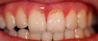
Color anomalies
Healthy, young teeth look snow-white due to a thick dentin layer and high-quality enamel, which has sufficient characteristics of whiteness, shine and transparency. Lifestyle, age, genetic factors, and ecology can slightly change the color of teeth from bluish to yellow. Small deviations are not considered pathology, although they indicate a change in the quality of the enamel.
Various pathological processes occurring in the body have a more significant impact on the condition of the enamel. The cause may be carious lesions, the use of medications, or a lack of certain microelements in the body. Teeth can take on a gray, pinkish, brown, purple and even black tint.
When starting to treat a patient, the dentist excludes the development of chronic pathologies. After a course of therapy and stable remission is achieved, other causes of discoloration of the enamel are eliminated. The final stage of treatment is a course of teeth whitening.
Specialists at the Consilium Dent choose the enamel lightening technique together with the patient. The selection criteria are age, condition of the enamel, and the wishes of the patient.
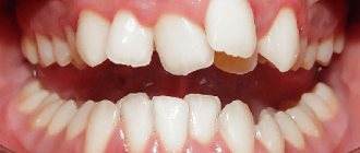
Position anomalies
Incorrect position of teeth always develops as a consequence of another pathology and forms an incorrect bite. May affect one or both jaws. Incorrect position of teeth makes chewing food and oral hygiene difficult. This can lead to digestive problems and the development of cavities. There are several options for the pathological position of teeth within or outside the dentition:
- External or vestibular position means that the tooth is located outside the dentition, closer to the vestibule of the mouth. This position is typical for canines and incisors and develops as a serious cosmetic defect.
- Oral or internal position. The teeth are deviated inward closer to the tongue. The disorder is typical for canines, incisors and premolars. May cause tongue injuries and dysfunction of the temporomandibular joint.
- Mesial and distal position. Dental units are displaced forward or backward along the dentition, which leads to its shortening.
- Supra and infraocclusion are high or low positions of teeth relative to the occlusal curve. The cause of the disorder is underdevelopment of the alveolar process. It appears due to the presence of some obstacle that interferes with the normal formation of the tooth germ. Effective therapy is surgery.
- Tortoanomaly. The tooth is rotated around a vertical axis. The disorder is typical for incisors, canines, and premolars. The anomaly can cause injury to adjacent teeth. Dental units are rotated into the correct position using orthodontic structures.
- Transposition. The teeth change their location with each other. The disease affects canines that exchange places with premolars or lateral incisors. The disorder develops at the stage of tooth germ formation. The therapy is as follows: problematic teeth are removed followed by prosthetics.
- Crowding of teeth occurs due to lack of space when tooth germs are located too closely. Crowded teeth are closely adjacent to each other and rotate around their axis. Occurs in combination with macrodentia or with an undeveloped basal part. To eliminate the violation, some dental units are removed.
- Diastemas and tremas are wide spaces between teeth. The disorder develops as a consequence of an abnormal shape and size. The problem is treated with orthodontic methods or by installing veneers, which helps restore the aesthetics of the smile.
Misaligned teeth cannot always be cured using braces. Severe pathology, which is characterized by a skeletal disorder of the maxillofacial region, is treated with surgical reconstruction. The pathological fragment is transferred to the desired position and then secured. A fragment containing part of the dentition or the entire dentition can be used.
Retention period
Tortoanomalies corrected with orthodontic appliances require a long period of retention as the periodontal and interdental ligaments strive to return the tooth to its previous abnormal position.
Retention methods are divided into orthodontic, surgical and physiotherapeutic:
- Orthodontic retention primarily involves the use of lingual retainers bonded to the inner surface of the teeth. The device used to correct the anomaly can also be used as a retainer.
- The surgical method of retention involves dissection of the interdental papillae with ligaments that tend to return the displaced unit to its previous position.
- The physiotherapeutic method consists of vibration stimulation of the rotated tooth with a frequency of up to 50 Hz. Vibrations are accompanied by repair of the bone tissue of the alveolar sockets, which helps maintain the tooth in a rotated position. Vibration stimulation sessions are carried out several times a week.
The retention time by orthodontic methods depends on the severity of the tortoanomaly and the correction method used, and can be several months.
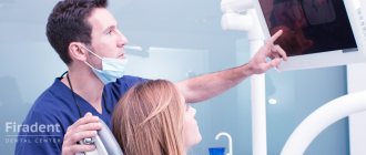
Diagnostics
An experienced dentist diagnoses dental abnormalities after an external examination and questioning of the patient. To clarify the diagnosis and identify the causes of the disease, a more detailed examination of the dental system is carried out:
- The construction of diagnostic plaster models and odontometric measurements make it possible to accurately identify changes in the size of dental units and their characteristics, which indicate the presence of pathologies.
- The color and transparency of the enamel are determined using a photographic method.
- Computed tomography or teleradiography makes it possible to obtain information about the condition of abnormal dental units in order to determine further treatment tactics.
- Panoramic radiography of the jaws is an important diagnostic method
- Electromyography of the jaws helps to assess the functional state of the facial muscular system.
To carry out differential diagnosis, the patient is referred for consultation to specialized specialists: endocrinologist, otolaryngologist, geneticist. Based on the diagnostic results, the attending physician determines treatment tactics. Depending on the type of disorder and the complexity of the disease, the patient may need the help of a dentist, surgeon, orthopedist, or implantologist.
Diagnostic measures
The pathology is easily diagnosed by examining the oral cavity. However, finding out its causes, assessing the position and condition of the roots and periodontium, necessary to draw up a treatment plan, usually requires additional research.
These include the following:
- X-ray (orthopantomography, targeted images). This is the main diagnostic method in dentistry, showing the condition of the periodontium and roots.
- CT scan. It is used when it is necessary to clarify the details of the subgingival part.
- Anthropometric and biometric studies. They allow you to determine the angle of rotation of the crown, the presence or absence of macro- and microdentia, and other features of the morphology of the elements of the dentofacial apparatus.
- Assessment of bite and occlusion using diagnostic models made from impressions of the jaws.
- T-scan is a computer method for analyzing occlusal contacts from Tekscan. Occlusion diagnostics T-scan is designed to identify problems with bite, accurately identify problem units, the closure of which causes improper distribution of the chewing load.
It is extremely rare that all or at least most of the methods listed above are necessary. However, it is useful to know about them in any case.
Prognosis and prevention
Consilium Dent clinic uses advanced methods of therapeutic, surgical, and orthopedic treatment of dental anomalies in patients. By skillfully combining them, experienced specialists are able to restore the aesthetics of a smile and ensure the normal functioning of the dental system, even if the degree of deviation is significant.
Prevention of anomalies in the formation and development of dental units begins during the period of intrauterine development of the fetus and is carried out throughout a person’s life. The main stages are as follows: monitoring the successful course of the prenatal period, caring for harmonious postnatal development, organizing a correct lifestyle while the baby is growing up, caring for dental health and general health throughout life. By conducting an annual preventive examination, the dentist identifies dental anomalies at an early stage, when the pathological process is still reversible.
Expert of the article you are reading:
Features of diagnosing dental anomalies
The diagnosis is made even if the tooth is rotated only a few degrees around its axis. Cases of turning at an angle of up to 45° are classified as minor, and all others are classified as significant. The maximum rotation angle of the dental crown is 180°.
To diagnose and develop a pathology correction program, the patient may be prescribed:
- X-ray of the problem area;
- orthopantomogram, which allows assessing the quality of tissue;
- anthropometric analysis, during which, using specially prepared models of the patient’s jaws, the parameters of the components of his dental system are determined and their relationship is analyzed.
Diagnostics makes it possible to identify the cause of tortocclusion and develop a treatment program that corresponds to the degree of its severity and the condition of the dental tissues. When the teeth are rotated by 10–20°, they resort to straightening the teeth with braces, a trainer or a plate. When the angle is 20–40°, special orthodontic devices based on a spring arch and elastic rings are used. In even more complex cases, orthodontic treatment is combined with surgical treatment.