In most cases, dental plaque is the cause of insufficient oral hygiene. But it also happens that people who are very sensitive to hygiene issues develop black plaque on their teeth.
The reasons for its appearance in adults and children differ : if in the former it is a consequence of drinking large amounts of tea and coffee, as well as smoking, then in the latter it may be associated with the activity of certain bacteria, the use of mouthwashes and the use of chewable vitamins.
Types of plaque
Plaque on teeth is divided into two large groups:
- the occurrence of which is associated with external contamination of the tooth;
- appearing due to the deposition of pigments.
If oral hygiene is not maintained, plaque may appear on the teeth:
- soft microbial;
- mineralized;
- hard, often called tartar.
If everything is in order with hygiene, then black plaque is a consequence of:
- regular consumption of tea, coffee and nicotine;
- use of medications with high iron content;
- vital activity of chromogenic bacteria;
- use of certain types of rinses and antiseptics.
Bacterial plaque on teeth: causes
Soft or, as it is also called, bacterial plaque has a soft consistency, so it is easy to get rid of it with the help of an ordinary toothbrush. The main place of its accumulation is the neck of the teeth.
Harder plaque that cannot be removed with a brush is called tartar. It occurs during the mineralization of soft plaque with phosphorus and calcium salts present in saliva.
Why does plaque form?
There are constantly a huge number of bacteria in the mouth, which, firstly , multiply, and secondly , leave behind waste products. Even if you brush your teeth regularly, plaque cannot be avoided: within 6 hours after thorough brushing, you can easily notice, without resorting to the use of specialized equipment, a bacterial mass on the surface of the tooth enamel. A surge in microbial activity is observed immediately after eating: leftover food in the mouth forces bacteria to begin intensive processing.
At the same time, even small pieces of food that are invisible to the human eye are enough for microorganisms: they can feed on a film consisting of carbohydrates and proteins that invariably remains on the surface of the teeth after eating, and on food debris that gets into the spaces between the teeth. In this regard, it is imperative to brush your teeth for 15 minutes after each meal, since the mass of bacterial plaque can increase several times in volume in 1-2 hours. Plaque accumulates especially intensely on the necks of teeth in people who like to snack on a bun or something sweet between main meals.
Almost immediately after the soft plaque appears, the process of mineralization begins, as a result of which it gradually hardens. The period of primary mineralization (i.e., when the plaque, in simple terms, “sets” but still remains loose) is 10-15 hours. When the plaque finally hardens, its surface becomes ideal for the formation of deposits.
How to use
Considering the different forms of microorganisms that chlorhexidine should affect, the concentration and treatment mechanism are selected by the dentist individually, depending on the overall picture of the disease. Examples of treatment can be viewed in the table below:
| Disease | Concentration of chlorhexidine solution | Reception scheme | Duration of therapy |
| For stomatitis | 0.05% | Apply after complete cleansing of the oral cavity. Rinse your mouth with the solution for 30-60 seconds without stopping, 2 times a day. | Up to 10 days |
| For gingivitis | 0.05% | Rinse twice a day immediately after brushing your teeth and using dental floss. | Up to 10-12 days |
| For gum inflammation | 0.05% | First, the patient rinses the mouth with chamomile decoction or iodine-saline solution. Then, treat the mouth with 1 tbsp. l. chlorhexidine. It is forbidden to eat food within 2 hours after the treatment procedure. To treat infants, the affected areas are moistened with a cotton pad soaked in chlorhexidine. | Up to 10 days |
| After tooth extraction | 0.05% | Treatment is carried out 1 hour after brushing your teeth, 2-3 times a day. The solution is poured into the mouth and held for 1-2 minutes, while the head is slightly tilted from side to side. Head movements should not be too sharp to avoid injury to the protective blood clot. It is forbidden to eat food within 1 hour after treatment. | The duration of therapy is determined by the doctor individually. |
| For toothache | 0.05% | Rinsing is carried out 3-5 times a day, holding the solution for 1-2 minutes. | 7 days |
| For periodontal disease | 0.05% | It is used at the time of removing plaque in the form of baths. The product must be in contact with the pockets of periodontal tissue and remain in effect for 2-3 minutes. | 5-7 days |
| For preventive purposes | 0.05% | Caress the oral cavity for 20 seconds, 2 times a day | 5 days |
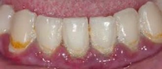
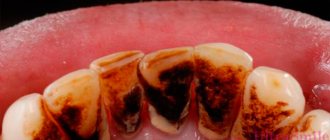
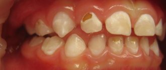
Pigmented dark plaque on teeth
The main reason for the appearance of such plaque in adults is smoking, drinking coffee and tea. Maintaining hygiene guarantees the absence of bacterial plaque and tartar, but if you neglect it, black plaque will not take long to appear. Pigments adhere very well to enamel covered with bacterial plaque formed as a result of infrequent brushing of teeth.
Types of pigment plaque include:
- brown coating that occurs from regular consumption of tea and coffee in large quantities;
- accumulation of nicotine deposits;
- darkening of the enamel as a result of taking medications containing iron;
- proliferation of bacteria that form dyes (they are called chromogenic);
- change in enamel color after using antibiotics;
- blackening due to diseases of internal organs.
If plaque is caused by bacterial factors, then regular thorough brushing of teeth, the use of rinses and floss reduce the likelihood of its occurrence to a minimum. However, if the reason for the appearance of plaque lies in the influence of dyes, maintaining hygiene to prevent it will not help.
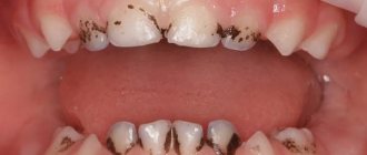
Black plaque on children's teeth , the causes of which are associated with the activity of chromogenic anaerobic bacteria, appears in the form of spots localized in the neck area. Actinomycete bacteria produce hydrogen sulfite, which reacts with iron (found in saliva and red blood cells). As a result, a form of black iron that cannot be cleaned off with a regular brush is deposited on the surface of the teeth. Chromogenic coloration can also be orange, green and brown.
Black plaque on a child’s teeth can also form for other reasons . It appears due to:
- the use of antiseptics that contain chlorhexidine, benzaclonium chloride, as well as essential oils (the latter are found in the popular Listerine product);
- consumption of foods rich in iron, as well as vitamins with a high content of iron.
Medical Internet conferences
The influence of chlorhexidine on the microbial composition of the bottom of a prepared carious cavity
Tverskova V.Yu.
Scientific supervisor: candidate of medical sciences, assistant Kazakova L.N.
GBOU VPO Saratov State Medical University named after. IN AND. Razumovsky Ministry of Health of the Russian Federation
Department of Pediatric Dentistry and Orthodontics
Relevance of the problem. Among oral diseases, caries ranks first in prevalence. Currently, caries is diagnosed in children under 2 years of age in 36% of cases, and in 3-year-old children in 53% of cases[1]. The incidence of deep caries in children is 23% of the total number of caries diseases. According to many authors, caries can be classified as an infectious disease [2], the leading factor in which is Streptococcus mutans. Str. mutans attaches to the smooth surfaces of teeth, participates in the formation of dental plaque, and this action is mediated by the synthesis of glucose polymers from sucrose present in food. Glucan formation causes intercellular aggregation of Str. mutans and other bacteria present in the plaque. The sticky glucan matrix of dental plaque prevents the diffusion of large amounts of acid produced by microorganisms, which prolongs its residence on the surface of the teeth and leads to demineralization of the enamel, causing dental caries. The emerging focus of demineralization of the surface layer of enamel is the “entry gate” for further infection of deeper located hard tissues and dental pulp, this is especially true immediately after eruption, at the first stage of secondary mineralization. The special structure of hard tissues, characterized by less mineralization at this stage of tooth development, creates a number of difficulties in the treatment of acute deep caries with a favorable outcome. Since it is impossible to create conditions close to sterile at the border with the pulp using standard irrigation of the prepared cavity.
Goals and objectives of the study. The purpose of our work was to study the effect of chlorhexidine bigluconate, used in different forms, on the qualitative microbial composition of the prepared carious cavity in acute deep caries.
Materials and methods of research. During the study, a group of children (20 people) was identified with a diagnosis of acute deep caries of permanent teeth at the stage of ascending development. To determine the microbiocinosis of the bottom of the carious cavity, a bacteriological study was carried out.
Acute deep caries was treated conservatively only in children with a good level of oral hygiene and with a compensated form of caries in two visits.
On the first visit, rational anesthesia was carried out, the carious cavity was prepared under a bath of the same antiseptic, this was done carefully, but with extreme caution. After the final preparation of the carious cavity with a sterile bur, a section of dentin was taken from the bottom and a bacteriological examination was carried out. Then the children were blindly divided into 2 groups of 10 people. In the first group of patients, a tampon moistened with 0.05% chlorhexidine was placed on the bottom of the carious cavity under a temporary dressing. In the second group, a “patch” of self-adhesive film “Diplen Denta” containing chlorhexidine bigluconate 0.01-0.03 mg/cm² was applied to the bottom of the carious cavity under a temporary dressing. Further research was carried out after 48 hours.
On the second visit, having previously isolated the tooth from saliva, a temporary bandage and a tampon with an antiseptic were removed in the first group; in the second group, the self-adhesive film dissolved on its own, and in both cases, a sterile spherical bur without pressure was taken from the bottom of the cavity for bacteriological examination. In patients, a calcium-containing preparation was applied to the bottom of the carious cavity, and the cavity was filled with permanent filling material “Vitremer”.
Further bacteriological examination was carried out in accordance with generally accepted rules of clinical microbiology.
Results. Microbiological examination of the bottom of the carious cavity in patients after complete preparation of the carious cavity revealed a significant degree of its contamination with pathogenic and conditionally pathogenic microflora. Streptococci, enterococci and yeast of the genus Candida were sown (Table No. 1). In the group of patients, when using 0.05% chlorhexidine during re-seeding from the bottom of the carious cavity, a decrease in the number of streptococci was observed, the effect of 0.05% chlorhexidine on enterococci was insignificant, the growth of yeast of the genus Candida was not detected (Table No. 1).
When using the film "Diplen Dent" a significant decrease in the colonization of cariogenic species Str. mutans, Str. Sanguis, Str. salivarius. The number of representatives of the resident group of bacteria - enterococci - decreased (Table No. 1).
We associate the higher efficiency of the Diplen Denta film containing chlorhexidine bigluconate in a concentration of 0.01-0.03 mg/cm² with its ability to adhere tightly to the bottom of the cavity, which ensures deeper penetration of the active component through weakly mineralized dentin and dentinal tubules.
Conclusions. Thus, the results of the study confirm that the application of chlorhexidine digluconate in the form of a film for 48 hours, as an intermediate stage in the treatment of acute deep caries in children, leads to a significant decrease in the level of streptococci, enterococci and yeast of the genus Candida in the peripulpal area.
How to remove plaque from teeth on your own
Many people do not like to visit the dentist again, especially when it comes to removing plaque, which does not cause much inconvenience. The reasons for this are clear: lack of free time and reluctance to pay for the procedure.
You can try to remove plaque from your teeth at home, but this will only work if there are no subgingival deposits and the plaque layer is thin.
The first method involves using a special toothpaste to remove plaque with abrasive substances. The label of such pastes must indicate the RDA indicator, indicating the degree of abrasiveness. In pastes that eliminate plaque, its value should exceed 100. But such pastes cannot be used on an ongoing basis. If the paste contains pyrophosphates, then it can help get rid of tartar, since they have the ability to dissolve its matrix.
The most effective of these pastes are:
- President White Plus. The abrasiveness index is 200 units; in addition, the composition contains silicon dioxide, which has abrasive and polishing properties. You should not use the paste more than once a week.
- Lacalut White. According to the abrasiveness index (RDA=120), it is more gentle than the previous sample. The composition contains pyrophosphates, which make the plaque loose.
The second method is to use a special toothbrush, which allows you to remove plaque yourself. Recommended use:
Plaque on a child’s teeth: treatment
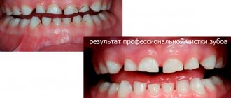
Removing bacterial and chromogenic plaque in a child will be no different from a similar procedure recommended for an adult: ultrasonic cleaning and AirFlow are used in the same way.
However, chromogenic staining of teeth may return over time. The only possible solution to the problem is regular professional cleaning by a dentist. To make visits to the dentist as rare as possible, your child should be taught to use an electric toothbrush, the pulsating circular movements of the head of which perfectly break up plaque.
Accelerated formation of deposits is promoted by blood in the oral cavity, so the child’s gingivitis must be treated. The use of chlorhexidine and antiseptics based on it should be abandoned.
Why do you need Chlorhexidine: properties and action
After a tooth is removed, an open wound remains in the gum and fills with blood. A blood clot forms in the hole, protecting the open area of the periosteum from the penetration of pathogenic bacteria and microbes. If the clot does not adhere tightly to the socket or is accidentally damaged during eating, the risk of infection of the bone tissue and the development of osteomyelitis of the jaw increases - a purulent-inflammatory pathology, which, if not treated in a timely manner, can lead to the death of bone structures (osteonecrosis).
And this is how a blood clot may look in real life - it is very important not to wash it out when rinsing your mouth
To avoid damaging the clot, the following recommendations must be followed:
- in the first 1-2 days after removal, you should not drink through a straw, spit saliva, or perform other actions that create a vacuum in the mouth;
- You can chew on the side on which the removal took place 72 hours after surgery;
- You need to rinse your mouth carefully, trying not to make too active movements;
- Do not touch the clot with your tongue or try to remove it from the hole.
Another common complication after tooth extraction is alveolitis. This is a disease in which inflammation of the socket occurs. The pathology is accompanied by fever, severe pain, which can radiate to the ear, eye area, jaw and cervical and submandibular lymph nodes. The gums at the extraction site swell, and severe swelling and redness appear.
- How to rinse your mouth with Chlorhexidine for gum inflammation
Development of alveolitis in the socket after tooth extraction
The main cause of alveolitis is non-compliance with the hygiene regime and an increase in the number of pathogenic flora in the oral cavity. Pathogenic microorganisms entering the alveoli cause inflammation of soft tissues and the development of a pathological process. To prevent alveolitis and other possible complications, the patient is prescribed local treatment of the oral cavity with antiseptic solutions, one of which is Chlorhexidine. The use of this drug ensures disinfection of the mucous membranes and hard tissues of the tooth and prevents the development of infectious and inflammatory processes.
Tooth extraction is a full-fledged surgical operation.