What types of caries are there?
There are several stages of the disease. They have a certain appearance and symptoms.
How to determine the stage of caries:
- In the spot stage: a white spot is observed on the surface of the enamel (focus of demineralization). The spot gradually turns brown. Enamel is the densest tissue in the human body, but due to various cariogenic factors it is gradually destroyed. Detecting the white spot is quite difficult.
- Medium caries: characterized by damage to the enamel-dentin junction in the form of a cavity, short-term sensitivity.
- Deep: extensive lesion affecting the deep layers of dentin. Pain appears from different types of irritants: mechanical, temperature, chemical.
Diagnosis of the disease is carried out using a visual examination and an x-ray. The image will show violations of the integrity of the enamel and dentin with uneven contours.
Radiation diagnostics of dental injuries
After an injury, radiation diagnostics of the damaged and adjacent teeth, and sometimes antagonists, is almost always prescribed. The scope of emergency dental measures depends on its results. The most common injuries are:
- tooth fractures - isolated or combined, that is, in combination with a violation of the integrity of the jaw (the same symptoms are noticeable in the picture as with jaw fractures);
- dislocations - mainly in the area of the incisors (the x-ray shows how the tooth has come out of the socket, and with subluxation, the asymmetry of the periodontal gap is noticeable).
Radiation diagnostics is also used for other dental diseases if the benefits from it outweigh the expected harm from radiation.
What does caries look like on teeth?
Not all patients know how to identify dental caries. To do this, you need to come to the dentist for an examination.
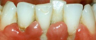
Black cavities of varying sizes are found on the chewing units. The pathological process develops in the natural depressions of the enamel: fissures, pits.
This disease leads to the destruction of hard tissues and cosmetic defects. There are black cavities on the front teeth, noticeable when talking or smiling:
Relevance.
The frequency of occurrence of dental caries in the population requires timely diagnostic measures, including objective x-ray examination, the significance of which becomes especially obvious in complex clinical situations.
Purpose of the study
— improving the quality of diagnosis of carious dental cavities by studying the features of their display using different X-ray examination techniques.
Material and methods.
In an experiment on skeletonized jaws with teeth and in clinical studies of patients, 453 intraoral periapical and interproximal radiographs, radiographs performed using a parallel technique, and 432 orthopantomograms were studied. Intraoral X-ray devices Trophy Radiologie (France) and Heliodent DS (Germany), orthopantomographs Proscan (Finland) and Orthophos XG5 DS Ceph (Germany) were used.
Results.
On periapical radiographs, carious cavities on the chewing surfaces of teeth projectedly increased in size vertically and often overlapped the tooth cavity, creating a false picture of perforation. At the same time, in the teeth of the upper jaw, carious lesions were displayed less clearly. On orthopantomograms, radiographs performed using a parallel technique, and especially on interproximal photographs, caries of the chewing surface of the tooth was more clearly visualized. Cavities usually become noticeable when they penetrate the dentin layer. Enamel caries was displayed less clearly, and in case of small lesions it was not detected at all due to the intense shadow of the preserved enamel layer being layered on the carious defect. Enamel defects varied when the enamel layer was affected by ½—¾ or more of its thickness. In all of the listed types of images, the shadows of carious cavities on the vestibular and oral surfaces in the natural fissures of the tooth crowns and in the cervical region, as a rule, were added to the gaps of the tooth cavity. Carious destruction was visualized when it reached a significant size, or when it did not overlap the shadows of the tooth cavity. Lesions of contact surfaces and the cervical part of different groups of teeth were detected on intraoral radiographs and orthopantomograms better than caries of other localizations. However, when the teeth were crowded, the image of the contact surfaces of the tooth crowns was summed up, especially on orthopantomograms. The additional use of intraoral imaging using a parallel technique or interproximal radiography made it possible to answer the question about the absence or presence of hidden carious cavities. It should be noted that during clinical instrumental examination, small carious lesions on the contact surfaces of tooth crowns, especially with close proximity and crowding of teeth, were difficult to access or inaccessible for detection. Cervical carious cavities on the distal surface of the second molars of the lower jaw, which arose at the site of contact of the crown of the dystopic and impacted third molar, were not detected during clinical examination and were clearly visualized on intraoral radiographs performed using a parallel technique, and on orthopantomograms. Small carious cavities under fillings and artificial crowns were not diagnosed on radiographs due to the summation with intense shadows of filling material, metal-ceramic or metal crowns. These cavities were identified only when they reached a size that allowed them to appear behind the contour of the radiopaque material. During periapical photography, the shadows of metal crowns were often combined with carious cavities located below the equator of the crown. These cavities were better visualized on interproximal films and radiographs obtained using a parallel technique. With digital radiography and orthopantomography, the difference in the density of blackening at the border between the intense shadow of a metal crown or filling and the shadow of the tooth created a picture of a decrease in the density of hard dental tissues, simulating carious destruction. A similar situation sometimes arose in the area of the enamel-dentin border due to the unequal X-ray density of the enamel and dentin layers.
Conclusion.
X-ray examination is of critical diagnostic importance in identifying carious lesions on tooth surfaces that are difficult to access for clinical examination. Moreover, in the event of a questionable picture of the presence of caries on an orthopantomogram, it is advisable to additionally use intraoral radiography using a parallel technique or interproximal radiography for clarifying diagnostics.
How does caries form?
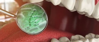
The causes of caries are poor hygiene and the presence of bacteria in the oral cavity.
There are many bacteria in the human oral cavity. These bacteria feed on food debris that remains on the surface of the enamel after eating. Bacteria “love” foods high in carbohydrates: sweets, flour. During their life, bacteria produce acid, which destroys teeth.
Caries occurs in both adults and children. This disease spreads in each person at a different rate.
Factors contributing to the rapid development of caries:
- insufficient or lack of oral hygiene;
- excess carbohydrate foods: flour products, sweets, carbonated drinks;
- low buffering capacity of saliva, when acids and alkalis are not sufficiently neutralized in the oral cavity;
- insufficient fluorine and calcium content in enamel;
- lack of vitamins and minerals in the body;
- severe somatic pathologies suffered in childhood (tuberculosis, rickets);
- reduced immunity.
Answering a frequent question from patients: “Why do caries form on teeth?”, we note that the most important factor is insufficient hygiene. All dentists strongly recommend brushing your teeth regularly and using floss to clean the gaps. Without careful hygiene, bacteria will destroy teeth one by one, leading to serious complications.
Causes
Caries can be either an independent disease or a concomitant one. The development of caries is promoted by:
- decreased general and local immunity;
- problems with the gastrointestinal tract;
- non-compliance with the recommended diet;
- unfavorable environmental conditions;
- low quality of drinking water;
- work in hazardous industries involving inhalation of acid fumes;
- insufficient oral hygiene (especially ignoring hard-to-reach places when brushing teeth);
- individual characteristics inherent at the genetic level;
- thinning of enamel associated with impaired mineral metabolism.
What happens if caries is not treated?
- For a long time, the disease may not bother the patient in any way. When the destruction of hard tissue has reached a significant extent, sensitivity appears during eating.
- If you do not consult a doctor at the first stage, the carious process will go further and reach the pulp (nerve). Spontaneous severe pain appears. Treatment of pulpitis will be required.
- If the patient does not seek help at this stage, the infection spreads beyond the root. Periodontitis occurs. The prognosis here is unfavorable, since such a tooth often needs to be removed to eliminate the infection.
Therefore, timely visit to the dental clinic will help to avoid acute pain and complications.
X-rays after therapy
X-ray is not only a method for diagnosing pathology, but also acts as a control over the effectiveness of therapy. Through research, the doctor determines:
- Quality of pulp removal. Incomplete removal leads to re-inflammation.
- The nature of the tooth canals, their physiological structure and depth.
- How well were the canals filled using medical cement or gutta-percha? In case of insufficient filling or excess filling material in the canals, the tooth is treated again.
After therapy, control radiography is recommended, usually every six months.
What to do if caries appears on teeth
First of all, you need to immediately contact your dentist. When a tooth is destroyed due to caries, treatment cannot be delayed. Otherwise, the complications described above arise. It is worth noting that no available means at home will relieve the patient of this problem. Only a dentist can cure this disease.
You should also pay attention to your oral hygiene:
- Brush your teeth at least twice a day.
- Use floss and mouth rinses.
- Visit your dentist for a checkup every six months.
- Come for professional hygiene every 6 months.
Dentists will help you select individual hygiene products (brushes, pastes, threads, rinses) at your appointment. The doctor will also determine why dental caries appears in your particular case and give the necessary recommendations.
Symptoms
Caries is diagnosed during a visual and instrumental examination. At the same time, the disease is characterized by the absence of spontaneous pain: pain appears only under the influence of irritants (sugar, cold or hot food). Spontaneous pain indicates that the process has already moved from caries to pulpitis.
- With deep caries, unpleasant sensations may also occur when mechanical pressure is applied to the bottom of the carious cavity. Food debris gets inside and presses, causing compression of the tooth pulp.
- The presence of caries can also be suspected due to an unpleasant odor in the mouth. Food debris that gets into the carious cavity begins to rot.
- If you feel sharp edges and irregularities with your tongue, this is a reason to seek advice from a dentist.
How is caries treated?
The treatment is carried out in professional dentistry by a general practitioner and is absolutely painless. This treatment is carried out in one visit, and several teeth can be treated in one visit.
Sequence of treatment stages:
- Examination of the oral cavity. Carrying out diagnostics: a targeted diagnostic image of one tooth or computed tomography of the jaws (if multiple caries is detected during the examination). This is done in order to make an accurate diagnosis and see the volume of destroyed tissue.
- Local anesthesia: selected individually according to health conditions. Only after the desired area of the oral cavity has become numb does the doctor proceed to the next manipulations.
- Placement of isolation: the doctor places a rubber dam on the desired area - this is a latex plate that isolates the tooth from the rest of the oral cavity. The rubber dam creates comfortable conditions for the doctor, the patient, and also allows you to create dry, sterile conditions for placing a filling. This is necessary for the filling to serve for a long time and with high quality.
- Preparation of enamel, dentin: necrectomy. The doctor uses a drill to drill out the destroyed tissue.
- Treating the cavity with antiseptic solutions, applying an adhesive - a special solution that, like glue, reliably bonds hard tissue to the filling.
- Filling with light-curing materials. The anatomical shape of the tooth is restored, and the color is carefully selected.
- Polishing and grinding so that the filling does not feel different from your own teeth. Also, using special guidelines, the doctor checks whether the filling is too high or does not interfere with the normal closure of the jaws. Excess is removed, sharp edges are sanded.
- The doctor gives recommendations after treatment. If a tooth has been severely damaged by caries, then in addition to a filling, an orthopedic onlay or crown may be required. This is due to the fact that the filling is not as strong as enamel. Over time, cracks and chips are possible. Orthopedic structures will help protect the treated tooth from these complications.
After these stages, the tooth is completely free of caries. Next, you will need to maintain regular hygiene, come for preventive examinations and professional cleaning.
Prevention
Fighting soft plaque helps prevent tooth decay. For this it is important:
- use high-quality toothbrushes and toothpastes;
- use dental floss (removes food debris between teeth);
- When brushing, pay attention not only to the teeth, but also to the tongue, gums, and the inner surface of the cheeks;
- regularly undergo professional oral hygiene procedures;
- limit your consumption of sweets (it is better to replace sweets with dried fruits);
- pay close attention to the temperature of food and drinks (the enamel may not withstand it and become covered with microcracks from temperature changes, for example, when eating ice cream with coffee).
People often do not think that harmless caries leads to serious consequences (including tooth extraction). Going to the dentist for preventive examinations is faster and cheaper than delaying the “X moment” for several years, and then ending up straight in the surgical chair.
Back
Timely diagnosis of caries using panoramic images
Problem: the patient came to the Family Dentistry with complaints of periodic pain in the teeth on the lower right. The patient could not indicate a specific tooth, but assumed that the wisdom tooth on which he saw a dark spot was to blame.
Solution: a panoramic photograph of the teeth was taken for diagnosis. Caries of three teeth was discovered and treated: one of the teeth was depulped, tooth canals were treated with a microscope and restored with a ceramic inlay, the other two teeth were restored with light fillings.
A situation similar to that considered in this work occurs quite often. The patient either cannot indicate the exact source of the pain, or points to a tooth that doctors do not consider problematic. Dial-Dent specialists have extensive experience in finding the cause of unclear pain in the teeth (see the work “Pain in a tooth, but not that one...”). A huge help in correct diagnosis is a modern digital orthopantomograph, with the help of which Dial-Dent takes a panoramic photograph of the teeth. A panoramic shot allows you to see the whole situation and find out where the source of pain lies. Correct diagnosis using modern digital equipment saves the patient time, effort and money. A digital panoramic image is clearer, and the doctor can enlarge individual fragments for a closer look. All this, as well as more than 10 years of experience of Dial-Dent specialists, makes it possible to conduct high-quality diagnostics, and as a result, precise targeted treatment, quickly relieving the patient of dental pain.