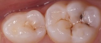
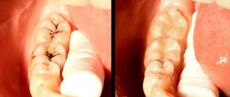
Cervical caries, which occurs at or under the gum, is considered one of the most dangerous forms of caries. In the cervical area, tooth enamel is more vulnerable to cariogenic microbes, so pathology develops very quickly. In the article we will consider the features of the course and methods of treatment of cervical caries.
The structure of a tooth, when it comes to its structure, includes three main elements: the root, the neck and the crown. The first two are hidden by soft tissue. The enamel on them is thinner, and there are fewer minerals in it than on the visible part, so it is more vulnerable to cariogenic bacteria. Caries that develops in the area where the tooth comes into contact with the gums and under them is called cervical or root caries. It occurs mainly in adults over 30 years of age, and can appear on both the front and chewing teeth.
In this article
- Why does cervical caries occur?
- Stages of cervical caries
- How is cervical caries diagnosed?
- How to treat initial cervical caries
- How is advanced cervical caries treated?
- How to treat cervical caries on the front teeth
- What you can do at home
The problem with cervical caries is that it is almost asymptomatic until the third stage. The person does not complain of pain or discomfort, there is no visible destruction, and therefore treatment begins late. At the same time, basal caries progresses rapidly. The disease does not pose a serious danger if you visit the dentist every six months. People who rarely visit dental offices are at risk of losing a tooth. Let's talk about the causes and symptoms of cervical caries.
Why does cervical caries occur?
The main cause of any type of caries is damage to the enamel by cariogenic microorganisms from the group of streptococci. They live in the oral cavity of almost all people. Under certain conditions, microbes begin to actively multiply and secrete acids that corrode the enamel, washing away minerals from it. Demineralization of the enamel surface causes exposure of hard tooth tissues. Bacteria penetrate microcracks in dentin and form a carious cavity. Without treatment, it spreads and leads to gradual tooth decay. However, microbes alone are not enough for the development of caries. They are dangerous only in the presence of other factors for the occurrence of carious lesions. Among them:
- Insufficient oral hygiene. If you brush your teeth twice a day, use dental floss and irrigators for cleaning, then the risk of caries is quite low. Otherwise, deposits and food debris will accumulate in the interdental spaces and gum pockets. They are a favorable environment for the spread of microbes.
- Poor nutrition. Cariogenic bacteria feed on carbohydrates. Immediately after eating sweet foods, which contain a lot of fast carbohydrates, active proliferation of microbes begins. This process is accompanied by a sour taste in the mouth. The more sweets a person eats, the more often he develops tooth decay.
- Nicotine addiction. Smoking contributes to the formation of plaque on the enamel. Over time, it mineralizes and hardens, leading to the formation of tartar, which contains many bacteria. They may also be covered with plaque, as a result of which caries begins to develop between the enamel and tartar.
- Disorders of the salivary glands. Saliva not only washes away food debris and microbes from the oral cavity, but also neutralizes the waste products of cariogenic bacteria. Some diseases are accompanied by insufficient saliva production, which can become a factor in the development of caries.
- Enamel abrasion. The enamel surface protects the hard tissues of the tooth from external factors. Some people's enamel is very thin and tends to wear away, resulting in more tooth decay.
These are the main factors in the development of caries. But there are many other reasons, including weak immunity, mechanical injuries to teeth, hormonal imbalances, endocrine pathologies, etc. If they are not eliminated, the disease will periodically recur.
Features of restoration of the roots of the frontal group of teeth destroyed below the gum level
The so-called all-ceramic designs of dentures (manufactured in various ways: slip casting, injection molding, layer-by-layer application of ceramic mass on a fire-resistant model, etc.) have become firmly established in practice, and today it is difficult to imagine a full range of work in orthopedic dentistry without them. The use of such structures has created new requirements for the tooth stump on which such a crown will be fixed (the minimum thickness of the crown should be 2.5 mm, requirements for transparency and polishing of the shoulder part of the stump). Some difficulties are presented by clinical cases when the tooth stump needs to be restored after a root fracture located just below the gum level. But even in such cases, adequate restoration of the destroyed tooth stump is possible. The main difficulty will be the requirements for surface drying when working with composite materials and dentin bonding agents used for core restoration, since drying the root surface located below the gum level is quite problematic. It is necessary to work with the obligatory use of a cofferdam and try to restore the initially destroyed wall at least to the level of the gum. You can then safely proceed with the adhesive fixation of the fiberglass pin in the root canal, since the transparency requirements complicate the use of metal pin structures for restoration. Using a clinical case, I would like to demonstrate a similar situation. A 22-year-old patient came to our clinic with a fracture of the crown of tooth 21 located below the gum level (Fig. 1).
rice. 1. Patient M., 22 years old, fracture of the crown of tooth 21 located below the level of the marginal gum.
At the same time, it was necessary to create the most “live”, natural work. We chose the treatment option using an all-ceramic crown. To do this, it was necessary to restore the destroyed tooth stump using materials close to the natural transparency of the tooth. For restoration, a composite material and a fiberglass pin were used, fixed in the canal using dual-curing cement (Fig. 3,4,5).
rice. 3. Using a cofferdam and contoured matrix strips, we restore the destroyed wall to the level of the gum.
rice. 4. We restore the cervical area of the stump while simultaneously preparing the canal for adhesive fixation of the pin.
rice. 5. The fiberglass pin is fixed.
Previously, endodontic treatment was carried out using the Thermafil system, as well as correction of the gingival margin on the medial side (Fig. 2).
rice. 2. Endodontic treatment of tooth 21 using the thermophil system.
The final preparation of the stump and especially its inferior part was carried out using carbide burs. We chose these burs due to a number of competitive advantages. Firstly, the sufficiently large length of the working surface of the bur allows processing the tooth stump to its entire height, which significantly facilitates the task performed (Fig. 6.).
rice. 6. A sufficiently large length of the working surface of the bur allows processing the tooth stump to its full height.
Secondly, carbide burs have horizontal notches located in a certain way on the working blades of the bur, which significantly speeds up the process of removing large volumes of hard tissue. Thirdly, the end of the bur head is increased in size and has a fairly large radius of curvature, with a complete absence of horizontal notches on the end of the bur head (Fig. 7.).
rice. 7. The end of the bur head is increased in size and has a fairly large radius of curvature, with a complete absence of horizontal notches on the end of the bur head.
This design of the bur makes it simply an ideal tool for forming the final ledge on the tooth stump and, at the same time, for finishing the ledge to a glossy surface. (Fig. 8.9.)
rice. 8. The formed ledge on the tooth stump is clearly visible.
Fig. 9. The ledge on the stump of the prepared tooth is polished to a glossy surface.
Using carbide burs, we can easily create a rational, semicircular ledge for a future all-ceramic crown. Subsequently, we only correct it with a bur that has a larger radius of curvature of the head, which allows us to level the surface of the ledge, removing sharp edges and elevations at the border of the ledge and the gums. It should be noted that the production of such work requires very careful preparation with a good centered tool. Since only a perfectly formed stump, without undercuts, will enable the technician to create a crown that fits exactly to the stump. Otherwise, there is a danger of the ceramic structure splitting due to microstresses arising in the ceramic during function, due to uneven contact with the stump. We carried out the taking of a double refined impression and the direct production of a provisional crown according to the generally accepted method. We carried out the final fixation of the finished artificial crown using dual-curing composite cement, since only this group of materials is capable of forming a chemical bond with both the tooth stump and the crown. In addition, these materials are quite strong, plastic and fluid, and also have a fairly wide range of colors and gradations of transparency, which ensures natural color rendition and transparency of the artificial crown (Fig. 10).
Fig. 10. Finished work in the oral cavity.
Stages of cervical caries
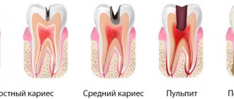
Cervical caries develops according to the same scenario as other types of this disease, but much faster. Let us describe the stages of development of root caries:
- Initial (white spot stage). It is asymptomatic. A demineralized area of enamel forms in or under the gum area. It looks like a white or chalky spot. Gradually it begins to fade, but this is difficult to notice with the naked eye, especially if the pathology is localized on the back surface of the tooth or on the molars and premolars. If it starts under the gum, then it can only be detected using hardware diagnostics.
- Superficial. The carious spot becomes larger and acquires a yellowish or brownish tint. Destructive processes begin on the tooth surface. At this stage, the pathological focus has not yet spread to hard tissue. The patient may experience discomfort when eating sweet foods. It disappears immediately after the stimulus is removed. Often people attribute this symptom to increased sensitivity of the gums.
- Average. Damage to hard tissues is observed. A person experiences severe discomfort when eating sweet, sour, cold or hot foods. Possible pain. These signs disappear as soon as you brush your teeth.
- Deep. The last stage is an advanced form of the disease. Deep damage to dentin occurs. The pathological process involves the tooth root and pulp. The tooth may split into several parts or break in the neck area. The patient almost constantly experiences pain, which painkillers do not help eliminate.
Treatment for caries can vary depending on the degree of damage to the tooth. The method is selected individually after diagnosis.
Possible consequences
Many people are very afraid to visit the dentist, but the consequences of such neglect of their health can be much more serious than it seems. Decay that begins on one tooth can eventually spread to neighboring units, gums and roots. Neglected dental problems can cause the development of diseases of other organs.
If the immune system stops coping with inflammation and decay, general intoxication of the body may begin. As a result, problems with the digestive system, respiratory organs will appear, cardiac activity may be disrupted, severe headaches will appear, and much more.
A person may feel constant fatigue and loss of appetite, hearing may deteriorate, arthrosis and polyarthritis may appear. The most dangerous complications include diseases such as sinusitis or otitis media, as well as serious brain damage. That is why you should not ignore the first signs of tooth decay, since the problem at the initial stage is much easier to eliminate. It will be much more difficult to deal with its consequences after a few years.
previous post
Do you need to brush your tongue when brushing your teeth?
next entry
How is cervical caries diagnosed?
If the carious lesion is localized on the visible part of the tooth, the doctor makes a diagnosis after an external examination. But to confirm it, a special test is performed using a dye solution. It is applied to the enamel and left for 3-4 minutes. The affected area turns blue. After a few hours, the dye is completely washed out of the mouth.
The doctor may also prescribe an x-ray to assess the nature of the spread of caries, the depth of the carious cavity, the condition of the nerve endings, etc. Based on the results of the examination, a treatment method is selected. It is determined, first of all, by the stage of the disease.
What factors can influence the development of caries?
Individual tooth structure : location of fissures and grooves on the surface. The most “dangerous” are flask-shaped fissures; it is almost impossible to clean plaque from the bottom yourself. We can recommend hermetic sealing of fissures.
Malocclusion: Dental hygiene is complicated by the position of the teeth, so plaque will provoke the development of caries (and other possible problems). A visit to the orthodontist will allow you to choose an individual bite treatment plan.
Eruption of wisdom teeth (eighth teeth): Since “eighth teeth” grow in at the age of 23-26 years, when the jaw is already formed, displacement of the remaining teeth may occur. And since they are often inaccessible for daily cleaning, carious bacteria accumulate on them.
Various general diseases : stomach diseases, diabetes, cardiovascular problems, etc.
Hereditary predisposition to carious lesions: for example, low mineral content in saliva, low level of the immune system.
Pregnancy and breastfeeding period.
How to treat initial cervical caries
At the white spot stage, caries can be treated using therapeutic procedures. It is necessary to restore the enamel structure by saturating it with minerals. First, the dentist will clean the affected tooth from plaque and tartar, and then apply a solution containing fluoride, magnesium, phosphorus and calcium to it. Under the influence of the active components, the enamel is remineralized and the pathological process is blocked. The patient is also prescribed mouth rinses with an antiseptic solution and toothpaste with fluoride.
How to treat superficial caries
If the carious area has managed to destroy the enamel, but has not penetrated into the hard tissues, remineralization is carried out, as in the first stage. The dentist grinds the affected area of the tooth, removes damaged tissue and applies a solution to the enamel to restore it. Several procedures may be required - 5-10 sessions. Throughout the treatment, the patient uses medicated toothpaste.
Treatment of cervical caries at the middle stage
As a rule, people consult a doctor when root caries has reached the third stage. At this stage, tooth filling is required. The doctor removes the affected tissue, carries out antiseptic treatment of the carious cavity and fills it with filling material. The procedure is performed under local anesthesia.
What to give up from food to prevent tooth decay
If you like to eat a lot of sweets, then the issue of preventing tooth decay may be more difficult than it seems. Everyone knows that pathogenic microflora feels very comfortable when a person eats sweets. It is the excessive, regular consumption of buns, sweets, cakes, chocolate that not only spoils the figure, but also the teeth.
Regular consumption of fried, too spicy or salty foods also has an adverse effect on teeth. Too acidic also negatively affects tooth enamel. Gradually destroying its structure, promoting the development of caries.
Particular attention should be paid to the following products:
- Various greens;
- fresh vegetables;
- fresh fruits.
It is better to eat food that has been subjected to gentle heat treatment; eating apples, carrots and similar products is very useful. If you are a fan of canned foods, then keep in mind that marinades also have a bad effect on tooth enamel.
How is advanced cervical caries treated?
Deep caries is often accompanied by damage to the pulp, so depulpation is required. After this, root canal treatment and filling are performed. In some cases, nerve removal can be avoided. Then treatment is carried out according to the standard algorithm:
- Anesthesia.
- Removal of deposits and tartar.
- Selecting the shade of the future filling.
- Retraction of the gingival margin.
- Preparation of a carious cavity.
- Tooth isolation.
- Grinding.
- Filling.
- Polishing the filling.
The procedure can be more complicated if the carious lesion is located under the gum. It has to be cut to open access to the neck and root. After the main treatment, the doctor applies sutures, which dissolve within a few days.
If the patient goes to the doctor late and the tooth is almost completely destroyed, it is removed. The question arises about installing an implant.
Surgical treatment of tooth root caries
Treatment is often complicated by gum disease. Inflammation of soft tissues is an obstacle to the filling procedure or tooth root removal. In such cases, plastic surgery or gum excision is required. Only after this a temporary filling is installed. When the tissue is completely restored, the doctor performs a permanent filling.
During the treatment of dental roots, a rubber dam is used - a latex plate that allows you to isolate one or more teeth from the area that is affected by the carious process.
In general, the procedure is carried out according to the standard algorithm:
- Application of anesthesia. It is impossible to do without an anesthetic drug during such an operation, because the root and basal areas are most sensitive to external influences.
- Cleaning the carious cavity from necrotic tissue. The doctor uses a drill to excise damaged areas of the tooth to prepare the cavity for filling.
- Treating the tooth with an antiseptic solution. If you apply a filling without disinfection, caries will begin to develop underneath it.
- Filling. Compomer materials that are placed on the root of the tooth are most resistant to the effects of saliva. In addition, they are the most wear-resistant and durable. They contain fluorides, which restore root cement and promote rapid tissue healing.
Tooth root caries develops more often in older people, who, as a rule, have crowns, dentures and other structures in the oral cavity. If they were installed incorrectly or worn out, they must be replaced with new ones. They must exactly match the bite, otherwise the likelihood of recurrent caries increases significantly.
How to treat cervical caries on the front teeth
Cervical caries of the anterior teeth causes the most discomfort. Firstly, it develops and progresses faster than on molars and premolars. Secondly, the front teeth fall into the smile zone, so a cosmetic defect also occurs. Treatment is carried out according to the usual scheme, but the material for the filling is selected more carefully. It should not differ in color from the rest of the tooth, otherwise it will look unaesthetic. The cost of such a filling is significantly higher than the standard one.
How do you know if a decayed tooth needs to be removed?
Unfortunately, there are often cases when a tooth is so damaged that classical treatment methods in the form of filling are simply impossible. If the root of the tooth is preserved and can be used as a basis for installing a crown, the dentist can make a verdict about preserving the root. However, in some cases, root removal is necessary.
It is carried out if:
- as a result of periodontitis in the final stages, significant tooth mobility became noticeable,
- damage to the tooth root causes significant pain, discomfort and swelling,
- deep damage reaches the level of soft tissue and cannot be restored,
- a cyst has formed that cannot be eliminated without removing the root,
- part of the tooth wall has broken off, and the second goes under the gum,
- wisdom teeth cause frequent severe pain and interfere with the normal development of other teeth,
- After the tooth was removed, part of the root remained in the gum.
What you can do at home
We described the features of caries treatment at different stages in the dentist's office. Many people are interested in the question of what to do if the disease develops at home, whether it can be stopped on their own. The only thing available to the patient is prevention. If a carious lesion has already begun, it is impossible to reverse it with the help of rinses or folk remedies.
However, you can slow it down or stop it if you follow a number of simple rules:
- brush your teeth twice a day and rinse your mouth after eating;
- give up sweet foods and sodas;
- quit bad habits, in particular smoking;
- use irrigators to clean the oral cavity.
In addition, do not forget to check your teeth every six months, even if you are not worried about caries symptoms.
Causes of the disease
Before you learn how to stop dental caries, it is worth understanding the reasons for its spread. The main factors include:
- Insufficient oral hygiene, improper brushing of teeth.
- Increased sensitivity of enamel to acids.
- The volume of saliva secretion (if there is insufficient saliva secretion, plaque will accumulate faster).
- Dental crowding.
- Presence of foods high in sugar in the diet.
Should a decayed tooth be removed for children?
In addition, a separate question is raised about whether to remove the roots of a decayed tooth (as well as the tooth itself) in children: as a rule, it is better to let milk teeth fall out on their own and not interfere with the natural course of development. However, there are situations when pathologies of dental tissue lead to the fact that as a result of caries, injury or damage to the gums, it is necessary to remove the tooth in the clinic. In other words, removal of the tooth and its root, both in children and adults, is the last option, which is resorted to only as a last resort, when other methods are no longer effective.
