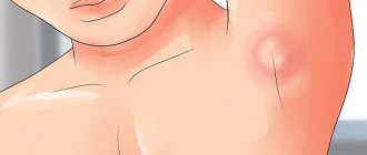
Mumps (mumps, behind the ears) is a contagious acute infectious disease of viral origin, in which the salivary glands behind the ears become inflamed in a child. Thus, when mumps occurs in children, treatment must be strictly carried out under the supervision of a doctor. At the SANMEDEXPERT clinic, the pediatrician will examine the little patient, tell you what medications should be taken and how to avoid complications.
This disease is called so because the child’s cheeks seem to puff out, and the little patient becomes like a pig. At risk are young unvaccinated children under seven years of age, most often boys. An adult suffers from the disease much worse than children.
Infants rarely suffer from a disease such as mumps - breast milk provides them with treatment and a kind of immunity from the disease; This immunity lasts up to 5 years. The peak of disease activity occurs in early spring (March, April).
The main cause of the disease is considered to be contact with a sick person, therefore most often they become infected through airborne droplets, but there are often cases when the disease affects a person through contaminated objects.
If a person has been ill with a disease such as mumps, the treatment has been completed, then he will never get sick again.
Symptoms of gum inflammation in a child
Symptoms of gum disease vary depending on the type and form of the disease. These include:
- bleeding and pain;
- unpleasant odor from the mouth;
- purulent discharge;
- deposits of plaque or tartar;
- hypersensitivity.
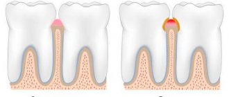
Most often, childhood gum disease is associated with the development of gingivitis. The inflammatory process occurs due to poor oral hygiene (bacteria multiply).
REFERENCE! If you have problems with your gums, a visit to the dentist is required. Only a specialist will select an individual treatment algorithm based on the symptoms.
What are adenoids and where do they come from?
Adenoid vegetations (nasopharyngeal tonsil) are lymphoid tissue in the vault of the nasopharynx. It is present in all children without exception and is a peripheral organ of the immune system, part of the lymphoid pharyngeal ring. The main function of this anatomical formation is to fight bacteria or viruses that enter the child’s body. Its main difference from other tonsils is that the surface is covered with a special epithelium that produces mucus. An increase (hypertrophy) of adenoid tissue is provoked by frequent allergic and respiratory diseases of viral or bacterial etiology. Therefore, the peak of hypertrophy of adenoid tissue occurs precisely at the age of 3-7 years. Then the lymphoid tissue is gradually reduced at the age of 10–12 years. By the age of 17, only fragments of tissue often remain; in healthy adults, adenoid tissue is absent. Hypertrophy of adenoid tissue is usually divided into several degrees according to its volume in the nasopharynx, from the first, where the adenoids close the nasal passages (choanae) by 1/3, to the third or fourth degree, when complete obstruction of the nasopharynx occurs with the impossibility of nasal breathing.
Causes of gum inflammation in children
The abundance of causes that cause gum problems can be divided into two large groups.
Common ones include:
- problems with the gastrointestinal tract;
- frequent colds;
- hormonal disorders.
Among the local causes, doctors identify:
- poor-quality fillings (they collapse and injure soft tissues);
- caries (infection from the tooth immediately spreads to the gum);
- incorrectly installed braces;
- crowding of incisors.
What are the reasons?
Among the infections that can cause mesadenitis in a child are bacteria such as:
- coli;
- staphylococci;
- streptococci;
- salmonella;
- Mycobacterium tuberculosis.
These may also be the following viruses:
- enterovirus;
- adenovirus;
- Epstein-Barr virus;
- cytomegalovirus.
Doctors often associate the development of mesadenitis in children with the presence of diseases such as:
- pneumonia;
- flu;
- Infectious mononucleosis;
- tonsillitis;
- bronchitis.
After severe infectious diseases, a child is often diagnosed with reactive mesadenitis. In addition, this pathology often occurs in children as a specific reaction to vaccinations or uncontrolled use of certain types of medications.
Treatment methods
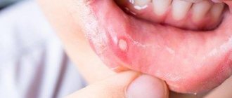
Some of the common childhood gum diseases include:
- stomatitis (inflammation of the mucous membrane due to a virus or fungus);
- periodontitis (advanced form of gingivitis);
- periodontal disease (associated with inflammation, rarely diagnosed);
- gingivitis (a consequence of injury or plaque, usually occurs in the catarrhal form, during the eruption or change of teeth).
For treatment, local administration of various medications and rinsing with antiseptics may be prescribed. Additional measures: sanitation of the oral cavity and removal of tartar. In the chronic form, physiotherapeutic methods are used.
Methods for studying adenoid vegetations
In its normal state, it is impossible to see this tonsil without additional optical devices. There are a number of studies that help determine the extent of adenoid vegetations: digital examination, posterior rhinoscopy with a mirror, radiography of the nasopharynx, endoscopy of the nasopharynx, three-dimensional X-ray examination or CT scan of the nasopharynx. The most modern methods today are:
- endoscopy of the nasopharynx and nasal cavity. The procedure is performed in our clinic under local anesthesia during an appointment with an ENT doctor. It is completely painless and allows you to assess not only the degree of adenoid vegetations, but also the nature of inflammation, the condition of the mouths of the auditory tubes, and also examine the posterior parts of the nasal cavity.
- three-dimensional X-ray examination / CT scan of the nasopharynx. The methods are significantly more informative than conventional X-rays of the nasopharynx, since they allow one to determine not only the size, but also the ratio of adenoid vegetations to other structures of the nasopharynx (the mouths of the auditory tubes, choanae, etc.). The radiation dose is almost 3 times less (0.009 m3v), and the duration of the study is no more than 2 minutes. You can undergo this study at the clinic on Usacheva.
Prevention
The main preventative rule is daily brushing of teeth twice a day (morning and evening). It is important not to forget about the palate and tongue. Please also pay attention to the following recommendations:
- caries is immediately healed (it is advisable not to start the disease in the early stages);
- it is also necessary to treat the disease that could provoke gum inflammation;
- malocclusion needs correction;
- with the help of good nutrition and taking multivitamin complexes according to age, they strengthen the immune system;
- during teething, use a special fingertip to massage the gums;
- food and drink for the baby cannot be too hot or, conversely, cold;
- They explain to their son or daughter that food is always experienced on both sides.
REFERENCE! It is the parents' responsibility to monitor how thoroughly and correctly the child brushes his teeth.
Our specialists at modern pediatric dentistry “Avesta” believe that timely preventive measures help minimize the development of a serious disease. This is the easiest way to keep your baby's gums and teeth healthy.
Mumps symptoms
So, the following symptoms indicate the need for treatment for mumps:
- the temperature rises sharply (sometimes up to 40 degrees),
- joint and muscle pain,
- headache,
- chills and fever (but this symptom does not always appear),
- pain behind the ears, in front and behind the earlobe,
- weakness and lethargy,
- sharp pain when swallowing and chewing food,
- increased salivation.
But the main marker of the disease is gumboil and swelling behind the ears, and sometimes under the jaw. The sublingual glands can also be affected by the disease. Inflammation persists for up to 10 days; flux usually reaches its maximum on the 3rd day. The child remains contagious until the 10th day of illness. If mumps symptoms are detected early, treatment is faster and without possible complications.
Complications
Treatment of mumps in children can be complicated by secondary diseases. Glandular organs are considered weak points when mumps is affected, so common complications are:
- pancreatitis (pancreas),
- oophoritis (ovaries),
- atrophy, orchitis, priapism, infertility (testicles),
Meningitis, encephalopathy, otitis media, hearing loss, and sometimes complete deafness also occur. These diseases are characterized by the following alarming symptoms:
- vomiting and stomach pain,
- apnea,
- convulsions,
- increased drowsiness,
- prolonged fever
- enlargement and pain in the testicles.
Advice from the doctor
As a generalization, I want to say that the well-known joke about curing a runny nose in 7 days and a week does not work with children! Those who treat a child’s runny nose as “ordinary snot that will go away on its own” most often face a whole bunch of complications in the future. Therefore, the sooner you consult an ENT doctor and begin competent treatment, the higher the likelihood that the problem of adenoids will bypass you!
Make an appointment with a pediatric otolaryngologist by calling the single contact center in Moscow, fill out the online appointment form, or contact the reception desk of the Family Doctor clinic.
Health to you and your kids!
Treatment of adenoiditis
Treatment of adenoiditis is usually divided into conservative and surgical. Conservative treatment requires from parents, first of all, a lot of patience (you need to teach the baby to blow his nose correctly, use the nasal cavity with him, sometimes several times a day!), attend procedures (rinsing the nose by an ENT doctor, physical therapy, etc.), and strictly follow all doctor's orders. This is far from a quick process, but if the parents and the doctor are at the same time, and act as a cohesive team, then the result will not be long in coming! But there are cases when conservative treatment is ineffective, then the doctor decides on surgical intervention, and this does not always depend only on the degree of the adenoids. Most often, indications for surgical treatment are: complete absence of nasal breathing, recurrent otitis (tubo-otitis), night apnea, persistent hearing loss.
“If they are involved in the immune response, why remove them? There is nothing superfluous in the body!”
Indeed, adenoid tissue is part of the lymphoid ring of the pharynx, as mentioned above, but only a part! Here it is important to assess the balance of harm and benefit to the body. In the case of chronic adenoiditis, the tonsil itself becomes a place of residence and reproduction of pathogenic microorganisms, which clearly does not benefit the child, and frequent exacerbations lead to an increase in adenoid tissue in size, causing parallel ear disease, followed by persistent hearing loss.
“If you remove them, they will grow back!”
At this stage of medical development, this opinion is erroneous. The adenotomy operation is performed under general anesthesia using endoscopic techniques. Modern equipment allows you to remove adenoid tissue completely under visual control, thereby ensuring the absence of relapses. With adenotomy under local anesthesia, as was previously performed everywhere, the risk of repeated adenotomies is really high, since most often part of the tonsil is not removed the first time, which causes a relapse.
Symptoms of gingivitis in babies
The course of the disease in children is most often observed in an acute form, which is easy to determine:
- When brushing your teeth, a lot of blood remains on the brush;
- soft tissues become bluish due to damage due to bacterial infection;
- painful sensations occur when biting food or drinking drinks;
- swelling, redness and discharge of serous contents.
If you have one or more symptoms, you should contact your dentist immediately. Only professional and high-quality treatment provided on time can prevent chronic inflammatory processes in the oral cavity.
In its extreme form, destruction of tooth enamel and damage to teeth by caries is possible, and, as a result, further complex therapy, up to extraction and prosthetics. Seeing a doctor as quickly as possible at the first symptoms of inflammation will help avoid complications.
Remember that mild gum disease responds quickly to treatment.
Survey
In adolescents and adults, laryngoscopy (examination of the larynx) and fibrolaryngoscopy (examination of the larynx with a flexible endoscope inserted through the nasal cavity) are the generally accepted standard for diagnosing epiglottitis.
In children, the diagnostic method depends on the age and severity of the disease. Laryngoscopy is not always possible or safe, so the diagnosis is often made based on the clinical picture. If necessary, it is confirmed by an X-ray examination of the neck in a lateral projection.
During laryngoscopy, the doctor identifies inflammatory changes in the larynx, swelling of the epiglottis, laryngeal cartilage, and vestibular folds. Palpation of the anterior neck may be tender, especially in the area of the hyoid bone.
Patients with epiglottitis (especially if Hib infection is suspected) should always be examined for the presence of extralaryngeal foci of infection: pneumonia, cervical lymphadenitis, septic arthritis, and less commonly, meningitis.
Laboratory assessment should include a complete blood count with leukocyte count, blood culture for sterility, and in intubated patients - bacteriological examination with sampling of material from the epiglottis area.
Differential diagnosis
True croup (diphtheria) . The clinical picture of diphtheria is sometimes similar to that of epiglottitis. Symptoms—sore throat, malaise, and low-grade fever—usually appear gradually. Diphtheria is extremely rare in countries with high levels of immunization against diphtheria, tetanus and whooping cough.
False croup . The main symptom is a barking cough. It is not observed with epiglottitis. Children with croup usually feel comfortable lying on their back.
Bacterial tracheitis . There is an acute onset, a rapid increase in upper respiratory tract obstruction, and fever, which is similar to epiglottitis. Upon examination, no changes in the laryngopharynx are revealed, and radiographs reveal irregularities in the tracheal wall, and there are no changes in the epiglottis.
Peritonsillar abscess . In children with peritonsillar abscess or other infections of the oropharynx (retropharyngeal abscess, acute tonsillopharyngitis), the development of the disease is slower, breathing at the onset of the disease is not difficult, and intoxication is less pronounced than with epiglottitis.
Foreign bodies in the larynx, trachea and esophagus can cause complete or partial airway obstruction, which requires immediate treatment.
Angioedema (Quincke's edema) . Characterized by rapid onset without previous symptoms of cold or fever. The main manifestations are swelling of the lips and tongue, urticaria, dysphagia without hoarseness.
Upper respiratory tract trauma , including thermal burns.
