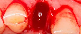
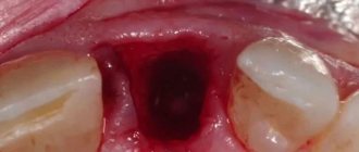
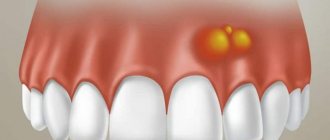
alveolar sockets or alveoli the holes in the bone tissue of the jaw in which the teeth are held. They are also called “the receptacle of the dental root system.” When a diseased tooth is removed, an empty socket is left and if its walls, dry socket or gum are injured, then alveolitis - suppuration of a blood clot . With alveolitis, the socket itself and the surrounding tissues are affected. But there are rare exceptions, when alveolitis is caused by chronic inflammation and infections (more details in the article “Alveolar osteitis: when a tooth was removed carelessly“)
Alveolus (alveolar socket) and its infection (alveolitis)
Description of the disease
Not all dental surgeon patients know that inflammation may develop after removal, so they often ask the question: “Alveolitis - what is it, and why does it occur?” Other names for the disease: “dry socket”, “alveolar local osteitis”. The pathogenesis of the condition is based on a violation of the formation of a physiological clot or its loss from the socket after tooth extraction, which most often occurs due to a violation of the postoperative regimen or low human immunoresistance.
Inflammation of the alveoli is possible only due to removal of the segment. Alveolitis cannot develop after tooth treatment, since the hole is formed only as a result of extraction of the unit.
Causes of alveolitis
The causes of alveolitis are as follows: a tooth has been removed, but a cyst (granuloma) remains inside, which introduces an infection into the bloodstream. The remains of a tooth (or its root) remain in the hole. Removal was difficult, or the tooth was removed due to purulent inflammation. After the tooth was removed, the hole did not fill with blood.
However, there are cases when the dentist is not to blame for the occurrence of alveolitis:
- when the patient rinsed his mouth and spat out the clot;
- the dentist’s recommendations were not followed, the treatment was not carried out according to the doctor’s regimen;
- the patient has a lot of bacteria and infection;
- the problematic tooth was removed due to chronic inflammation;
- weak immunity.
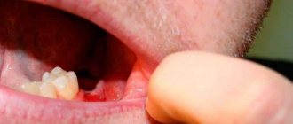
Sometimes the alveolitis is not externally visible. You can tell that you have alveolitis by a bad smell from the socket, swelling of the gums, pain that increases over time: these are signs of alveolitis, and if these alarming symptoms occur, you should immediately contact your dentist.
The main reasons for the appearance of alveolitis after tooth extraction
Tooth extraction is a surgical procedure that is accompanied by the formation of a wound. A blood clot forms in it, protecting it from the penetration of microorganisms. Inflammation of the tooth socket after extraction occurs due to disruption of the process of formation or displacement of a blood clot. The most common causes of pathology:
- Difficult extraction. The more traumatic the operation, the more the tissue’s ability to regenerate decreases and the likelihood of infection increases.
- Wisdom tooth removal. The structure of the root system of third molars is complex, so their extraction is always traumatic. In addition, the bone in the figure eight area has increased density and is less vascularized compared to other areas, which also creates conditions for the formation of a dry socket.
- Concomitant somatic diseases of the patient (diabetes mellitus, immunodeficiency states).
- History of pericoronitis (inflammation of the soft tissues around the tooth).
- The presence of foci of infection in the mouth before surgery. Penetrating into the wound with saliva, pathogenic flora causes alveolitis.
- Inadequate sanitation of the alveoli. After removal, careful curettage of the wound is necessary to remove fragments of bone tissue, roots, pieces of filling, and granulations. Foreign fragments disrupt healing and lead to alveolitis.
- Exceeding the dose of anesthetic. Leads to a narrowing of the lumen of the capillaries, the development of local ischemia, which disrupts the filling of the hole with blood.
- Mechanical damage to the clot. Failure to follow the doctor’s recommendations during the postoperative period (rinsing the mouth, carelessly brushing teeth, etc.).
- Impaired hemostasis. Due to poor blood clotting, a clot does not form.
Alveolitis can be provoked by smoking, taking hormonal contraception, and insufficient oral hygiene. Age plays a certain role in the development of alveolitis after tooth extraction: the older the patient, the greater the likelihood of developing pathology. This is due to a slowdown in metabolic processes and a decrease in the regenerative ability of tissue cells.
Dry socket after tooth extraction: causes
There are many reasons why alveolitis develops. It can arise due to the fault of the doctor, the fault of the patient, and for reasons beyond anyone’s control. If we talk about the patient’s responsibility, then alveolitis can occur when -
- poor oral hygiene,
- the presence of untreated carious teeth,
- due to smoking after removal,
- when ignoring doctor's recommendations,
- if you rinse your mouth vigorously and simply rinse the blood clot out of the hole.
Alveolitis can also occur in women due to increased levels of estrogen in the blood during the menstrual cycle or as a result of taking oral contraceptives (birth control pills). A high concentration of estrogen leads to fibrinolysis of the blood clot in the socket, i.e. to degradation and destruction of the clot.
It is because of fibrinolysis that a blood clot is destroyed both with poor oral hygiene and with carious teeth. The fact is that pathogenic bacteria, which live in large numbers in dental plaque and in carious defects, secrete toxins, which, like estrogens, lead to fibrinolysis of the blood clot in the socket.
When alveolitis occurs due to the fault of the doctor –
- If the doctor left a tooth fragment, bone fragments, or inactive fragments of bone tissue in the socket, which lead to injury to the blood clot and its destruction.
- A large dose of a vasoconstrictor in an anesthetic - alveolitis can occur if, during anesthesia, the doctor injected a large volume of an anesthetic with a high content of a vasoconstrictor (for example, adrenaline). Too much of the latter will result in the hole simply not filling with blood after the tooth is extracted. If this happens, the surgeon must scrape the bone walls with an instrument and cause socket bleeding.
If the doctor left a cyst/granulation in the socket, when removing a tooth with a diagnosis of periodontitis, the doctor must necessarily scrape out the cyst or granulations (Fig. 10), which might not come out with the tooth, but remain in the depths of the socket. If the doctor did not inspect the socket after extracting the tooth root and left a cyst in the socket, the blood clot will fester.
- Due to the large trauma to the bone during removal, this usually happens in two cases: firstly, when the doctor cuts out the bone with a drill without using water cooling of the bone at all (or when it is not cooled sufficiently).
Overheating of the bone leads to its necrosis and the start of the process of destruction of the clot. Secondly, many doctors try to remove a tooth for 1-2 hours (using only forceps and elevators), which causes such trauma to the bone with these instruments that alveolitis is simply bound to develop. An experienced doctor, seeing a complex tooth, will sometimes immediately cut the crown into several parts and remove the tooth fragment by fragment (spending only 15-25 minutes), and thereby reduce the trauma caused to the bone.
- If, after a complex removal or removal against the background of purulent inflammation, the doctor did not prescribe antibiotics, which in these cases are considered mandatory.
Conclusions: thus, the main causes of destruction (fibrinolysis) of a blood clot are pathogenic bacteria, excessive mechanical trauma to the bone, and estrogens. Reasons of a different nature: smoking, loss of a clot while rinsing the mouth, and the fact that the hole did not fill with blood after the tooth was extracted. There are also reasons that do not depend on either the patient or the doctor, for example, if a tooth is removed due to acute purulent inflammation - in this case it is stupid to blame the doctor for the development of alveolitis.
Symptoms of alveolitis of the socket
Alveolitis goes through several stages of development, each of which has its own clinical picture:
| Stage | Description |
| Serous inflammation | The first clinical symptoms appear the next day or after 3 - 4 days after surgery. Non-purulent inflammation is manifested by the appearance of episodic pain and swelling of the soft tissues around the alveoli. |
| Purulent alveolitis | If measures were not taken at the previous stage, a purulent process develops within a few days. The pain becomes constant, radiating to the auricle, jaw, and neighboring teeth. The infection spreads to nearby lymph nodes, causing them to enlarge. A gray-green coating appears inside the hole. The temperature rises. |
| Purulent-necrotic stage | The pain intensifies and becomes throbbing. A putrid odor appears from the mouth. Progression leads to disruption of microcirculation, ischemia, and the development of necrosis. The patient’s health deteriorates significantly, and signs of intoxication appear. |
| Hypertrophic tissue change | Lack of medical intervention within 14 days leads to chronicity of the process. Soreness and swelling become less pronounced. Soft tissues grow and fill the hole. When pressure is applied to the granulation, pus is released. The mucous membrane is swollen, bluish in color. |
If left untreated, alveolitis has an unfavorable outcome. The main complications after alveolitis:
- phlegmon (extensive purulent inflammation);
- acute periostitis (inflammatory process of the periosteum);
- osteomyelitis (purulent-necrotic inflammation of the jaw bone);
- sepsis (purulent-septic blood disease).
Alveolitis of the tooth: treatment
The treatment of alveolitis after tooth extraction must be taken seriously, since the disease will not go away on its own, and its consequences can be very serious:
- blood poisoning
- abscess
- osteomyelitis
- phlegmon
- periostitis
Only an experienced dental surgeon will create an effective treatment plan. It is better to start it when the first signs of the disease appear. The treatment process may include:
- numbing the affected area using anesthesia
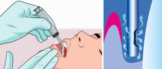
- washing the hole with warm antiseptic solutions
- removing particles of tissue decay that remain after washing using a sharp surgical spoon
- treatment with a sterile cotton swab
- applying a gauze bandage with iodoform impregnation or an anesthetic and antiseptic bandage
- impregnation of soft tissues at the site of inflammation with anesthetic and lidocaine
- application of antiseptic tampons, hemostatic sponge with kanamycin
- removal of dead tissue
- microwave therapy, fluctuarization, infrared laser beams, ultraviolet irradiation
- prescribing vitamins, analgesics and sulfa drugs
- antibiotic therapy
With proper treatment of serous alveolitis, the inflammatory process subsides after 3–4 days.
A week after the pain disappears, the walls of the socket begin to heal and become covered with new mucous tissue. After 14 days, the swelling subsides and the mucous membrane takes on a normal pink color. After treatment of purulent alveolitis, the wound heals within 1–2 months, and tissue restoration takes about six months.
Alveolitis after wisdom tooth removal
Indications for the removal of eighth segments in a row can be different: deep caries, improper eruption, orthodontic correction of jaw bite, dystopia, etc. Extraction of third molars is always associated with trauma to nearby tissues, which increases the development of inflammation. The pathological process is observed in 45% of cases due to the removal of lower wisdom teeth.
Symptoms of alveolitis after wisdom tooth removal are similar to the general symptoms of the disease. However, with such localization of inflammation, a sore throat and impaired functionality of the temporomandibular joint on the affected side may occur (difficulty and painful opening of the mouth).
Why does alveolitis begin?
A slight inflammation in the socket is an inevitable process, since during tooth extraction tissue is injured and an open wound appears, and the environment in the mouth is unsterile. But alveolitis does not occur in everyone. So why does an infection get into the hole that the human body cannot cope with on its own?
Causes of infectious inflammation in the socket
The natural protective barrier is destroyed
A blood clot forms at the site of the extracted tooth; it closes the wound and protects it from infection. If this natural barrier is destroyed, for example by rinsing the mouth after tooth extraction, then the infection can penetrate into the tissue of the hole and provoke inflammation.
Poor oral hygiene
Bacteria live in the oral cavity. During the doctor’s manipulations, as well as after them, particles of tartar or soft plaque can get into the wound. This is a very good environment for bacteria, so they multiply quickly. Severe inflammation begins.
Poor sterilization of surgical instruments
Paradoxically, an infection in the socket can also be caused by a dental surgeon who uses poorly sterilized instruments.
Violation of the rules for processing the tooth socket
After surgery, especially if the extracted tooth had a granuloma, the doctor must carefully treat the wound so that it fills with blood to form a clot, and apply sterile cotton wool. Neglecting these rules can provoke alveolitis.
Violation of doctor's recommendations after removal
Even if you did not make a mistake in choosing a dental surgeon and the removal went well, an infection can get into the wound. This happens if you violated the doctor’s recommendations for caring for the hole. For example, they disturbed the wound with a tongue or some object and introduced an infection into it.
Decreased immunity
The cause of alveolitis may be decreased immunity or exhaustion of the body. This happens after a serious illness.
Caries
If there are teeth in the oral cavity affected by caries, then there is always a possibility that after removal you will encounter alveolitis.
To avoid the occurrence of alveolitis after tooth extraction, choose a dental surgeon responsibly and follow all his recommendations. A good doctor will definitely advise you to undergo professional oral hygiene before removal. This will reduce the risk of infection getting into the socket.
Methods for diagnosing alveolitis
How long the alveolitis will take to heal depends on timely diagnosis and treatment. Therefore, at the first signs of illness, you should consult a dentist. Identification of pathology occurs after listening to complaints, collecting anamnesis (where there is recent tooth extraction) and examining the socket cavity.
Sometimes the doctor prescribes an x-ray examination, which helps to detect tooth fragments and other foreign fragments in the alveolus. In rare cases, when alveolitis of the tooth socket , computed tomography is prescribed for the purpose of differential diagnosis of other diseases (trauma, tumor, etc.).
Tooth structure
The main function of teeth is to chew food. Also, teeth, as part of the chewing-speech apparatus, are involved in creating sounds during communication.
Teeth are part of a complex complex of interconnected and interacting organs. This complex is responsible for chewing, breathing, speech formation and includes the jaws, teeth themselves, masticatory muscles, cheeks, palate, tongue and salivary glands.
The structure of the tooth as part of the dentofacial segment is determined by the function it performs. Thus, teeth with a cutting edge (incisors) perform the function of biting food. Fangs – tearing off pieces. Small and large molars – chopping and grinding food.
The dentofacial segment (a section of the jaw with a tooth located on it) includes the tooth, the dental alveolus and the part of the jaw adjacent to it; ligamentous apparatus, blood vessels and nerves.
Upon closer examination, the dentofacial segment consists of the following elements:
- periodontal fibers;
- alveolar wall;
- dentoalveolar fibers;
- alveolar-gingival branch of the nerve;
- periodontal vessels;
- arteries and veins of the jaw;
- dental branch of the nerve;
- bottom of the alveoli;
- tooth root;
- neck of the tooth;
- crown of the tooth.
The first teeth to appear in a person are baby teeth. Twenty primary teeth erupt in a specific sequence; The first pair, as a rule, appears first. By the age of two, the eruption of baby teeth is completed, and at the age of five to six years, baby teeth begin to gradually fall out and be replaced by permanent (molar) teeth. The change of dentition is completely completed by the age of 12-15 years.
Anatomically, a tooth consists of three parts - crown, neck and root.
The crown of a tooth is its visible part. It is the condition of the crowns that is responsible for the aesthetic beauty of a smile. The crown consists of natural dentin and enamel, which together provide the hardness of the tooth, and is located in the alveolus (the cavity of the jaw). The formed crown does not increase in size over time, but can wear off and darken. As a rule, loss of the whiteness of a smile is a consequence of poor nutrition, lack of care and damage to the crown enamel.
The neck of a tooth is the connecting link between its crown and root. With healthy gums and good condition of teeth, the neck is not visible. Exposing the neck of a tooth indicates the presence of oral diseases and can provoke increased tooth sensitivity (reaction to cold, hot, etc.).
The root provides immobility for the tooth, serving as natural cement, and also serves as protection for the neurovascular bundle located in the canal. The root of the tooth is located in the socket of the jaw. At the root there is a pulp, the inflammation of which is called “pulpitis”. In most cases, pulpitis is a complication of untreated caries.
To maintain the health of your teeth and the beauty of your smile, make it a rule to attend preventive dental examinations from a specialist at the Neo-Dent clinic once every six months.
How to treat alveolitis after tooth extraction
The main task of correction is aimed at eliminating the cause that caused the pathology and stopping the inflammatory process. The doctor decides how to treat alveolitis after tooth extraction. Treatment tactics depend on the stage of the pathological process. It may include revision (if necessary, curettage) of the hole, drug treatment, physical therapy (laser, magnet, fluctuarization, ultraviolet radiation). All manipulations are performed under local anesthesia.
Treatment program:
- Local anesthesia.
- Antiseptic treatment of the wound (removal of saliva, food debris, pus).
- Freeing the cavity from a disintegrated blood clot, foreign inclusions (tooth fragments, root remains, etc.), granular tissue.
- Repeated treatment with an antiseptic, application of a turunda soaked in an anesthetic drug.
- Applying a medicated antiseptic bandage.
When treating alveolitis, the doctor can use proteolytic enzymes (trypsin, chymotrypsin), which break down necrotic tissue, thereby helping to cleanse the socket. To reduce pain, a blockade is made with local anesthetics, which are injected into the soft tissue near the inflamed alveoli. To suppress the infectious process, a course of antibiotic therapy is prescribed.
Stages of alveolitis
All these symptoms of alveolitis cannot appear at the same time. Signs of the disease accumulate, alternate, or overlap as inflammation progresses. Symptoms help determine the stage of the disease.
Serous alveolitis
It develops 72 hours after tooth extraction. The main symptom is aching pain that intensifies while eating. Body temperature is not elevated, regional lymph nodes are not enlarged. Upon examination, pieces of food and saliva are found in the hole, but there may not be a blood clot there, or it may be partially destroyed. Serous alveolitis continues for a week, and if left untreated, it turns into a purulent form.
Purulent alveolitis
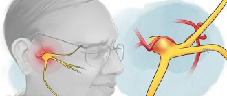
Occurs 10 days after tooth extraction. By this time, the pain from alveolitis has already become so intense and constant that it is impossible to eat. In this case, unpleasant sensations spread along the branches of the trigeminal nerve. The soft tissues of the affected area swell, and mouth opening is limited. The patient feels weak and unwell, his temperature rises (up to 38 degrees) and a putrid taste and smell appears in the mouth. Upon examination, you can see redness, swelling, a dirty gray coating, and the alveolar process is thickened on both sides of the socket.
Chronic purulent (hypertrophic) alveolitis
As the disease becomes chronic, the pain begins to gradually subside, body temperature normalizes, and the patient’s general condition noticeably improves. The soft tissue in the area of the inflamed hole grows, and pus is released from it. The gums at the site of inflammation are swollen and have a bluish tint.
Alveolitis can occur not only in adults, but also in children. If a child has had a permanent tooth removed, parents need to carefully monitor compliance with the doctor’s recommendations.
