What are blockades
Therapeutic blockades are the relief of acute or chronic pain and spasms. It is performed by injection into the area where the nerve trunks and plexuses pass. Severe pain causes physical and psychological suffering, incapacitates the patient and can cause disability. Depression that occurs against this background often leads to suicide attempts.
Therapeutic blockades quickly relieve acute pain. After some time, swelling, inflammation, spasm of muscles and blood vessels decrease. This leads to a lasting improvement in the condition. At the 100med medical center, blockades are performed by neurologists and anesthesiologists. Several groups of drugs are used for the procedure:
- local anesthetics;
- corticosteroid hormones;
- chondroprotectors.
The effect of drug administration lasts from several days to several weeks. After the medicine stops working, the pain may return. However, its severity will be less by about 50%.
During the procedure, the patient may feel some discomfort associated with the injection. However, the discomfort quickly passes. There is no need to be afraid of blockades, as this method is one of the safest. Our specialists strictly follow all the rules for performing the procedure, as well as asepsis and antiseptics. This guarantees the effectiveness and safety of blockades in our clinic.
Nerve block
A nerve block is a treatment in which a local anesthetic is injected into the soft tissue where the peripheral nerve passes. The procedure allows you to relieve pain, spasm of muscles and blood vessels, swelling and inflammation. Indications for manipulation:
- neuritis;
- neuralgia;
- tunnel syndrome;
- muscular-tonic syndrome;
- diseases of the spine;
- pain after injury or surgery;
- chronic pain syndrome;
- oncological diseases.
Nerve blocks do not require special preparation. After a thorough examination and examination of the patient, the doctor determines the injection site and treats the skin with an antiseptic. An injection of pain medication is given into the soft tissue. After a few minutes, numbness, warmth and a feeling of heaviness appear in this place. The course of injections (up to 3 sessions) can be repeated every year.
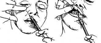
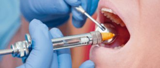
Anesthesia on the lower jaw
Mandibular anesthesia. Methodology
Intraoral palpation method.
- The anterior edge of the mandibular ramus is felt at the level of the distal edge of the crown of the 3rd molar. By moving the finger inward, the temporal ridge is determined. The finger is fixed in the retromolar fossa.
- Having positioned the syringe at the level of the premolars of the opposite side, insert the needle medially from the temporal ridge and 0.75-1.0 cm above the crown of the 3rd molar. Advance the needle outward and backward. They reach the bone at a depth of 0.5-0.75 cm and release 0.5-1.0 ml of anesthetic (switching off the lingual nerve).
- The syringe is moved to the level of the central incisors and the needle is advanced another 2.0 cm, where 2-3 ml of anesthetic is injected (switching off the lower alveolar nerve).
Intraoral apodactyl method. Having placed the syringe at the level of the premolars of the opposite side, a needle is inserted into the outer slope of the pterygomandibular fold, in the middle of the distance between the chewing surfaces of the crowns of the upper and lower molars. The needle is advanced outward and posteriorly 1.5-2.0 cm until it contacts the bone and 2-3 ml of anesthetic is injected (switching off the lower alveolar and lingual nerves). Extraoral method. A needle is inserted into the area of the base of the lower jaw, moving 1.5 cm anterior to the angle of the lower jaw, the needle is advanced 3.5-4 cm parallel to the posterior branch, and 2 ml of anesthetic is released.
Pain relief zone:
- all teeth of the corresponding half;
- bone tissue of the alveolar process;
- gums on the vestibular and lingual sides;
- mucous membrane of the sublingual region;
- anterior 2/3 of the tongue;
- skin and mucous membrane of the lower lip;
- skin of the chin of the corresponding side.
Onset time: 10-20 minutes.
Torusal anesthesia. Methodology
- The syringe is placed on the molars of the opposite side. The needle is inserted into the groove formed by the lateral slope of the pterygomandibular fold and the cheek, 0.5 cm below the chewing surface of the upper 3rd molar. The needle is advanced to the bone by 0.25-2 cm and 1.5-2 ml of anesthetic is injected (switching off the lower alveolar and buccal nerves).
- The needle is withdrawn a few millimeters in the opposite direction and 0.5-1.0 ml of anesthetic is injected (switching off the lingual nerve).
Pain relief zone:
- all teeth of the corresponding half;
- bone tissue of the alveolar process;
- gums on the vestibular and lingual sides;
- mucous membrane of the sublingual region;
- anterior 2/3 of the tongue;
- skin and mucous membrane of the lower lip;
- skin of the chin of the corresponding side;
- mucous membrane and skin of the cheek.
Onset time: 10-20 minutes.
Anesthesia in the area of the mental foramen (mental). Methodology
Extraoral method.
- A needle is inserted 0.5 cm above and posterior to the projection of the mental foramen, which is located 12-13 mm above the base of the body of the lower jaw in the area of the 2nd premolar.
- Move the needle down, inward and anteriorly until it comes into contact with the bone, and inject 0.5 ml of anesthetic.
- A needle is inserted into the mental foramen, advanced into the canal 3-5 mm and 1-2 ml of anesthetic is injected.
Intraoral method.
- The soft tissues of the cheek are moved to the side. A needle is inserted at the level of the middle of the crown of the 1st molar, moving a few millimeters outward from the transitional fold.
- Move the needle 0.75-1.0 cm down, anteriorly and inward to the mental foramen and inject 0.5 ml of anesthetic.
- A needle is inserted into the mental foramen, advanced into the canal 3-5 mm and 1-2 ml of anesthetic is injected.
Pain relief zone:
- soft tissue of the chin and lower lip;
- premolars, canines and incisors;
- bone tissue of the alveolar part;
- mucous membrane of the alveolar process within the anesthetized teeth.
Onset time: 5 minutes.
Anesthesia in the area of the lingual nerve. Methodology
Using a spatula, the tongue is retracted in the opposite direction and a needle is inserted into the mucous membrane of the maxillo-lingual groove at the level of the middle of the crown of the lower 3rd molar, where the nerve lies very superficially. 2 ml of anesthetic is injected.
Pain relief zone:
- mucous membrane of the sublingual region;
- anterior 2/3 of the tongue.
Onset time: 3-5 minutes.
Anesthesia in the area of the buccal nerve. Methodology
A needle is inserted into the area of the anterior edge of the coronoid process at the level of the chewing surface of the upper molars into the mucous membrane of the cheek, directing the syringe from the opposite side. Advance the needle 1.0-1.5 cm to the anterior edge of the coronoid process and inject 1-2 ml of anesthetic.
Anesthesia zone: mucous membrane and skin of the cheek.
Onset time: 10 minutes.
Anesthesia according to Bershe. Methodology
The needle is inserted perpendicular to the skin under the lower edge of the zygomatic arch, moving 2 cm anteriorly from the tragus of the auricle. The needle is advanced through the mandibular notch by 2.0-2.5 cm and 3-5 ml of anesthetic is injected.
Pain relief zone: relaxation of the masticatory muscles.
Onset time: 5-10 minutes.
Anesthesia according to Egorov. Methodology
A needle is inserted under the lower edge of the zygomatic arch 0.5-1.0 cm anterior to the articular tubercle at an angle of 60-75° to the skin. The needle is advanced up to the outer surface of the temporal bone and the needle is removed 0.5-1.0 cm. Then, at a right angle to the surface of the skin, the needle is immersed in the soft tissue to the extracted distance and 2 ml of anesthetic is injected.
Anesthesia zone: blockade of all motor branches of the mandibular nerve.
Onset time: 5-10 minutes.
Joint blockade
Joint blockade is a method of introducing a drug into the joint cavity or adjacent soft tissues (articular capsule). The method not only relieves pain, but eliminates inflammation, relieves muscle and vascular spasm, and restores range of motion in the joint. The procedure is indicated for the following diseases:
- arthritis of joints of various sizes;
- arthrosis;
- arthralgia;
- synovitis;
- bursitis;
- tendovaginitis;
- periarthritis;
- facet syndrome;
- Reiter's disease;
- Bekhterev's disease.
The injection is performed without additional preparation after a thorough examination. The patient is seated or laid on a couch and the skin over the joint is treated. The injection is made into the cavity of the joint capsule, into the area of the periarticular ligaments or bursa. The effect occurs within a few minutes and lasts up to 3 weeks. The duration of the course depends on the severity of the lesion and can be up to 15 procedures.
Celiac plexus block
With this type of blockade, the medicine is injected into the solar plexus area. The injection is done under the control of an X-ray machine or ultrasound.
The procedure is effective for:
- chronic pancreatitis, pancreatic necrosis;
- abdominal adhesions;
- Crohn's disease;
- tumors of the abdominal organs;
- ineffectiveness of painkillers.
You should prepare for the blockade in advance. A few days before the procedure, the doctor at our clinic recommends stopping taking anticoagulants and diuretics and adjusting the dose of glucose-lowering medications. The patient is placed on his stomach with a bolster or pillow. First, the doctor anesthetizes the soft tissue at the injection site. Then, under X-ray control, a special long needle is inserted. Through it, a local anesthetic or a drug that destroys nerve cells is supplied to the solar plexus. The patient feels warmth in the abdomen and a decrease in pain. After the procedure, observation is required in our hospital for 24 hours.
Paravertebral blockade
Paravertebral blockade is performed to reduce pain in the spine. The effect is achieved by blocking the ganglia or spinal nerves on one or both sides. Indications for this are:
- osteochondrosis;
- radiculitis, lumbago;
- neuralgia;
- myositis;
- hernias and protrusions of intervertebral discs;
- spinal injuries.
To carry out the manipulation, the patient is placed on a flat couch. Local soft tissue anesthesia is first performed. The medicine is injected into the segment of the spine where the pain is felt the most. The injection is given on one or both sides depending on the patient’s condition.
Iatrogenic trigeminal neuropathy.
This nosological entity arose due to the fact that treatment of trigeminal neuralgia in most cases began with neurodestructive operations (alcohol-lidocaine blockades, neuroexeresis, destruction of the trigeminal ganglion). As a result, a significant number of patients experienced iatrogenic traumatic or toxic-traumatic neuropathies of the trigeminal nerve. The maxillary and mandibular nerves were most often affected.
In many textbooks on neurology, in case of trigeminal neuralgia, it is proposed to carry out alcohol-novocaine and alcohol-lidocaine blockades of its peripheral branches or node - the so-called alcoholization . The analgesic effect in this case is achieved on average after the second or third procedure, but it is explained by the fact that numbness occurs due to the development of destructive changes in the nerve trunk. Over time, toxic-traumatic neuropathy develops, practically resistant to treatment, so the patient must continue to undergo blockades, the effectiveness of which decreases in proportion to their number.
Thus, neurodestructive operations that are performed in the treatment of neuralgia lead to the development of toxic-traumatic neuropathy. This determines the nature of the pain syndrome.
Clinic.
The clinical picture is usually represented by the presence of constant aching, burning or dull neuropathic pain in the area of innervation of the affected nerve, against the background of which neuralgic paroxysms occur with irradiation of pain, respectively, to the segmental areas of the face (Zelder segments). Patients experience various types of paresthesia (numbness, crawling, burning) and sensitivity disorders (hypesthesia with symptoms of hyperpathia or hyperesthesia), which sometimes spread beyond the innervation of one of the branches of the trigeminal nerve.
In many cases, vegetative fibers are drawn into the process, which leads to trophic changes in the oral mucosa (gingivitis), the dental system (progressive periodontitis) and facial skin (pigmentation or depigmentation, dryness, peeling, soft tissue atrophy). In such cases, the pain becomes unbearably burning, tearing, boring, and is accompanied by vegetative reactions (redness and swelling of the facial skin, local increase in body temperature, lacrimation, salivation).
During neurodestructive manipulations on the mandibular nerve, painful contraction of the teeth may occur (patients are forced to eat through a straw and cannot speak or open their mouth). With each subsequent alcoholization, the nature of the pain syndrome changes: neuralgic paroxysms become longer, more frequent, a neuralgic status can form, and slightly pronounced trigger areas appear on the skin of the face. Pain is provoked by weather conditions (cold or heat), exacerbation of somatic pathology, food consumption, and physical activity. The exit points of the trigeminal ditch are painful during palpation in approximately 2/3 of patients.
The data presented convincingly indicate that neurodestructive manipulations are not the method of choice for the treatment of trigeminal neuralgia, since in most cases a short-term effect is achieved. However, such therapy leads to the development of toxic neuropathy, disease progression and the development of resistance to conservative treatment methods. Only when all the methods used to treat neuralgia are not effective, and the intensity of the pain syndrome remains severe, can neurodestructive operations recently developed by neurosurgeons be used.
Epidural block
An epidural block is the injection of medication into the space between the dura mater and the periosteum of the spinal canal. Depending on the injection site, caudal, translumbar, and transforaminal blockades are distinguished. A good effect of blockades is observed when:
- damage to the radicular nerves;
- periduritis;
- intervertebral hernias;
- spondylolisthesis;
- narrowing of the spinal canal;
- back pain after injury or surgery.
The procedure must be carried out on an empty stomach. The patient is placed on a couch, and the doctor applies local anesthesia to the desired area. Then, under the control of an X-ray machine, a needle is inserted through which the medicine is administered. When performed correctly, the patient feels a slight burst in the back.
Indications for therapeutic blockade
- pain in the neck, back;
- osteochondrosis;
- neuralgia, neuritis;
- pain in the spine due to intervertebral hernia;
- rheumatism;
- osteoarthritis;
- pain in the postoperative period;
- phantom pain;
- Minière's syndrome;
- neuropathy;
- spasticity;
- amputation pain;
- pain and spasticity after a stroke;
- radiculitis;
- migraine;
- sciatica;
- tunnel syndrome.