An x-ray is the dentist’s main tool in making the correct diagnosis. However, a conventional orthopantomogram or targeted photograph has limited diagnostic potential and does not provide complete data on the condition of the teeth and maxillofacial area. But technologies are constantly being modernized and today, more informative technology has come to the aid of conventional radiography - dental computed tomography (CT).
What does a 3D dental x-ray show?
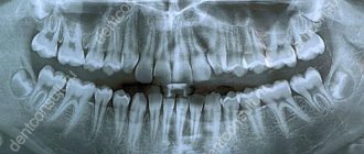
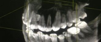
3D dental tomography is a highly accurate diagnostic method that makes it possible to obtain a three-dimensional image of the dental system in different projections. Volumetric images obtained with CT allow the specialist to enlarge, rotate and examine the area of interest from all sides and at different depths:
- The entire maxillofacial apparatus.
- A dentition or an individual tooth.
- Paranasal sinuses.
- Bone and periodontal tissues.
Dental CT allows you to detect inflammation, assess the homogeneity of the filling material and check the quality of installation of a filling, crown or implant, see the number of dental roots and their fragments, identify neoplasms, assess the degree of curvature of teeth, determine the exact parameters of bone tissue (height, width, density, etc.). d.). The information obtained allows the doctor to optimize treatment measures and predict the result.
Indications for 3D computed tomography
The main indications for such an examination are various diseases of the oral cavity. The most common of them include the following: the presence of partially or completely impacted units, for example, eights growing in the wrong place of the teeth, bite pathologies, disruption of the mandibular joint, fractures after injuries, as well as photographs are necessary before complex surgical interventions.
In terms of information content and quality of images, CT is significantly superior to X-rays and orthopantomograms. This is very important for full preparation for the installation of implants, that is, titanium tooth root replacements. Diagnostics makes it possible to detect the presence of cysts, cracks, tumors even at the initial stage. Using CT, the doctor can examine existing tissue diseases and assess their severity.
It is also necessary to carry out this procedure before increasing bone tissue using any of the available methods. Of course, a classic X-ray image shows the bone well, but a CT scan can also tell you everything about soft tissues, blood vessels, nerve endings and mucous membranes. The slightest changes will be reflected immediately.
Why are dental x-rays prescribed?
A 3D photograph of teeth is performed if the following indications exist:
- Injuries of the maxillofacial area.
- Preparation for endodontic treatment (structure of root canals, pathological processes in the periodontium, degree of pulp damage, etc.).
- Diagnosis of neoplasms (cysts, abscesses, granulomas, tumors).
- Anomalies of development and deformation of the maxillofacial apparatus.
- Quality control of filling and implant installation.
- Planning of orthodontic treatment (identification of impacted and dystopic teeth, analysis of the condition of the tissues around each tooth, etc.).
- Detection of hidden periodontal cavities and pockets.
- Implantation planning (assessment of jaw bone parameters, indications for sinus lift or osteoplasty, modeling the result of implantation).
- Endogenous pathologies of the maxillary sinuses.
Three-dimensional x-ray examination is the gold standard when planning any complex dental procedure or surgery. CT allows you to quickly make an accurate diagnosis, competently plan treatment or dental prosthetics, and monitor the results.
Before and after 3D computed tomography
Often, the dentist prescribes a tomography along with a classic x-ray; this is required to obtain the most complete picture of what is happening in the oral cavity. Diagnostics may be needed not only by the surgeon before a major intervention, but also by the implantologist and orthodontist who is going to install braces to correct the bite. In the volumetric image obtained during the study, you can see the smallest details and deviations from normal indicators.
On an imaginary section of tissue, all layers are clearly visible; the presence of benign and malignant seals, cysts, and foci of inflammation can be determined. In addition, based on the results obtained, the doctor can easily determine the cause of a particular pathology. A competent approach allows you to prescribe the optimal treatment plan and avoid unwanted consequences.
Due to the fact that CT scans do not require any incisions or punctures, the traumatic nature of the procedure is minimized. After visiting a dental clinic, a person does not feel pain, he does not need to recover, because there are no wounds on the soft periodontal tissues. In this case, the appearance does not change in any way, there are no disturbances in diction, there is no feeling of being “frozen”, because anesthesia is not required.
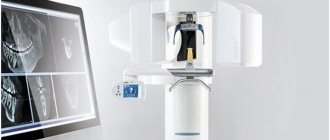
What equipment is used
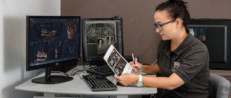
To carry out 3D diagnostics, a three-dimensional computed tomograph SOREDEX Scanora 3D with advanced functionality is used. This is the latest generation equipment, which allows you to obtain three-dimensional images of the anatomical structures of the maxillofacial region in a few seconds, with the least radiation exposure for the patient.
The program analyzes the obtained multiplanar sections and builds them into a 3D model, thanks to which the specialist is able to accurately assess the condition of the dental system, detect all pathological processes occurring in this area and competently plan a treatment regimen.
A virtual 3-dimensional model of the scanned area can be recorded on any digital media (CD, flash drive), which allows the attending physician, if necessary, to view diagnostic data or involve related specialists in the analysis of the received information.
On-site 3D scanning from Star Smile company
While on-site scanning
operates in Moscow and St. Petersburg. Thanks to this service, all our clients - clinics or doctors - can call a Star Smile employee with an intraoral scanner, who, by agreement, will come to your clinic at the appointed time and scan the patient.
How much does on-site 3D scanning cost?
By the way, the on-site scanning service is free when ordering Star Smile aligners. It increases the accuracy of aligners, speeds up the process of their production, and eliminates the possibility of errors when taking impressions. And as an added bonus: your clinic will look more advanced, high-tech to your patients.
For now, this service is available in Moscow and St. Petersburg. Since we Star Smile operates in more than 70 cities of Russia, we are already considering other cities in which on-site scanning will be in demand by orthodontists and dentists.
Possible harm
Cone beam dental computed tomography is the safest and fastest diagnostic method. Thanks to the use of a conical X-ray beam, the radiation dose received during the study is 10 times less than when using spiral CT. And the pulsating mode of the X-ray beam further reduces the radiation dose. The three-dimensional computed tomograph SOREDEX Scanora 3D is one of the safest devices in terms of X-ray radiation dose - only 0.035 m3v.
However, despite the safety of the study, CT also has contraindications. If we just talk about dental tomography, it is not performed during pregnancy (in the 1st trimester). 3D dental x-rays with contrast are prohibited for pregnant and lactating women, patients with endocrine disorders (diabetes mellitus, thyroid pathologies), renal failure and intolerance to iodine-containing drugs.
Rehabilitation after 3D computed tomography
As after a classic X-ray, there are no special recommendations for rehabilitation after a CT scan. This is a safe and easy way to find out about the presence of diseases, after which a person can immediately return to his usual lifestyle. In the rarest cases, a tomography with the introduction of a contrast agent may be prescribed. This is done extremely rarely for dental treatment; the recovery plan will be the same as after a CT scan of any other part of the body.
Experts strongly recommend drinking as much liquid as possible on the first day after the manipulations, and also giving preference to liquid foods, such as soups. You can also drink milk, but you can increase your usual portions. If the doctor has not prescribed antibiotics and other medications, you can drink a glass of red, dry wine (no more than 250 ml). Many doctors recommend eating natural honey, drinking freshly squeezed juices with pulp, and if there is a lack of hemoglobin, adding beets and pomegranates to the diet.
If you act correctly, the contrast is eliminated from the body within the next day (up to 90%). However, if there are problems with the functioning of the kidneys, the volume of fluid consumed should not be sharply increased. If a person remains in the hospital after a CT scan, he may be treated with saline and glucose intravenously. General recommendations after CT include eating plenty of nuts, fruits, parsley and beans. It is important to note that this procedure does not affect the possibility of conception in any way.
Online consultation with a doctor
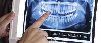
If you are concerned about the condition of your teeth. Painful sensations arise, gums bleed for a long time, and seals have appeared on the jaw. You are concerned about previously installed dental implants. And there was a need to take a 3D photo of the teeth. Then, after receiving three-dimensional visualization, it is better to go for an examination or consultation with a dentist to interpret the images and compare the results with your current complaints. It is impossible to independently understand the nuances of a 3D image, much less make a diagnosis. The specialist will explain the situation and give recommendations before the in-person appointment.
Price
The cost of 3D computed tomography of teeth in Moscow averages 2500-5000 rubles, depending on the scope of the diagnosis.
The price for a dental photograph at the Moscow Center for Dental Implantology RUTT starts from 1,300 rubles - depending on the scope of the examination.
Interpretation, assessment or marking for implantation, regardless of the size of the area under study, during treatment in our clinics is carried out free of charge.
CT scans can be taken on any day, including weekends and holidays. You can make an appointment for diagnostics by phone or by leaving a request online.
Free online consultation with a dentist
| Service | Price |
| Description of the radiologist (assessment and/or marking for implantation) regardless of the size of the area being examined during treatment in our clinics | from 0 rub. |
| CT scan | from 1,300 rub. |
| 3D tomography of 1 to 3 teeth (6*6 cm) with recording on CD | from 4,000 rub. |
| CBCT area 7.5*14.5 cm (both jaws) + recording on CD | from 4,000 rub. |
| CBCT of both TMJs + recording on CD and/or sending by email | from 4,000 rub. |
| 3D tomography of 1 jaw with recording on CD | from 5,000 rub. |
| 3D tomography of the temporomandibular joint with recording on CD | from 5,000 rub. |
| Evaluation and marking for implantation of 3 teeth with printing on film | from 5,000 rub. |
| Assessment and marking for implantation of 1 jaw or 2 jaws (on 1 side) with printing on film | from 7,500 rub. |
| 3D tomography of the paranasal sinuses with recording on CD | from 10,000 rub. |
| Linear tomography of the temporomandibular joint with recording on CD | from 12,000 rub. |
| Assessment and marking for implantation of 2 jaws with printing on film | from 15,000 rub. |
| 3D tomography of the 2nd jaw, TMJ, including sinuses (13.00*14.5 cm) with recording on CD | from 20,000 rub. |
Consultation and diagnostics are free!
All prices Promotions
Sign up for a consultation
three ROOTT specialists + diagnostics as a gift
Advantages of the method
- The ability to rotate, enlarge, and examine images in any projection and section, which is impossible with conventional 2-dimensional scanning.
- The examination lasts only a few seconds (8-20 seconds).
- Complete diagnostic information.
- Maximum security.
- Digital information format.
- Detection of any pathological processes at an early stage.
- No prior preparation required.
- 3D reconstruction without distortion or artifacts.
- A wide range of purposes - from endodontic dental treatment and implantation to maxillofacial operations.
Is there an alternative to CT
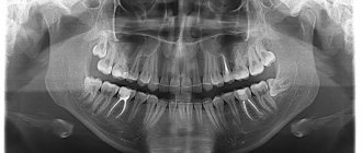
There are many other diagnostic imaging methods (x-ray, orthopantomogram, ultrasound, etc.), but only CT provides the possibility of highly accurate, separate images of all types of tissue at different angles and to different depths. Although a panoramic dental photograph remains an equally important diagnostic tool for a dentist today, it can only provide a general overview. In turn, a 3D tomogram allows you to obtain not a single flat image of the jaw, but a whole series of sequential multiplanar images in different projections and without the distortions inherent in a panoramic image.
Example:
due to the different density of bone structures exposed to X-ray radiation, it is impossible to see less dense bone in a 2-dimensional image; accurate information is provided by a 3- D image of the teeth.
How does the procedure and decoding work?
To take a 3D photograph of teeth, a standing or sitting patient needs to bite a special plate and fix his position in the device using a fixing stand. During the entire scanning time, you must remain absolutely still.
The tomograph sensor makes a series of revolutions around the patient’s head for 8-20 seconds, producing about 200 images in different projections. Processing digital data takes 5-15 minutes, after which the information is written to a disk or flash drive. No preparation is required, you just need to remove all metal jewelry from your neck, ears, and hair before the procedure.