Lately I have received several different questions, one way or another relating to the use of x-rays in dentistry. It must be said that there have always been many different misconceptions, myths and conjectures around this topic, mixed with a pronounced phobia in our country towards everything that is somehow associated with “radiation”. Therefore, I decided not to write answers separately to each question, but to combine them into one note.
What is a visiograph and how does it differ from an x-ray?
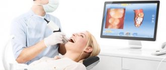
This one of the frequently asked questions is akin to the difference between a car and a traffic light... It seems that both concepts have some kind of connection, but it is somehow difficult to compare them. It's the same here. A radiovisiograph is a system that receives x-ray radiation, transforms it into digital form and displays the image on a computer screen. Roentgen (who is Wilhelm Conrad) is a long-dead German physicist who gained worldwide fame for his discovery of short-wavelength rays with enormous penetrating power. The physicist himself called these rays X-rays (in English today they are called exactly that - X-ray), but now we often call them X-rays, and in everyday life simply “X-rays”. The unit of radiation power was also called the x-ray. Now it is clear that a visiograph and an x-ray are completely different things. If we compare the visiograph with anything, it is with x-ray film, which it is universally replacing from all areas of medicine.
Sensitivity to radiation in various parts of the body
Physicists call this the tissue weight factor. For example, the ovaries, which would be in the X-ray field on many studies of the lower back, are much more susceptible to radiation damage than the skin in the cheek.
Skin (cheek) and bone (teeth) have a weighting factor of 0.01, and genitals have a weighting factor of 0.08, which is eight times greater. The colon has a weight factor of 0.12, which is much larger than the skin and teeth. This is a bigger difference than just the difference in thickness between these body parts.
Thyroid
Know that the thyroid gland has a tissue weight factor of 0.04 - that's five times that of teeth and skin. During intraoral examinations, the thyroid gland itself will not be included in the radiation field, but it will receive some scattered radiation, which is generated in the patient in the form of scattered x-rays from bone and some soft tissue.
Despite the fact that this dose is extremely small, this problem has attracted a lot of public attention and is currently a very pressing problem.
Lens of the eye with dental x-ray
Another radiosensitive organ that we never think about, but is all important to us, is the lens of the eye.
The International Atomic Energy Association recently lowered its threshold for dose to the lens of the eye from occupational exposure to 20 mSv per year. According to the results of the study, although the international association reduced the dose rate for the lens to 20 mSv per year, it did not establish a direct relationship with eye cataracts.
Is it true that a visiograph is safer than a regular film photograph?
When asked about such a comparison, they mean the radiation exposure that the patient receives when using different techniques. In this sense, indeed, a visiograph is preferable, since its sensor is much more sensitive than the best film. Therefore, to obtain a high-quality image using a visiograph, much shorter shutter speeds are needed. To take a picture on film, the shutter speed is 0.5-1.2 seconds. To obtain the same image using a visiograph sensor – 0.05-0.3 sec. Those. 10 times shorter. As a result, the radiation exposure received by the patient when using a visiograph is reduced to an insignificant minimum.
3-D Dental Scanning
The advent of volumetric imaging (3-D scanning) has changed everything when it comes to the radiation dose in a 3D dental image. As we saw above, the real reason why the dose of X-rays has always been considered very low is that it is projected onto a small X-ray.
But 3D dental scanners use fan beam technology and their detectors are different from the technology used for hospital CT scanners. This provides some dose reduction compared to traditional CT machines, but many of the technical parameters are the same. It is difficult to directly calculate and compare the dose of dental 3-D scanners with traditional X-ray scanning in dental clinics.
It is important to train staff and inform them that low doses will change if a positioning error is detected or unusual anatomy is identified in the patient and unintended body parts are also irradiated during the scan.
It must be remembered that manufacturers of dental 3D scanners may take into account the salivary glands and other anatomical structures that are in the radiation field, but the lenses of the eye or the thyroid gland are usually not exposed to radiation.
The truth is that there's nothing magical about the X-rays used to create stunning 3D images in new dental scanners. Detectors have improved significantly over the years and require less radiation than in the past to reconstruct these 3D images.
Background radiation
The most common way dentists explain the dose from their x-rays to interested patients is by relating it to background radiation. If you just look at the numbers, you can say that one oral X-ray equals only 20 minutes of background radiation. At first glance this may seem correct, but it is not a direct comparison. One concentrated burst of X-ray radiation on a small area of skin in one tenth of a second is not the same as the whole body being bathed in a regular stream of radiation for 20 minutes. Technically it may be the same amount of radiation if it is listed as a dose in the table, but it is applied to the body differently.
Cancer risk
On the Visited Cancer website you can even enter the effective equivalent dose of radiation you receive from a dental examination and it will calculate it will give you the increased risk of cancer as a result of that examination. Because the risk calculation is based on the main biological effect of ionizing radiation.
No one really knows whether one small x-ray from a dental examination can be extrapolated to the whole body dose received by atomic bomb survivors.
Our discounts and promotions
Is it possible to do x-rays for pregnant women?
I will not expand on the topic that it would be better to prepare for pregnancy in advance, including “preparing” your own teeth at the dentist in advance. Yes, so as not to run away later with acute pain and be killed by doubts whether this or that manipulation will harm the developing baby... Therefore, let’s leave the lyrics and look at the bare facts and common sense. Without phobias, prejudices, speculations and myths. So, is it possible to do x-rays for pregnant women? Here's what they write to us about this in the documents (SanPiN 2.6.1.1192-03):
7.16. Pregnant women are prescribed for X-ray examination only according to clinical indications. Studies should, if possible, be carried out in the second half of pregnancy, with the exception of cases when the issue of termination of pregnancy or the need for emergency or emergency care must be decided. If pregnancy is suspected, the question of the admissibility and necessity of an x-ray examination is decided based on the assumption that there is a pregnancy...
7.18. X-ray examinations of pregnant women are carried out using all possible means and methods of protection so that the dose received by the fetus does not exceed 1 millisievert for two months of undetected pregnancy. If the fetus receives a dose exceeding 100 mSv, the doctor is obliged to warn the patient about the possible consequences and recommend terminating the pregnancy.”
In general, the conclusion from these two main points is simple and clear. In the first half of pregnancy, it’s definitely not worth taking pictures, but in the second half - 1 mSv for a visiograph - this is practically unlimited.
I would also like to add here that I have often encountered the militant obstinacy of this opinion: an x-ray at the dentist during pregnancy is an absolute evil. It’s better, they say, to screw up a tooth, to cure crooked canals... there are a lot of teeth, pregnancy is more important. Moreover, such sermons are given not only by lay patients who have little understanding of the essence of things, but also often by dentists themselves, who have forgotten their school physics course. To resolve this doubt, we must understand that sources of ionizing radiation are not only found in medical offices. And you don’t have to live next to Chernobyl (and now Fukushima) to receive some doses from the environment around us every day. After all, every second we are affected by both natural sources (sun, water, earth) and man-made ones. And the doses received from them are much greater than those received from an x-ray of a tooth. For clarity, we can give one simple example. As you know from a school physics course, the sun emits electromagnetic energy in a wide range, not only in infrared (heat), visible (light), ultraviolet (tan), but also in x-rays and gamma radiation. Moreover, the higher you are from the surface of the earth, the more rarefied the atmosphere is and, therefore, the weaker the protection from sufficiently strong radiation from the sun. And after all, while “fighting” radiation at the dentist, the same people often calmly fly south to bask in the sun and eat fresh fruit. Moreover, during a 2-3 hour flight “for a healthy” climate, a person receives 20-30 μSv, i.e. the equivalent of approximately 10-15 images on a visiograph. In addition, 1.5-2 hours in front of a cathode ray monitor or TV gives the same dose as 1 picture... How many pregnant women, sitting at home, watching TV series, hanging out on the Internet, think about how many pictures they “took” while watched another program, and then discussed it with friends on the forum and social networks? Almost no one, because the average person does not associate all this with ionizing radiation, unlike an image in a doctor’s office.
And yet, dear expectant mothers, prepare for pregnancy in advance. For many people, visiting the dentist still remains stressful. And it’s not so much that anesthesia or x-rays can be harmful during this period, but what is important is your peace of mind and the absence of unnecessary worries (of which many already have more than enough during this period).
Is it dangerous to take pictures?
According to SanPiN1, when performing X-ray procedures for preventive purposes, radiation exposure should not exceed 1000 microsieverts (µSv) per year. We are talking specifically about prevention, since for medicinal purposes the indicator can be much higher.
In terms of the number of shots it looks like this:
- 500 targeted shots (1-3 µSv),
- 80 OPTG (13-17 µSv),
- 20 digital CT scans (50-60 µSv).
For comparison, here is a table that shows the radiation doses that a person receives during diagnostic procedures in dentistry, in other areas of medicine and in life in general. The last table shows parameters that are truly dangerous to the body and can be fatal.
| In dentistry | In other areas of medicine | In life | Dangerous indicators |
| 1-3 μSv – one targeted shot | 30-60 µSv – one digital fluorography, 150-250 µSv – old type film FOG | 5 µSv – 3 hours in front of a computer or TV | 750 thousand µSv – minor changes in blood composition |
| 13-17 μSv – one panoramic image (OPTG) | 500-700 µSv – one mammography procedure | 20-30 µSv – one 2-3 hour flight | 1 million µSv – mild radiation sickness |
| 50-60 μSv – one CT procedure in dentistry | 2000 μSv – one head CT procedure | 2000-3000 μSv – natural dose of radiation per person per year (food, solar radiation, air) | 7 million µSv – lethal dose of radiation |
As can be seen from the table, irradiation can cause harm (minor and short-lived) only when a dosage of 750 thousand µ3V is reached, while only one thousand µ3V is allowed for diagnosis. Therefore, 20 CT images or 80 panoramic images will not cause any harm to the body.
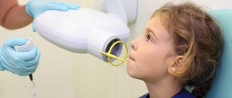
Examination of children
Dental x-rays are often prescribed for young children. You should not be afraid of this, since children sometimes need such examination more than adults. Baby teeth are often subject to carious problems, and caries appears in inaccessible places. As with an adult, the dentist cannot fully assess the condition and depth of the problem in a child. To accurately assess the situation, treat and prevent diseases, X-rays are needed. Also, a photo of baby teeth helps determine the process of eruption of molars, the quality of bone tissue, and the formation of a bite. In childhood, it is easiest to prevent improper formation of the dentition. If such problems are not resolved in time, in later life this will happen longer and more severely.
For small patients, only minimal doses of radiation are used. The doctor builds treatment and diagnostic tactics so as not to expose the child to radiation again. The procedure is the same as for adults: the rest of the body is covered with a protective apron, the session lasts a couple of minutes, no pain or discomfort occurs. In addition, some clinics use a collimator - a tube that is attached to the device. This device shrinks the X-ray beam and changes its contour, so children receive even less radiation.
Description of the procedure
There is a certain algorithm that describes how to properly take a dental x-ray:
- the patient must remove metal jewelry;
- then he is brought to the X-ray machine and asked to bite down on the photosensitive film so that the tooth being examined is between the film and the machine;
- a picture is taken.
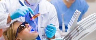
If required, the picture can be taken in a different projection. In cases where x-rays are performed using a computer radiovisiograph, the patient puts on a special apron, and then a sensor connected to the device is placed on the area of the dental system being examined. The photo is displayed on the computer.
Another option for x-rays is using an orthopantomograph. The subject stands at the device and places his chin on a special support for complete fixation. Next, he bites the block with his teeth, which will prevent the jaws from closing. Pictures are taken while the device rotates around the patient's head.
Usually the procedure takes only a few minutes, after which the finished images are described and transferred to the patient.
Dental Imaging
Extraoral imaging allows the dentist to see a complete picture of the inside of the patient's mouth. Panoramic and 3D imaging assist in diagnosis, treatment planning and evaluation of certain dental conditions.
Dental systems using flat panel detectors have the advantage of providing highly detailed images for more accurate diagnosis. Modern high-speed, high-sensitivity flat panel detectors are an attractive option for dentists and are used for imaging in:
- Periodontal bone loss or disease detection
- Extraoral exams
- Planning a dental implant
- Visualization of abnormal teeth
- Jaw and face assessment
- Cleft palate assessment, dental cavity diagnosis
- Endodontic diagnostics, including root canals
- Diagnosis of dental trauma
And also offers beam computed tomography, also known as CBCT. Dental CBCT systems allow manufacturers to optimize their system to provide fast images while reducing radiation dose to patients. CBCT provides three-dimensional information and images compared to the two-dimensional image obtained with conventional x-ray images. 3D imaging can be especially useful in diagnosis, treatment planning, and evaluation of certain dental conditions.
Our discounts and promotions